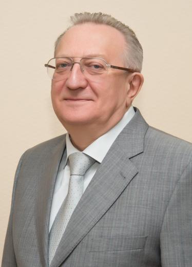
In October, our University will celebrate the 95th anniversary; we are just one step away from its centennial. The great legacy created by the students, faculty, and staff of the University lies behind it!
For almost a century, the University, like a lighthouse, has shone for those who studied, taught, created, loved, dreamed, and grew up here.
Over the course of 95 years, the University has trained top-notch professionals who make a significant contribution to the development of world science and the practice of food production technologies, the development, transportation, and processing of food resources, and in recent years to a wide range of biotechnological, veterinary, and medical science and practice.
Over the years, the University has trained about 45 thousand engineering technologists, mechanics, designers, economists, over 420 Doctors and about 3300 Candidates of Science, and more than 3600 specialists for other countries. The professors and faculty of the University are the authors of more than 600 textbooks, monographs, and fundamental scientific works, as well as a great numberof handbooks and educational manuals.
The formation and development of the University is connected with the names of outstanding scientists and educators, organizers of higher education and scientific research who have received Russian and international recognition. Among them there are the creators of prominent scientific schools that have determined the paths of science and technology development of in their fields.
Thanks to our shared determination, creative search, high professionalism, and the ability to carefully preserve and develop established traditions, we conquer new heights in Russian and international rankings. Our scientists and educators become laureates of various awards, and our students achieve great success not only in their studies but also in the social life and in the spheres unrelated to their educational profile.
Today, the University consists of seven institutes and several scientific and educational centers, where about ten thousand students, graduate students, and residents study every year.
Our Institute, the Medical Institute of Continuing Education, was established within the University structure in March 2011, by a decision of the University Academic Council, supported by the Ministry of Science and Higher Education. Currently, the Medical Institute of Continuing Education is fully integrated into the educational, scientific, international, social, and public activities of the University.
The institute coordinates the educational, methodological, tutoring, and research activities of 18 departments, and annually provides education to about one thousand residents, doctors, pharmacists, and specialists with secondary medical and pharmaceutical education.
Today, Russian Biotechnological University (ROSBIOTECH) faces new, even more ambitious challenges in personnel training and conducting scientific research aimed at ensuring biotechnological and technological leadership. The best thorough preparation for celebrating our first centenary is the major achievements outlined in the Institute’s development strategy. I wish all of us and our dear University continued prosperity, and may these 95 years of history become a bright milestone in the many future centennial chapters of its fruitful life.
Best regards, Editor-in-Chief, MD, PhD Professor V.V. Gladko
METHOD OF EXTENDED DESYMPATHIZATION WITH SIMULTANEOUS AMPUTATION IN ATHEROSCLEROTIC GANGRENE OF THE FOOT CAUSED BY RESTENOSIS OF THE VASCULAR REVASCULARIZATION ZONE
1 City Clinical Hospital No. 29, Moscow, Russia 2 Kabardino-Balkarian State University, Nalchik, Russia 3 Sechenov First Moscow State Medical University, Moscow, Russia 4 JSC "Concern GRANIT", Moscow, Russia
ABSTRACT
Background. Currently, it is relevant to evaluate the results of extended desympathization with simultaneous amputation of the lower limb with restenosis of its main arteries at various levels.
Materials and methods. A prospective, non-randomized study involved 30 people who underwent repeated vascular reconstruction. All patients had gangrene of the toes without spreading to the tarsal. Group 1 (n=18) underwent extended desympathization after repeated reconstructive or endovascular surgery on the arteries of the lower limb. Group 2 (n=12) received repeated arterial reconstruction of the restenosis zone, with delayed amputation on the 6th to 7th postoperative day allowing for the formation of a clearly demarcated line with necrotic tissues. The average age of the patients was 58.3±0.5 years. The follow-up period was 6 months.
Results. When performing amputation at various levels of the foot, its border was separated from the necrosis zone in the first group by an average of 5.01±0.11 cm; while in the second group it was 3.21±0.25 cm (p<0.05). In the experimental group, the early postoperative period was uneventful. In the control group, suppuration of the amputation stump was noted in 5 patients (41.6%), which in 3 cases (25%) required amputation at the shin level. The number of re-amputations in the groups was comparable according to the results of the 6-month follow-up. The time to complication onset was: group 1–136.3±0.5 days; group 2–98.1± 0.6 days; p<0.05. Purulent complications and repeated hospitalizations were more common in the control group (group 1–3.4±0.1 hospitalizations; group 2–6.5±0.2 hospitalizations; p<0.05). The lumen diameter of small arteries at the resection edges in the experimental group was 0.83±0.01 mm2, whereas in the control group it was 0.44±0.02 mm2 (p<0.05).
Conclusions. In case of repeated vascular interventions caused by restenosis of primary revascularization zones, it is effective to perform extended desympathization of the lower limb in combination with simultaneous amputation of the toes with their atherosclerosclerotic gangrene.
CORRESPONDENCE: Alexey G. Vaganov, e-mail: aleksejvaganov4@gmail.com
FOR CITATIONS: Vaganov A.G., Aslanov A.Sh., Kuznetsov M.R. [et al.] Method of Extended Desympatization with Simulteneous Amputation in Atherosclerotic Gangrene of the Foot Caused by Restenosis of the Vascular Revascularization Zone // Bulletin of the Medical Institute of Continuing Education. — 2025. — Vol. 5, No. 3. — P. 8–13. — DOI 10.36107/2782–1714_2025–5-3–8-13
FUNDING SOURCE: The authors declare no funding for the study.
DECLARATION OF COMPETING INTEREST: The authors declare no apparent or potential conflicts of interest related to the publication of this article.
DATA AVAILABILITY STATEMENT: Data from the current study are available from the corresponding author upon reasonable request.
LITERATURE
Гаибов А.Д. и др. Эндоваскулярные методы реканализации в лечении хронической ишемии нижних конечностей: современное состояние проблемы // Российский медико-биологический вестник имени академика И. П. Павлова. — 2023. — Т. 31 — № 2. — С. 305–316.
Малаев Д.У., Таркова А.Р., Прохорихин А.А. и др. Современное состояние проблемы эндоваскулярного лечения бедренно-подколенного сегмента // Патология кровообращения и кардиохирургия. — 2020. — Т. 24 — № S3. — С. 10–17.
Шумаков Д.В., Шехян Г.Г., Зыбин Д.И. и др. Рестеноз стента: клиника, гемодинамические проявления, механизмы развития и возможности коррекции // Кардиологический вестник. — 2021. — Т. 16 — № 1. — С. 20 27.
Altin S.E., Kim Y.G., Aronow H.D. et al. Seasonal variation in U.S. hospitalizations for chronic limb-threatening ischemia // Catheter Cardiovasc Interv. — 2020. — Vol. 96. — № 7. — P. 1473–1480. doi: 10.1002/ccd.29261.
Закеряев А.Б., Виноградов Р.А., Сухоручкин П.В. и др. Отдаленные результаты нового способа бедренно-подколенного шунтирования // Российский кардиологический журнал. — 2022. — Т. 27. — № 8. — С. 32–41. https://doi.org/10.15829/1560–4071-2022–4843
Abou-Zamzam А., Gomez N., Molkara A. A prospective analysis of critical limb ischemia: factors leading to major primary amputation versus revascularization // Annals of vascular surgery. — 2007. — Vol. 21. — № 4. — Р. 458–463.
Ткаченко А.Н., Бахтин М.Ю., Жарков А.В. и др. Прогноз летальных исходов при проведении ампутаций нижней конечности у больных пожилого и старческого возраста // Фундаментальные исследования. — 2011. — № 9–2. — С. 304–308.
Чайковский В.В. Хроническая ишемия нижних конечностей у лиц старше 70 лет после инфраингвинального рентгенэндоваскулярного вмешательства // Вестник медицинского института «Реавиз»: реабилитация, врач и здоровье. — 2022. — Т. 2. — № 56. — С. 143–143.
Гавриленко, А. В., Котов, А. Э., Шаталова, Д. В. Механизмы развития осложнений эндоваскулярного лечения артерий нижних конечностей и их хирургическая коррекция // Анналы хирургии. — 2013. — № 5. — С. 27–35.
Переходов С. Н., Карпун Н. А., Сницарь А. В. и др. Опыт применения стентирования артерий голени в лечении критической ишемии нижних конечностей // Кремлевская медицина. Клинический вестник. — 2021. — Т.4. — С. 70–77.
Липатов К.В., Пермяков С.В., Асатрян А.Г., Бородин А.В., Гостищев В.К. Рентгеноэндоваскулярная хирургия при критической ишемии нижних конечностей с гнойно-некротическим поражением стоп // Хирургия. Журнал им. Н.И. Пирогова. — 2017. — № 9. — С. 4 16.
Ahn J., Raspovic K.M., Liu G.T. Lower Extremity Necrotizing Fasciitis in Diabetic and Nondiabetic Patients: Mortality and Amputation // Int. J. Low. Extrem Wounds. — 2019. — Vol.18. — № 2. — Р.114–121. doi: 10.1177/1534734619836464.
Zambetti B.R., Stiles Z.E., Gupta P.K. Analysis of Early Lower Extremity Re-amputation.// Ann Vasc Surg. — 2022. — № 81. — P. 351–357. doi: 10.1016/j.avsg.2021.10.030.
Червяков Ю.В., Ха Х.Н., Климов А.Е., Гавриленко А.В. Оценка результатов комплексного консервативного лечения пациентов с декомпенсированной ишемией нижних конечностей в сроки до 1 года с использованием системной классификации WifI // Кардиология и сердечно-сосудистая хирургия. — 2020. — Т. 13. — № 4. — С.347 354.
Никольский, В. И., Микуляк, А. И., Темников, В. А. Сравнительная оценка параметров микроциркуляции после поясничной ганглионарной симпатэктомии и расширенной десимпатизации. Известия высших учебных заведений // Поволжский регион. Медицинские науки. — 2012. — № 2. — С. 56–61.
Шор Н.А., Жаданов В.И. Показания к проведению поясничной симпатэктомии и прогнозирование ее исхода у пациентов с облитерирующим атеросклерозом артерий нижних конечностей при хронической критической ишемии тканей // Клиническая хирургия. — 2009. — № 10. — С.25–28.
Яриков А.В., Леонов В.А., Шпагин М.В. и др. Симпатэктомия и нейромодуляция в лечении критической ишемии нижних конечностей // Журнал им. Н. В. Склифосовского «Неотложная медицинская помощь». — 2022. — Т. 11. — № 1. — С. 158–167.
Ваганов А.Г., Ночной М.С., Лисицкий Д.А. и др. Результаты проксимальной гибридной артериальной реконструкции в сочетании с одномоментной ампутацией при сухой атеросклеротической гангрене пальцев стопы // Российский медико-биологический вестник им. академика И.П. Павлова. — 2024. — Т. 32. — № 3. — C. 389–400. doi: 10.17816/PAVLOVJ627546
ABOUT THE AUTHORS
Ваганов Алексей Геннадьевич — к.м.н., врач-хирург, хирургическое отделение № 1, ГБУЗ "ГКБ им Н.Э. Баумана" ДЗМ, SPIN-код: 2202-0746, ORCID: 0000-0001-8191-2551
Асланов Ахмед Дзонович — д.м.н., профессор, профессор кафедры медицинской академии ФГБОУ ВО «КБГУ им. Х.М. Бербекова». Россия, Нальчик, ORCID: 0000-0002-7051-0917, SPIN код: 1452-7020.
Кузнецов Максим Робертович — д.м.н., профессор, заместитель директора Института кластерной онкологии имени Л.Л. Левшина, ФГАОУ ВО Первый Московский государственный медицинский университет имени И.М. Сеченова. Россия, Москва, ORCID: 0000-0001-6926-6809; mrkuznetsov@mail.com.
Косенков Александр Николаевич — д.м.н., профессор кафедры госпитальной хирургии Института клинической медицины им Н.В. Склифосовского, ФГАОУ ВО Первый Московский государственный медицинский университет имени И.М. Сеченова. Россия, Москва, ORCID: 0000-0002-6975-5802;
Бетуганова Алина Латифовна — ассистент кафедры нормальной и патологической анатомии человека медицинской академии ФГБОУ ВО «КБГУ им. Х.М. Бербекова». Россия, Нальчик, ORCID: 0000-0002-5228-870Х.
Галкина Екатерина Анатольевна — начальник лаборатории электробиофизических и химических исследований АО«Концерн ГРАНИТ», ORCID:0000-0003-3824-2577
AUTHOR CONTRIBUTIONS
А.Г. Ваганов, А.Д. Асланов, А.Н. Косенков — концепция статьи
А.Г. Ваганов, М.Р. Кузнецов, А.Д. Асланов — концепция и дизайн исследования
А.Г. Ваганов — написание текста
А.Л. Бетуганова, Е.А. Галкина — сбор и обработка материала
А.Н. Косенков, А.Л. Бетуганова, Е.А. Галкина — обзор литературы
А.Г. Ваганов, А.Д. Асланов, М.Р. Кузнецов — анализ материала
INFORMED CONSENT FOR PUBLICATION: authors received written consent from patients for publication of medical data and photographs.
COMPLIANCE WITH ETHICAL PRINCIPLES: Проведенное исследование соответствует стандартам Хельсинкской декларации (Declaration Helsinki) в редакции 2024 г. Соответствие протокола исследования этическим принципам было подтверждено Локальным этическим комитетом ФГБОУ ВО "Кабардино-Балкарский государственный университет им. Х.М. Бербекова" от 04.03.2024, протокол № 05/24
1 Medical Institute of Continuing Education, Russian Biotechnology University (ROSBIOTECH), Moscow, Russia 2 EVA CLINIC, Moscow, Russia 3 New Medicine (VIP-clinic), Moscow, Russia 4 V.I. Vernadsky Crimean Federal University, Simferopol, Russia
ABSTRACT
Background. Rectus abdominis diastasis is an urgent problem affecting both the aesthetic and functional state of the anterior abdominal wall.
Purpose. To analyze modern methods of the correction of diastasis of the rectus abdominis muscles (DRAM).
Main content. The review provides a detailed analysis of the current approaches to the surgical treatment of DRAM. Special attention is given to:
comparative analysis of open and minimally invasive techniques
criteria for choosing the correction method
specifics of useing mesh implants
prevention of postoperative complications
The material is based on the latest clinical guidelines (EHS 2023, the Russian Society of Herniologists, 2022) and evidence-based medicine data obtained from the analysis of foreign and Russian scientific articles.
Conclusions. The variety of modern approaches makes it possible to individualize the tactics of treating rectus abdominis diastasis depending on the clinical situation. The most predictable results are achieved when using combined and minimally invasive techniques in accordance with current clinical guidelines.
CORRESPONDENCE: Larisa I. Folomeeva, e-mail: dr.folomeeva@mail.ru
FOR CITATIONS: Folomeeva L.I., Pakhomova R.A., Zatsarinny V.V. [et al]. Modern Methods of Surgical Correction of Rectus Abdominis Diastasis: a Complete Systematic Review // Bulletin of the Medical Institute of Continuing Education. — 2025. — Vol. 5, No. 3. — P. 14-18. — DOI 10.36107/2782-1714_2025-5-3-14-18.
FUNDING SOURCE: The authors declare no funding for the study.
DECLARATION OF COMPETING INTEREST: The authors declare no apparent or potential conflicts of interest related to the publication of this article.
DATA AVAILABILITY STATEMENT: Data from the current study are available from the corresponding author upon reasonable request.
LITERATURE
Mommertz G., Klink C.D., Henze J. Treatment options for abdominal rectus diastasis: a comprehensive review // Plast Reconstr Surg Glob Open. — 2020. — Vol. 8. — № 11. — P. e3263.
Hidalgo D.A., Sufyan Y. Aesthetic and functional repair of abdominal wall deformities // Clin Plast Surg. — 2022. — Vol. 49. — № 1. — P. 135–147.
Gammal H.E., Khedr A.M., Saad R.A. Integrating aesthetic and reconstructive surgery: a case report // Int J Health Med Res. — 2021. URL: https://ijhmr.com/ (дата обращения: 20.06.2025).
Dur A.H.M., de Vries Reilingh T.S., Broeders I.A.M.J., Hazebroek E.J. Could abdominoplasty with diastasis recti correction improve quality of life? // Plast Reconstr Surg Glob Open. — 2023. — Vol. 11. — № 12. — С. e5302.
Nahas F.X., Ferreira L.M., Ghelfond C. Management strategies for diastasis recti // Clinics. — 2005. — Vol. 60. — № 2. — P. 201–208.
Muysoms F.E., et al. EHS guidelines for rectus diastasis repair // Br J Surg. — 2023. — Vol. 110. — № 4. — P. 345–359.
Timmermans L., et al. Abdominal wall reconstruction: new frontiers // Ann Surg. — 2023. — Vol. 278. — № 3. — P. 560–567.
Nahabedian M.Y. Management of abdominal wall laxity and contour deformities. // Aesthet Surg J. — 2021. — Vol. 41. — № 5. — P. 553–560.
Мноян А.Х., Мударисов Р.Р., Вторенко В.И. Актуальные вопросы лечения диастаза прямых мышц живота в сочетании с грыжами передней брюшной стенки // Московский хирургический журнал. — 2022. — № 1. — С. 90–98. doi.org/10.17238/2072–3180-2022–1-90–98
Зимин А.В., Чурбанов А.В., Назаров И.А. Алгоритмы хирургического лечения диастаза прямых мышц живота // Пластическая хирургия и косметология. — 2023. — № 2. — С. 15–21.
Сергеев С.Н., Морозов А.П. Хирургия диастаза: текущие подходы // Хирургия. Журнал им. Н.И. Пирогова — 2023. — № 8. — С. 18–22.
Демидов В.Н., Петрова И.А. Абдоминопластика: показания и техника // Вестник хирургии. — 2021. — Т. 180. — № 3. — С. 43–47.
Малюков А.А., Егоров В.И. Хирургическая тактика при диастазе прямых мышц живота у женщин после родов // Вестник хирургии. — 2021. — Т. 180. . — № 5. — С. 66–71.
Иванов П.Л., Мирошниченко С.И. Эстетическая абдоминопластика: новые возможности и ограничения // Российский журнал пластической хирургии. — 2023. — Т. 25. — № 2. — С. 34–41.
Агапов В.А. и соавт. Опыт применения техники REPA // Эндоскопическая хирургия. — 2023. — № 4. — С. 33–39.
Palanivelu C., et al. Laparoscopic management of rectus diastasis // JSLS. — 2021. — Vol. 25. — № 2. — P. e2021.00022.
Schmitt A., et al. Comparison of open vs laparoscopic repair // J Am Coll Surg. — 2020. — Vol. 231. — № 5. — P. 451–459.
Köckerling F. Endoscopic techniques in abdominal wall surgery // Surg Endosc. — 2020. — Vol. 34. — № 7. — P. 2966–2972.
Юрасов А.В., Ракинцев В.С., Матвеев Н.Л. и др. Методы коррекции изолированного диастаза прямых мышц живота и его сочетания с первичными срединными грыжами. Эндоскопическая хирургия. — 2020. — № 26. — № 1. — С.49–55. doi.org/10.17116/endoskop20202601149
Moore J.H., Feliciano L., Harrell A.G. Robotic-assisted repair of rectus diastasis: technique and outcomes // Aesthet Surg J. — 2024. — Vol. 44. — № 3. — P. NP275-NP283.
Belyansky I., et al. Robotic-assisted repair of rectus diastasis // Surg Endosc. — 2024. — Vol. 38. — № 1. — P. 122–130.
Berger D. Robotic hernia surgery: an overview // J Robot Surg. — 2021. — Vol. 15. — № 4. — P. 465–472.
Cuccurullo D, Guerriero L, Mazzoni G, Sagnelli C, Tartaglia E. Robotic transabdominal retromuscular rectus diastasis (r-TARRD) repair: a new approach. Hernia. — 2022. — № 26. — № 6. — P. 1501–1509. doi.org/10.1007/s10029–021-02547-w
Козлов И.Л., Степанов С.В. Применение сетчатых имплантатов при диастазе: сравнительный анализ // Российский журнал герниологии. — 2022. — №1. — С. 25–30.
Rosen M.J., et al. Mesh selection in ventral hernia repair // Hernia. — 2020. — Vol. 24. — № 6. — P. 1257–266.
Bellows C.F., et al. The evolution of mesh technology // Surg Clin North Am. — 2021. — Vol. 101. — № 1. — P. 35–49.
Pawlak M., et al. Sublay mesh placement technique: meta-analysis // World J Surg. — 2023. — Vol. 47. — № 3. — P. 595–604.
Parker S.G., et al. Long-term outcomes after diastasis recti repair // Plast Reconstr Surg. — 2022. — Vol. 149. — № 5. — P. 987–995.
Chiarello C.M., Falzone D. Diastasis Recti Rehabilitation // StatPearls- Treasure Island (FL): StatPearls Publishing. — 2024. URL: https://www.ncbi.nlm.nih.gov/books/NBK573063/
Olsson A., Kiwanuka O., Sandblom G., Stackelberg O. Long-term follow-up after surgical repair of abdominal rectus diastasis // Scandinavian Journal of Surgery. — 2021. — Vol. 110. — № 1. — P. 101–107.
Parker S.G., Mallett S., Quinn A. Early complications, pain, and quality of life after reconstructive surgery of diastasis recti // Plast Reconstr Surg. — 2014. — Vol. 134. — № 6. — P. 1230–1238.
Сергацкий К.И., Никольский В.И., Титова Е.В. и др. Диастаз прямых мышц живота: истинные вопросы диагностики и лечения неистинной грыжи // Ульяновский медико-биологический журнал. — 2022. — № 2. — С. 22–37. doi: 10.34014/2227–1848-2022–2-22–37.
Клинические рекомендации Ассоциации герниологов России. Диастаз прямых мышц живота. — 2022. — 42 с.
Muysoms F.E., Miserez M., Berrevoet F. et al. European Hernia Society guidelines on management of rectus diastasis // Br J Surg. — 2021. — Vol. 108. — № 10. — P. 1189–1197.
Belyansky I., Daes J., Radu V.G. A new minimally invasive technique for the repair of diastasis recti // Surg Endosc. — 2021. — Vol. 35. — № 4. — P. 1675–1681.
ABOUT THE AUTHORS
Фоломеева Лариса Игоревна — к.м.н., доцент кафедры пластической ФГБУ МИНО «Росбиотех», http://orcid.org/0000–0002-0134–7183.
Пахомова Регина Александровна — д.м.н., доцент, заведующая кафедрой пластической хирургии ФГБУ МИНО «Росбиотех». http://orcid.org/ 0000–0002-3681–4685
Зацаринный Владимир Викторович — к.м.н, доцент кафедры пластической хирургии ФГБУ МИНО «Росбиотех». http://orcid.org/ 0009–0001-1154–2126
Анеликов Андрей Андреевич — врач пластический хирург; ООО «Новая медицина» (VIP-clinic). http://orcid.org/0000–0003-2064–3217
Ильченко Федор Николаевич — д. м. н., профессор, заведующий кафедрой хирургии № 2, Крымский федеральный университет имени В.И. Вернадского, Медицинский институт имени С.И. Георгиевского, http://orcid.org/ 0000–0003-3703–6595
Гривенко Сергей Геннадиевич — д.м.н., профессор, кафедра хирургии № 2, Крымский федеральный университет имени В.И. Вернадского, Медицинский институт имени С.И. Георгиевского, http://orcid.org/ 0000–0003-2602–0504
Честных Виктория Викторовна — врач пластический хирург — ООО «ЕВА-КЛИНИК»
AUTHOR CONTRIBUTIONS
Анеликов А.А., Фоломеева Л.И., Ефимова Н.М., Гривенко С.Г., Ильченко Ф.Н., Зацаринный В.В., Честных В.В. — обработка и систематизация материала, подбор литературы, написание текста статьи
Пахомова Р.А. — обработка и систематизация материала, подбор литературы, редактирование текста статьи
1 Main Military Clinical Hospital named after academician N.N. Burdenko, Moscow, Russia 2 Medical Institute of Continuing Education, Russian Biotechnology University (ROSBIOTECH), Moscow, Russia 3 National Medical Research Center of Endocrinology. Moscow, Russia
ABSTRACT
Background. Cerebral vascular injury (CVI) covers various forms of intra- and extracranial vascular damage to the brain resulting from head and neck trauma or injuries. This condition is considered the most severe complication of craniocerebral injuries and injuries with a high mortality rate.
Purpose. To examine through a clinical case, the possibility of using combined endovascular and endoscopic surgical methods for treating a wounded patient with cerebral vascular injury.
Materials and methods. A 45-year-old patient was admitted to the Neurosurgical Center of the N.N. Burdenko Main Military Clinical Hospital through in May 2024 via medical evacuation. He had a shrapnel craniofacial blind penetrating wound of the midface with a fracture of the facial bones and a fragment in the posterior wall of the sella turcica. According to selective angiography, the wounded patient had a pinpoint injury to the intracavernous part of the left internal carotid artery (ICA).
Results. The patient underwent endovascular trapping occlusion of the cavernous part of the left ICA due to bleeding. Then, endoscopic transnasal removal of a metal fragment from the anterior part of the left cavernous sinus was performed. The measures taken allowed to stabilize the patient's condition for a short time, however, due to the development of ischemic and secondary infectious complications, the patient died on the 33rd day after the injury.
Conclusions. Based on the presented clinical case, we would like to focus on the most important aspects of urgent neurosurgical care for injuries accompanied by damage to the cerebral vessels.
FOR CITATIONS: Gizatullin Sh.Kh., Isengaliev I.N., Kovzel V.A. et al. Combined Treatment of a Patient with a Gunshot Cranifacial Injury: a Clinical Case // Bulletin of the Medical Institute of Continuing Education. — 2025. — Vol. 5, No. 3. — P. 19-24. — DOI 10.36107/2782-1714_2025-5-3-19-24
FUNDING SOURCE: The authors declare no funding for the study.
DECLARATION OF COMPETING INTEREST: The authors declare no apparent or potential conflicts of interest related to the publication of this article.
DATA AVAILABILITY STATEMENT: Data from the current study are available from the corresponding author upon reasonable request.
LITERATURE
Begum M. et al. Management of Head and Neck Pseudoaneurysms: an Institutional Review //Stroke: Vascular and Interventional Neurology. — 2024. — Vol. 4. — P. e12984_324. https://doi.org/10.1161/SVIN.04.suppl_1.324
Cai M. J. et al. Treatment strategies for traumatic cervico-cranial pseudoaneurysms: a single institution experience //Neurology India. — 2018. — Vol. 66. — № 1. — P. 105–114. https://doi.org/10.4103/0028–3886.222873
Dawoud F. M. et al. Traumatic cerebrovascular injuries associated with gunshot wounds to the head: a single-institution ten-year experience // World neurosurgery. — 2021. — Vol. 146. — P. e1031-e1044. https://doi.org/10.1016/j.wneu.2020.11.078
Станишевский А.В. и др. Травматическая окклюзия задней мозговой артерии с ишемией затылочной доли // Журнал неврологии и психиатрии им. C.C. Корсакова. — 2021. — Т. 121. — № 12. — С. 50–55. https://doi.org/10.17116/jnevro202112112250
Гизатуллин, Ш. Х. Боевые огнестрельные ранения черепа и головного мозга // Вопросы нейрохирургии им. Н.Н. Бурденко. — 2021. — Т. 85. — № 5. — С. 124–131. DOI 10.17116/neiro202185051124. — EDN OHHKSH.
Bell R. S. et al. The evolution of the treatment of traumatic cerebrovascular injury during wartime: A review //Neurosurgical focus. — 2010. — Vol. 28. — № 5. — P. E5. https://doi.org/10.3171/2010.2.FOCUS1025
Гизатуллин, Ш. Х. Особенности огнестрельных черепно-мозговых ранений в современных войнах и вооруженных конфликтах // Медицинский вестник ГВКГ им. Н.Н. Бурденко. — 2021. — № 1(3). — С. 52–59. — EDN CCFLGF.
Santos G. et al. Traumatic middle cerebral artery aneurysm secondary to a gunshot wound //Journal of Neuroimaging. — 2013. — Vol. 23. — № 1. — P. 115–117. https://doi.org/10.1111/j.1552–6569.2010.00570.x
Гизатуллин Ш. Х. и др. Комбинированное лечение раненого с травматической аневризмой задней соединительной артерии, осложненной массивным внутрижелудочковым кровоизлиянием //Нейрохирургия. — 2024. — Т. 26. — № 3. — С. 103–111. DOI 10.17650/1683–3295-2024–26-3–103-111. — EDN VEKRBC.
Das A. S. et al. Cerebrovascular injuries in traumatic brain injury //Clinical neurology and neurosurgery. — 2022. — Vol. 223. — P. 107479. https://doi.org/10.1016/j.clineuro.2022.107479
Гизатуллин Ш.Х. и др. Опыт лечения травматических аневризм при огнестрельных черепно-мозговых ранениях // Организация оказания медицинской помощи раненым и пострадавшим: от поля боя до высоких технологий // М.: РАН, 2024. — С. 234–247.
Tunthanathip T. et al. Traumatic cerebrovascular injury: prevalence and risk factors //The American Journal of Emergency Medicine. — 2020. — Vol. 38. — № 2. — P. 182–186. https://doi.org/10.1016/j.ajem.2019.01.055
Mansour A. et al. Cerebrovascular complications in early survivors of civilian penetrating brain injury //Neurocritical care. — 2021. — Vol. 34. — № 3. — P. 918–926. https://doi.org/10.1007/s12028–020-01106-y
Abutarboush R. et al. Exposure to blast overpressure impairs cerebral microvascular responses and alters vascular and astrocytic structure // Journal of neurotrauma. — 2019. — Vol. 36. — № 22. — P. 3138–3157.https://doi.org/10.1089/neu.2019.6423.
Nicolai T. et al. Pseudoaneurysms in a Penetrating Head and Neck Injury: a Case Series // International Journal For Multidisciplinary Research. — 2023; n. pag. https://doi.org/10.36948/ijfmr.2023.v05i06.10968.
Chen K., Hu P. How I do it? Microsurgical treatment of traumatic anterior cerebral artery pseudoaneurysm with cerebrospinal fluid rhinorrhea //Acta Neurochirurgica. — 2025. — Vol. 167. — № 1. — P. 153. https://doi.org/10.1007/s00701–025-06540–5
Aljuboori Z. et al. Endovascular treatment of a traumatic middle cerebral artery pseudoaneurysm with the pipeline flex embolization device // World neurosurgery. — 2020. — Vol. 133. — P. 201–204. https://doi.org/10.1016/j.wneu.2019.10.008
Fry L. et al. Endovascular management of pediatric traumatic intracranial pseudoaneurysms: a systematic review and case series //World Neurosurgery. — 2023. — Vol. 176. — P. 213–226. https://doi.org/10.1016/j.wneu.2023.04.028
Evans L. et al. Surviving sepsis campaign: international guidelines for management of sepsis and septic shock 2021 //Critical care medicine. — 2021. — Vol. 49. — № 11. — P. e1063-e1143. https://doi.org/10.1007/s00134–021-06506-y
ABOUT THE AUTHORS
Гизатуллин Шамиль Хамбалович — д.м.н, доцент, профессор кафедры хирургии повреждений с курсом военно-полевой хирургии МИНО ФГБОУ ВО «РОСБИОТЕХ» , начальник нейрохирургического центра ГВКГ им. Н.Н. Бурденко, https://orcid.org/0000–0002-2953–9902.
Исенгалиев Ильяс Нуртуганович — врач-нейрохирург 22 нейрохирургического отделения ГВКГ им. Н.Н. Бурденко, https://orcid.org/0000–0002-6444–4757.
Ковзель Виктор Алексеевич — клинический ординатор нейрохирургического центра ГВКГ им. Н.Н. Бурденко.
Чернов Валерий Евгеньевич — начальник 22 нейрохирургического отделения нейрохирургического центра ГВКГ им. Н.Н. Бурденко.
Ким Эдуард Анатольевич — начальник 66 нейрохирургического отделения нейрохирургического центра ГВКГ им. Н.Н. Бурденко.
Григорьев Андрей Юрьевич — профессор, д.м.н., заведующий нейрохирургическим отделением ФГБУ ЭНЦ МЗРФ.
Бадулина Александра Николаевна — клинический ординатор нейрохирургического центра ФГБУ «ГВКГ им. Н.Н. Бурденко». ORCID: 0009-0004-9612-3079
AUTHOR CONTRIBUTIONS
Гизатуллин Ш.Х., Исенгалиев И.Н., Ковзель В.А., Чернов В.Е., Ким Э.А., Григорьев А.Ю. — концепция и дизайн исследования, редактирование
INFORMED CONSENT FOR PUBLICATION: authors received written consent from patients for publication of medical data and photographs.
COMPLIANCE WITH ETHICAL PRINCIPLES: авторы заявляют, что все процедуры, описанные в данной статье, соответствуют этическим стандартам учреждений, проводивших исследование, и соответствуют Хельсинкской декларации в редакции 2024 г. Статья одобрена Локальным этическим комитетом ФГБОУ ВО «РОСБИОТЕХ» (Москва, Волоколамское шоссе, дом 11) протокол №11/6-5 от 9.09.2025.
OPEN STENTING OF THE PANCREATIC DUCT AS A KEY TO UNDERSTANDING THE GENESIS OF CYSTIC-INFLAMMATORY TRANSFORMATION OF THE DUODENUM IN CHRONIC PANCREATITIS
1 Medical Institute of Continuing Education, Russian Biotechnological University (ROSBIOTECH), Moscow, Russia 2 Saint Petersburg State Pediatric Medical University, Saint Petersburg, Russia. 3 Saint Petersburg City Hospital №15, Saint Petersburg, Russia. 4 Mytishchi Regional Clinical Hospital, Mytishchi, Moscow Region, Russia.
ABSTRACT
Background. In the study, analyzing the genesis of duodenal passage disorders in chronic pancreatitis (CP), primarily cystic-inflammatory transformation (CIT) of the duodenum, a parallel is drawn with duodenostasis, which has a similar genesis of duodenal passage disorders.
Purpose. Establishing the genesis of CIT of the duodenum, which has not yet been determined, allows to develop its pathogenetically justified surgical treatment and avoid the currently predominating extremely aggressive surgical approaches.
Materials and methods. The key to understanding the genesis of CIT of the duodenum in CP was the results of open pancreatic duct stenting and follow-up observation of patients who underwent this procedure, as well as a study of the literature related to the surgery, anatomy, embryology, physiology, pathophysiology, and endocrinology of the pancreas and duodenum.
Results. The principal result of this study is a hypothesis of the origin and development of CIT of the duodenum in CP and its pathogenetically justified surgical treatment based on this hypothesis.
Conclusions. The conclusion of the study allows to avoid aggressive surgical treatment of CIT of the duodenum in favor of an organ-preserving approach significantly improving the results of surgical treatment for complicated forms of CP.
KEYWORDS: chronic pancreatitis, cystic-inflammatory transformation of the duodenum, open pancreatic duct stenting, diffuse endocrine system, immune-neuroendocrine dysfunction and dysregulation of the duodenum
CORRESPONDENCE: Nina M. Zvolskaya,e-mail: nina-zvolskaja@mail.ru
FOR CITATIONS: Zubritskiĭ V.F., Zvolskaya N.M., Khanevich M.D. [et al.] Open Stenting of the Pancreatic Duct as a Key to Understanding the Genesis of Cystic-Inflammatory Transformation of the Duodenum in Chronic Pancreatitis // Bulletin of the Medical Institute of Continuing Education. — 2025. — Vol. 5, No. 3. — P. 25-37. — DOI 10.36107/2782-1714_2025-5-3-25-37.
FUNDING SOURCE: The authors declare no funding for the study.
DECLARATION OF COMPETING INTEREST: The authors declare no apparent or potential conflicts of interest related to the publication of this article.
DATA AVAILABILITY STATEMENT: Data from the current study are available from the corresponding author upon reasonable request.
LITERATURE
Слобожанкин А.Д. Хроническая дуоденальная непроходимость (клинико-экспериментальное исследование): дисс. докт. мед. наук // Л. — 1989. — С. 476.
Максименков А. Н. Хирургическая анатомия живота // Л. — Медицина — 1972. — С. 688.
Смирнов А. В. Кистозно-воспалительная трансформация двенадцатиперстной кишки у больных хроническим панкреатитом: дисс. канд. мед. наук // Москва. — 2017. — С. 110.
Tomioka A. et al. Duodenal obstruction due to chronic pancreatitis of the pancreas tail treated by surgical intervention: A case report // Medicine. — 2019. — Vol. 98. — № 27. — P. e15856.
Stolte M. et al. A special form of segmental pancreatitis:" groove pancreatitis" // Hepato-gastroenterology. — 1982. — Vol. 29. — № 5. — P. 198–208.
Chatelain D. et al. Groove pancreatitis and pancreatic heterotopia in the minor duodenal papilla // Pancreas. — 2005. — Vol. 30. — № 4. — P. e92-e95.
Levenick J. M. et al. A comprehensive, case-based review of groove pancreatitis // Pancreas. — 2009. — Vol. 38. — № 6. — P. e169-e175.
Becker V., Mischke U. Groove pancreatitis // International journal of pancreatology. — 1991. — Vol. 10. — № 3. — P. 173–182.
Зубрицкий В.Ф., Левчук А.Л., Звольская Н.М. Хирургическое лечение нарушения дуоденальной проходимости при панкреатогенной кистозно-воспалительной трансформации двенадцатиперстной кишки // Медицинский вестник МВД. — 2019. — № 5. — С. 33–37.
Egorov VI., Petrov R., Schegolev A. et al. Pancreas-preserving duodenal resections vs pancreatoduodenectomy for groove pancreatitis. Should we revisit treatment algorithm for groove pancreatitis? // World J Gastrointest. Surg — 2021. — January — Vol. 13. — № 1. — P. 30–49.
Кригер А.Г., Смирнов А.В., Берелавичус С.В. [и др.]. Кистозная трансформация двенадцатиперстной кишки при хроническом панкреатите // Исследования и практика в медицине. — 2016. — Т. 3. — № 3. — С. 49–58.
Зубрицкий В.Ф., Звольская Н.М., Ширяев Ю.Н. [и др.]. Анализ хирургической тактики при нарушениях дуоденальной проходимости у больных хроническим панкреатитом // Вестник Медицинского института непрерывного образования. — 2024. — Т. 4. — № 4. — С. 109–114. — DOI 10.36107/2782–1714_2024–4-4–109-114.
Potet F, Duclert N. Cystic dystrophy on aberrant pancreas of the duodenal wall // Arch Fr Mal App Dig. — 1970 — Vol. 59. — № 4. — Р.223–238.
Трубицына И.Е., Чиркунова Б.З., Ткаченко Е.В. [и др.]. Патофизиология гормональных, иммунных, метаболических изменений при остром и хроническом панкреатите // Экспериментальная и клиническая гастроэнтерология. — 2008. — Т. 7. — P. 40–44
Зубрицкий В.Ф., Левчук А.Л., Звольская Н.М. Открытое стентирование панкреатического протока в лечении осложненных форм хронического панкреатита // М. — 2019. — 111 с.
Литвицкий П.Ф. Воспаление // Вопросы современной педиатрии. — 2006. — Т 5. — № 3 — С. 46–51.
Алексеева Н.Т., Никитюк Д.Б., Кварацхелия А.Г. [и др.]. Макро-микроскопические характеристики собственных желез двенадцатиперстной кишки у людей разного возраста // Журнал анатомии и гистопатологии. — 2016. — Т. 5. — № 4. — С. 12–15.
Яглов В.В., Яглова Н.В. Новые концепции биологии диффузной эндокринной системы: итоги и перспективы ее изучения // Вестник РАМН. — 2012. — № 4. — С. 74–81.
Zvolskaya N. M., Zubritskiĭ V.F., Sachechelashvili G.L. et al. Open internal stenting of the main pancreatic duct as life-saving surgery in a critically ill patient with chronic frequently relapsing pancreatitis and pancreatic ductal
ABOUT THE AUTHORS
Зубрицкий Владислав Феликсович — д.м.н., профессор, заведующий кафедрой хирургии с курсом военно-полевой хирургии МИНО ФГБОУ ВО «РОСБИОТЕХ», главный хирург МВД России. ORCID 0000-0003-4894-2796; SPIN-код 1724-1877; Author ID 328509.
Звольская Нина Михайловна — к.м.н., преподаватель кафедры хирургии с курсом военно-полевой хирургии МИНО ФГБОУ ВО «РОСБИОТЕХ». ORCID 0000-0002-7279-4888; SPIN-код 4605-358; Author ID 980772.
Ханевич Михаил Дмитриевич — д.м.н., профессор, заведующий кафедрой госпитальной хирургии ФГБОУ ВО «СПбГПМУ» Минздрава РФ, Санкт-Петербург. Author ID 464792.
Ширяев Юрий Николаевич — к.м.н., врач-хирург 5-го хирургического отделения Городской больницы № 15 г. Санкт-Петербург. ORCID 0000-0001-8204-7774; SPIN-код 4896-0772; Author ID 481972.
Юрьев Роман Алексеевич — врач-рентгенолог, ГБУЗ Московской области «Мытищинская областная клиническая больница». ORCID 0009-0004-7287-2163.
AUTHOR CONTRIBUTIONS
Зубрицкий В.Ф., Звольская Н.М. — концепция и дизайн исследования, написание текста
Звольская Н.М., Юрьев Р.А. — сбор материала
Ханевич М.Д., Ширяев Ю.Н. — редактирование, перевод
ИНФОРМИРОВАННОЕ СОГЛАСИЕ НА ПУБЛИКАЦИЮ: авторы получили письменное согласие пациентов на публикацию медицинских данных и фотографий.
СООТВЕТСТВИЕ ПРИНЦИПАМ ЭТИКИ: авторы заявляют, что все процедуры, описанные в данной статье, соответствуют этическим стандартам учреждений, проводивших исследование, и соответствуют Хельсинкской декларации в редакции 2024 г. Статья одобрена Локальным этическим комитетом ФГБОУ ВО «РОСБИОТЕХ» (Москва, Волоколамское шоссе, дом 11) протокол №11/6-5 от 9.09.2025.
INFORMED CONSENT FOR PUBLICATION: authors received written consent from patients for publication of medical data and photographs.
COMPLIANCE WITH ETHICAL PRINCIPLES: авторы заявляют, что все процедуры, описанные в данной статье, соответствуют этическим стандартам учреждений, проводивших исследование, и соответствуют Хельсинкской декларации в редакции 2024 г. Статья одобрена Локальным этическим комитетом ФГБОУ ВО «РОСБИОТЕХ» (Москва, Волоколамское шоссе, дом 11) протокол №11/6-5 от 9.09.2025.
DIAGNOSTIC VALUE OF SOME INFLAMMATORY MARKERS OF AS EARLY PREDICTORS OF ACUTE PERITONITIS IN PATIENTS WITH HIV INFECTION
A.P. Faller1,2, A.V. Kurnosov1
38-47
1 Infectious Clinical Hospital №2, Moscow, Russia 2 Medical Institute of Continuing Education, Russian Biotechnological University (ROSBIOTECH), Moscow, Russia
ABSTRACT
Background. The course of peritonitis in HIV-infected patients is blurred and its diagnosis is not always easy, which determines the urgency of searching for potential laboratory predictors.
Purpose. Assessment of the diagnostic value of leukocyte levels, neutrophil-lymphocyte ratio (NLR), procalcitonin and C-reactive protein as predictors of acute peritonitis in patients with HIV infection.
Materials and methods. The study included 158 HIV-infected patients with a preliminary diagnosis of acute peritonitis. Before surgery, leukocytes, procalcitonin, and C-reactive protein, as well as the immune status and viral load of HIV RNA, were determined in the peripheral blood of patients, and NLR were calculated. All patients underwent diagnostic laparoscopy, according to the results of which they were divided into the main and control groups. 83 patients were selected in the main group, in whom the diagnosis of acute peritonitis was confirmed. The control group included 75 patients whose diagnosis of acute peritonitis was not confirmed. Depending on the results of the immune status, the patients in each group are divided into four subgroups. The analysis of the diagnostic value of leukocyte levels, NLR, procalcitonin and C-reactive protein indicators as predictors of acute peritonitis in patients with HIV infection was carried out and the dependence of these inflammatory markers on immune deficiency and the severity of the viral load of HIV RNA was studied.
Results. There is a direct dependence of the leukocyte level on the immune status of the studied category of patients. The sensitivity and specificity of this laboratory test in the diagnosis of acute peritonitis are of low importance. The indicators of procalcitonin and C-reactive protein when compared in the main and control groups did not correlate with the severity of immunodeficiency. When studying the diagnostic test data, excellent quality of the data analysis model, high sensitivity and specificity indicators were obtained. The study shows that NLR do not correlate with the level of immune status, however, only a satisfactory quality of the data analysis model was obtained, and sensitivity and specificity have the lowest values in the presented study. The thresholds of this indicator in the study cohort are also not completely clear, since half of the patients in the control group exceeded the cut-off point obtained as a result of the ROC-analysis.
Conclusions. The level of peripheral blood leukocytes in the diagnosis of acute peritonitis in people living with HIV is of minor importance. The role of the NLR indicator in the studied category of patients is not entirely clear, and additional research is needed for its effective use in the diagnosis of acute peritonitis in patients with HIV infection. Procalcitonin and C-reactive protein can be used as laboratory predictors of acute peritonitis in HIV-positive patients.
KEYWORDS: leukocytes, neutrophil-lymphocyte ratio, procalcitonin, C-reactive protein, peritonitis, HIV infection
CORRESPONDENCE: Aleksei P. Kurnosov, e-mail: dr-kurnosov@mail.ru
FOR CITATIONS: Faller A.P., Kurnosov A.V. Diagnostic Value of Some Inflammation markers as Early Predictors of Acute Peritonitis in Patients with HIV Infection // Bulletin of the Medical Institute of Continuing Education. — 2025. — Vol. 5, № 3. — P. 38-47. — DOI 10.36107/2782-1714_2025-5-1-38-47
FUNDING SOURCE: The authors declare no funding for the study.
DECLARATION OF COMPETING INTEREST: The authors declare no apparent or potential conflicts of interest related to the publication of this article.
DATA AVAILABILITY STATEMENT: Data from the current study are available from the corresponding author upon reasonable request.
LITERATURE
Фаллер А.П., Курносов А.В. Особенности клинической картины острого перитонита у пациентов с ВИЧ-инфекцией // Вестник Медицинского института непрерывного образования. — 2024. — Т. 4. — № 3. — С. 92–100. https://doi.org/10.36107/2782–1714_2024–4-3–92-100.
Фаллер А.П., Курносов А.В., Сундуков А.В. Роль некоторых маркеров воспаления в диагностике острого перитонита у пациентов с ВИЧ-инфекцией // Хирургия. Журнал им. Н.И. Пирогова. — 2024. — Т.12. — С. 29–37. https://doi.org/10.17116/hirurgia202412129
Основы медицинской статистики. Дизайн биомедицинских исследований: практическое руководство. Жильцов И.В., Семенов В.М., Зенькова К.С. // Витебск: ВГМУ. — 2014. ISBN 978–985-466–677-8
Bone R. C. et al. Definitions for sepsis and organ failure and guidelines for the use of innovative therapies in sepsis //Chest. — 1992. — V. 101. — № 6. — P. 1644–1655. https://doi.org/10.1378/chest.101.6.1644.
Buonacera A. et al. Neutrophil to lymphocyte ratio: an emerging marker of the relationships between the immune system and diseases //International journal of molecular sciences. — 2022. — V. 23. — № 7. — P. 3636. https://doi.org/10.3390/ijms23073636.
Zahorec R. Neutrophil-to-lymphocyte ratio, past, present and future perspectives //Bratisl Lek Listy. — 2021. — V. 122. — № 7. — P. 474–488. https://doi.org/10.4149/BLL_2021_078.
Saridas A. et al. Comparison of the ability of newly inflammatory markers to predict complicated appendicitis //Open Medicine. — 2024. — V. 19. — № 1. — P. 20241002. https://doi.org/10.1515/med-2024–1002.
Seyedi S. A. et al. Neutrophil to lymphocyte ratio and spontaneous bacterial peritonitis among cirrhotic patients: a systematic review and meta‐analysis //Canadian Journal of Gastroenterology and Hepatology. — 2022. — V. 2022. — № 1. — P. 8604060. https://doi.org/10.1155/2022/8604060.
Song M. et al. Neutrophil-to-lymphocyte ratio and mortality in the United States general population //Scientific reports. — 2021. — V. 11. — № 1. — P. 464. https://doi.org/10.1038/s41598–020-79431–7
Karakonstantis S. et al. Pitfalls in studies of eosinopenia and neutrophil-to-lymphocyte count ratio //Infectious diseases. — 2018. — V. 50. — № 3. — P. 163–174. https://doi.org/10.1080/23744235.2017.1388537.
Серебровская Л. В. и др. Изучение показателей врожденного и приобретенного клеточного иммунитета периферической крови пациентов с вич-инфекцией //Клиническая лабораторная диагностика. — 2020. — Т. 65. — № 1. — С. 24–28.
Deng L. et al. Higher Neutrophil‐to‐Lymphocyte Ratio (NLR) Is a Preoperative Inflammation Biomarker of Poor Prognosis in HIV‐Infected Patients with Colorectal Cancer: A Retrospective Study //Canadian Journal of Gastroenterology and Hepatology. — 2023. — V. 2023. — № 1. — P. 7966625. https://doi.org/10.1155/2023/7966625.
Перитонит и абдоминальный сепсис: руководство для врачей. Лебедев Н.В., Климов А.Е., Персов М.Ю. // М.: ГЕОТАР-Медиа. — 2024. ISBN 978–5-9704–8069-4. https://doi.org/10.33029/9704–8069-4-PAS-2024–1-168
Arbutina D. D. et al. Significance of biomarkers in early diagnosis of abdominal sepsis //Chirurgia (Bucur). — 2022. — V. 117. — № 1. — P. 30–36. https://doi.org/10.21614/chirurgia.2660. PMID: 35272752.
Гараев М. Р. и др. Эндоваскулярная терапия в комплексном лечении распространенного гнойного перитонита //Креативная хирургия и онкология. — 2024. — Т. 14. — № 1. — С. 36–41. https://doi.org/10.24060/2076–3093-2024–14-1–36-41.
Sartelli M. et al. Duration of antimicrobial therapy in treating complicated intra-abdominal infections: a comprehensive review // Surgical Infections. — 2016. — V. 17. — № 1. — P. 9–12. http://doi.org/10.1089/sur.2015.130.
Dolin H. H. et al. A novel combination of biomarkers to herald the onset of sepsis prior to the manifestation of symptoms //Shock. — 2018. — V. 49. — № 4. — P. 364–370. http://doi.org/10.1097/SHK.0000000000001010.
ABOUT THE AUTHORS
Фаллер Александр Петрович — д.м.н., профессор кафедры хирургии повреждений МИНО ФГБОУ ВО РОСБИОТЕХ, заведующий хирургическим отделением ГБУЗ «Инфекционная клиническая больница №2 Департамента здравоохранения города Москвы». ORCID: 0009-0008-2304-9743, eLIBRARY SPIN-код: 1783-1364, eLIBRARY AuthorID: 1183722
Фаллер А.П. — концепция и дизайн статьи, редактирование статьи, одобрение окончательной версии статьи.
Курносов А.В. — анализ литературы, сбор и обработка материала, статистические расчеты, написание текста статьи.
ИНФОРМИРОВАННОЕ СОГЛАСИЕ НА ПУБЛИКАЦИЮ: авторы получили письменное согласие пациентов на публикацию медицинских данных и фотографий.
СООТВЕТСТВИЕ ПРИНЦИПАМ ЭТИКИ: авторы заявляют, что все процедуры, описанные в данной статье, соответствуют этическим стандартам учреждений, проводивших исследование, и соответствуют Хельсинкской декларации в редакции 2024 г. Статья одобрена Локальным этическим комитетом ФГБОУ ВО «РОСБИОТЕХ» (Москва, Волоколамское шоссе, дом 11) протокол №11/6-5 от 9.09.2025.
INFORMED CONSENT FOR PUBLICATION: authors received written consent from patients for publication of medical data and photographs.
COMPLIANCE WITH ETHICAL PRINCIPLES: авторы заявляют, что все процедуры, описанные в данной статье, соответствуют этическим стандартам учреждений, проводивших исследование, и соответствуют Хельсинкской декларации в редакции 2024 г. Статья одобрена Локальным этическим комитетом ФГБОУ ВО «РОСБИОТЕХ» (Москва, Волоколамское шоссе, дом 11) протокол №11/6-5 от 9.09.2025.
1 National Medical Research Center named after V.A. Almazov, Saint Petersburg, Russia 2 Saint Petersburg State Pediatric Medical University, Saint Petersburg, Russia 3 Medical Institute of Continuing Education, Russian Biotechnological University (ROSBIOTECH), Moscow, Russia. 4 Сlinical and Research Institute of Emergency Pediatric Surgery and Trauma – Dr. Roshal's Clinic (CRIEPST), Moscow, Russia. 5 State Research Institute of Applied Problems, Saint Petersburg, Russia
ABSTRACT
Background. Varicose veins of the pelvic veins remain an insufficiently studied disease and the causes of its development are not always obvious. Anomalies in the structure, shape and location of the renal vessels occur during the embryonic development of the vascular system. Congenital changes in the renal veins are most often associated with developmental disorders of the inferior vena cava into which they flow. Compression stenosis of the left renal vein (LRV) can exist in two clinical and morphological forms: the Nutcracker phenomenon and syndrome. Nutcracker syndrome is a rare disease, and according to clinical manifestations, it is not diagnosed enough to determine the difference between the "syndrome" and the "phenomenon". In addition to laboratory and urological diagnostics of the disease, biomedical imaging methods play a significant role in diagnosing the disease.
Purpose. To acquaint medical specialists with the clinical and diagnostic aspects of the Nutcracker phenomenon and syndrome using biomedical imaging methods.
Materials and methods. The Internet search was used to select literature. The analysis included 21 articles, 11 of which belong to foreign authors.
Results. Upper-level arteriovenous conflicts, in particular the Nutcracker syndrome, can lead to pelvic venous congestion and the development of varicose veins of the pelvis. Timely detection of arteriovenous compression syndromes using biomedical imaging, as well as an assessment of their hemodynamic and clinical significance, will allow for bypass surgery, which will prevent the development of complications, improve the quality of life of patients and reduce the burden on the healthcare system.
Conclusions. Using biomedical imaging methods, it is possible to identify congenital anomalies of the renal vessels, determine the relative position of the LPV relative to the aorta and superior mesenteric artery (SMA), and measure the angle of departure of the SMA. Currently, the following biomedical imaging methods are used to diagnose and assess the significance of arteriovenous conflicts, in particular, the Nutcracker syndrome:
ultrasound angioscanning (including with contrast enhancement, as well as intravascular ultrasound);
CT and CT angiography;
MRI and MR angiography;
direct X-ray phlebography (including with phlebotonometry).
KEYWORDS: Nutcracker symptom and syndrome, pathology, biomedical imaging, diagnosis, treatment
CORRESPONDENCE: Valery P. Kutsenko, е-mail: val9126@mail.ru
FOR CITATIONS: Kovaleva D.D., Kutsenko V.P., Postanogov R.A., Vlasenko A.V., Nosov A.V., Klimenko P.Yu. Clinical and Diagnostic Aspects of the Nutcracker Phenomenon and Syndrome using Biomedical Visualization Methods // Bulletin of the Medical Institute of Continuing Education. — 2025. — Vol. 5, No. 3. — P. 48-58. — DOI 10.36107/2782-1714_2025-5-3-48-58
FUNDING SOURCE: The authors declare no funding for the study.
DECLARATION OF COMPETING INTEREST: The authors declare no apparent or potential conflicts of interest related to the publication of this article.
DATA AVAILABILITY STATEMENT: Data from the current study are available from the corresponding author upon reasonable request.
LITERATURE
Берлизева О.Ю., Абоян И.А., Усенко Е.Е. [и др.] Значение ультразвукового исследования при варикозной болезни вен малого таза на фоне артерио-венозных конфликтов у мужчин // Урология. — 2020. — № 5. — С. 297–298.
Берлизева О.Ю., Абоян И.А., Амосов А.В. [и др.]. Комплексное ультразвуковое исследование в диагностике и дифференциальной диагностике типов варикозной болезни таза у мужчин с СХТБ (Материалы «XXI конгресса Российского общества урологов») // Урология. — 2021. — № 5. — С. 455.
Прокопьева Э.Д., Козловская О.В., Лопатина С.А. [и др.]. Синдром щелкунчика в практике детского нефролога //Children's medicine of the North-West. — 2021. — Т. 9. — № 4. — С. 71–76.
Мухтарулина С.В., Каприн А.Д., Асташов В.Л. [и др.]. Клиническое значение вариантов строения нижней полой вены и брюшного отдела аорты в хирургии забрюшинного пространства // Андрология и генитальная хирургия. — 2014 . — Т. 3. — С. 52–61.
Стюарт Б.Х., Рейман Г. Левопочечная венозная гипертензия при синдроме щелкунчика. Проводится путем прямой ренокавальной реимплантации // Урология. — 1982. — Т. 20. — С. 365–369.
Kim S.H., Cho S.W., Kim H.D. [et al.] Nutcracker syndrome: diagnosis with Doppler US // Radiology. — 1996. — Vol. 198. — № 1. — P. 93–97.
Felton B.M., White J.M., Racine M.A. An uncommon case of abdominal pain: superior mesenteric artery syndrome // West J Emerg Med. — 2012. — Vol. 13. — № 6. — P. 501–502.
Calado R., Braz M., Hobo L. [et al.]. Sindrome de Nutcracker: hematuria sem diagnostico // Acta Med Port. — 2011. — Vol. 24. — P. 695–698.
Ариеси А., Нагасе К. Почечная гематурия, вызванная феноменом «щелкунчика»: более логичное хирургическое лечение // Урология. — 1990. — Т. 35. — С.168–170.
Ишидоя С., Тиба Ю., Сакаи К. [и др.]. Феномен щелкунчика: случай с хирургическим лечением и его диагностические критерии // Хинекика Кие. — 1994. — Т. 40. — С. 135–138.
Рудлофф У., Холмс Р.Дж., Прем Дж.Т. [et al.]. Мезоаортальная компрессия левой почечной вены (синдром щелкунчика): отчеты о случаях и обзор литературы // Энн Васк Хирург. — 2006. — Т. 20. — С. 120–129.
Чжан Х., Ли М., Цзинь В. [et al.]. Синдром защемления левой почки: диагностика и лечение // Энн Васк Хирург. — 2007. — Т. 21. — С. 198–203.
Gozzo C., Giambelluca D., Cannella R. [et al.] CT Imaging Findings of Abdominopelvic Vascular Compression Syndromes: What the Radiologist Needs to Know // Insights Imaging. — 2020. — Vol. 17. — № 1. — P. 48
Али-Эль-Дейн Б., Осман Ю., Шехаб Эль-Дин А.Б. [и др.]. Передний и задний синдром щелкунчика: отчет об 11 случаях. Процедура трансплантации. — 2003. — Т. 35. — С. 851–853.
Ким Джи, Джох Дж.Х., Чхве Х.И. [и др.]. Транспозиция левой почечной вены при синдроме щелкунчика // Eur J Vasc Endovasc Surg. — 2006. — Т. 31. — С. 80–82.
Lamba R., Tanner D.T., Sekhon S. [et al.]. Multidetector CT of vascular compression syndromes in the abdomen and pelvis // Radiographics. — 2014. — Vol. 34. — № 1. — P. 93–115.
Охки Т., Мияхара М., Накадзима Ю. [и др.]. Синдром Щелкунчика при транспозиции левой почечной вены: клинический случай // Хинекика Кийо. — 1999. — Т. 45. — С. 183–186.
Simsek M., Burak F., Taskin O. Влияние микронизированной очищенной фракции флавоноидов на тазовую боль у женщин с лапароскопически диагностированным синдромом тазовой гиперемии: рандомизированное перекрестное исследование // Clin Exp Obstet Gynecol. — 2007. — Т. 34. — С. 96–98.
Kurklinsky A.K., Rooke T.W. Nutcracker phenomenon and nutcracker syndrome // Mayo Clin. Proc. — 2010. — Vol. 85. — P. 552–9. DOI: 10.4065/mcp.2009.0586.
Ananthan K., Onida S., Davies A.H. Nutcracker Syndrome: An Update on Current Diagnostic Criteria and Management Guidelines // Eur. J. Vasc. Endovasc. Surg. — 2017. — Vol. 53. — P. 886–94. DOI: 10.1016/j. ejvs.2017.02.015.
ABOUT THE AUTHORS
Ковалева Дарья Дмитриевна — ординатор 2-го года ФГБУ «НМИЦ им. В.А. Алмазова». ORCID: https://orcid.org/0000-0002-6236-3. SPIN-код: 9878-4125, AuthorID: 1148509.
Куценко Валерий Петрович — к.м.н. наук, доцент кафедры современных методов диагностики и радиолучевой терапии им. профессора С.А. Рейнберга ФГБОУ ВО Санкт-Петербургский государственный педиатрический медицинский университет. ORCID: https:// 0000-0001-9755-1906, SPIN код: 5760-0218, AuthorID: 841498.
Постаногов Роман Анатольевич — ассистент кафедры современных методов диагностики и радиолучевой терапии им. профессора С.А. Рейнберга ФГБОУ ВО «Санкт-Петербургский государственный педиатрический медицинский университет». ORCID:.org/0000-0002-0523-9411. SPIN: 8686-1597. AuthorID: 1181274.
Власенко Александр Владимирович — к.м.н., член-корр. МАНЭБ, профессор кафедры терапии с курсом фармакологии и фармации Медицинского института непрерывного образования ФГБОУ ВО «РОСБИОТЕХ», врач-эпидемиолог ГБУЗ «Научно-исследовательский институт неотложной детской хирургии и травматологии – Клиника доктора Рошаля» Департамента здравоохранения города Москвы. ORCID: https://orcid.org/0000-0003-4843-621X, SPIN-код: 9401-4883, AuthorID: 740391.
Носов Андрей Викторович — д.м.н., доцент, начальник лаборатории ФГУП «ГосНИИПП». SPIN код: 4910-8267, AuthorID: 176.117.129.6.
Клименко Полина Юрьевна — студентка 5 курса ФГБОУ «Санкт-Петербургский государственный педиатрический медицинский университет». ORCID: https://orcid.org/0009-0003-7055-8608. SPIN-код: 6184-4810, AuthorID: 1289404.
AUTHOR CONTRIBUTIONS
Ковалева Д.Д., Клименко П.Ю. — сбор и обработка материала, анализ литературы, подготовка и написание текста статьи.
Куценко В.П., Носов А.В. — определение концепции, цели и задачи статьи, анализ литературы, редактирование, одобрение окончательной версии статьи.
MANAGEMENT STRATEGY FOR A PATIENT WITH THE PHYMATOUS ROSACEA
E.G. Perevalova1, I.A. Lamotkin2,3
59-64
1 Clinic of Dermatology and Cosmetology "MDElena", Moscow, Russia 2 Main Military Clinical Hospital named after N.N. Burdenko, Moscow, Russia 3 Medical Institute of Continuing Education, Russian Biotechnological University (ROSBIOTECH), Moscow, Russia
ABSTRACT
Background. Patient N., 43 years old, complained of facial redness, rashes, swelling, burning of the face, hot flashes, pronounced skin hypersensitivity to stress, sun exposure, the pre-ovulation phase of her menstrual cycle. The redness was constant, intensifying in hot environments and during emotional stress. Rashes appeared periodically, the main trigger was hormonal changes, the beginning of the cycle. On examination: there was total lesion of the entire face, diffuse erythema over the forehead, cheeks, nose, chin; multiple papular, pustular, infiltrative elements; soft tissues edema of the face with an emphasis in the middle third of the face; xerosis, pronounced hyperkeratosis, signs of inflammation of the outer eye; multiple cicatricial atrophic deformities in the lateral cheeks; positive the symptom of white glasses (white foci around the eyes), a positive symptom of a leaf (a rustling sound of dry foliage when stroking the skin of the face) which is characteristic of severe skin dehydration. The more pronounced the skin xerosis is, the more pronounced the test is. On palpation, the patient was diagnosed with a sharply positive palpation test (a sharp increase in erythema at the site of palpation). Examination of the outer surface of the thighs reveals pronounced fibrosis of subcutaneous fat tissue, lymphostasis of tissues, cyanosis; on palpation, these areas were firm, fainful and cold to the touch. The therapy was performed in accordance with Invention Patent No. 2814360, as well as No. 2810361 in combination with home care. The presented clinical case of Patient N. with the phymatous rosacea is complex, demonstrates the unpredictability of the course of the disease and its variability in clinical manifestations. In case of torpid skin angioedema, it is important to enhance home care therapy for the body to achieve lymphatic drainage effect and for the face to restore the lipid mantle and relieve skin xerosis.
Purpose. To analyze a patient with the phymatous subtype of rosacea, to analyze the complexities of the clinical course of the disease, to develop a strategy for managing the patient in the clinic and at home, to determine the tactics of correcting home care products based on the dynamics of the skin condition.
Materials and methods. To analyze the treatment strategy for the phymatous subtype of rosacea, Patient N., 43 years old, was observed and managed. The patient provided information about treatment with cosmetologists, dermatologists, gastroenterologists, gynecologists before the initial medical treatment at the Dermatology and Cosmetology Clinic “MDElena”. Anti-inflammatory, seborrheic, and regenerating therapy was performed in accordance with Patent No. 2814360, as well as No. 2810361. A protocol was conducted for ophthalmorosacea to restore the eyes. The tactics of prescribing home care products were determined based on the links in the pathogenesis of the phymatous subtype of rosacea.
Results. The patient was observed regularly for 3 months at short intervals: every or two weeks, then the interval was increased to 2, 3, 6, 8 and 12 months. For the first 3 months, an active inflammatory process was observed, resolution of deep inflammatory elements was noted 5 months after the start of pathogenetic treatment (torpid clinical form), by 8 months the skin's resource and the state of the lipid mantle were fully restored, skin sensitivity to external and internal environmental factors was normalized, including hormonal changes in the body (the main trigger of the patient).
Conclusions. After anti-inflammatory, seborrheic, regenerating therapy in accordance with Patents No. 2814360 and No. 2810361, dynamic in the clinical picture of home care, all signs of a deep inflammatory process in the skin of the face and in the eye area were stopped, the lipid mantle was restored, the skin became intact.
KEYWORDS: phymatous subtype, rosacea, infiltrates, skin xerosis, facial edema, home care, rosacea treatment, pathogenetic approach, cosmeceuticals Passaggio
CORRESPONDENCE: Elena G. Perevalova, e-mail: mdelena@bk.ru.
FOR CITATIONS: Perevalova E.G., Lamotkin I.A. Management Strategy for a Patient with the Phymatous Rosacea // Bulletin of the Medical Institute of Continuing Education. — 2025. — Vol. 5, No. 3. — P. 59-64. — DOI 10.36107/2782-1714_2025-5-3-59-64
FUNDING SOURCE: The authors declare no funding for the study.
DECLARATION OF COMPETING INTEREST: The authors declare no apparent or potential conflicts of interest related to the publication of this article.
DATA AVAILABILITY STATEMENT: Data from the current study are available from the corresponding author upon reasonable request.
LITERATURE
Возианова С. В. Розацеа: патогенез, лечение и перспективы новых исследований //Актуальные проблемы транспортноймедицины. — 2008. — № 2. — С. 127– 132.
Юсупова Л. А. и др. Клинико-диагностические аспекты и варианты терапии больных розацеа //Лечащий врач. — 2017. — № 8. — С. 64–68.
Юсупoвa Л. A. и др. Особенности клиники терапии розацеа // Лечащий врач. — 2016. — № 9. — С. 35–44.
Rainer B. M., Kang S., Chien A. L. Rosacea: epidemiology, pathogenesis, and treatment //Dermato-endocrinology. — 2017. — Vol. 9. — № 1. — P. e1361574.
Григорьев Д. В. Розовые угри и родственные заболевания //РМЖ. — 2016. — Т. 24. — № 10. — С. 627–635.
Скрипкин Ю. К. и др. Дерматовенерология. Национальное руководство //М.: Гэотар-Медиа. — 2013. — 1024 с.
И.В. Майбородин, В.В. Нимаев, А.И. Шевела и др. Морфология кожи при лимфостазе и лимфоррее // Бюллетень СО РАМН. — 2005. — №1 (115). — С.132–137.
Перевалова Е.Г., Ламоткин И. А. Патогенетический метод лечения гранулематозного подтипа розацеа // Вестник Медицинского института непрерывного образования. — 2025. — Т.5. — №1. — С.99–103.
ABOUT THE AUTHORS
Перевалова Елена Геннадьевна — к.м.н., создатель клиники дерматологии и косметологии MDElena, SPIN-код: 1445-5020.
Ламоткин Игорь Анатольевич — д.м.н., заведующий кожно-венерологическим отделением ФГБУ «ГВКГ им. Н.Н.Бурденко»; профессор кафедры кожных и венерических болезней с курсом косметологии ФГБОУ ВО «Российский биотехнологический университет (РОСБИОТЕХ)», SPIN-код: 7153-3703, https://orcid.org/0000-0001-7707-441X.
AUTHOR CONTRIBUTIONS
Перевалова Е.Г. — генерация идеи исследования, выполнение рутинной работы, подготовка текста статьи, обработка статистических данных
Ламоткин И.А. — оформление статьи, одобрение окончательной версии
ИНФОРМИРОВАННОЕ СОГЛАСИЕ НА ПУБЛИКАЦИЮ: авторы получили письменное согласие пациентов на публикацию медицинских данных и фотографий.
СООТВЕТСТВИЕ ПРИНЦИПАМ ЭТИКИ: авторы заявляют, что все процедуры, описанные в данной статье, соответствуют этическим стандартам учреждений, проводивших исследование, и соответствуют Хельсинкской декларации в редакции 2024 г. Статья одобрена Локальным этическим комитетом ФГБОУ ВО «РОСБИОТЕХ» (Москва, Волоколамское шоссе, дом 11) протокол № 6/1 от 21.01.2025
INFORMED CONSENT FOR PUBLICATION: authors received written consent from patients for publication of medical data and photographs.
COMPLIANCE WITH ETHICAL PRINCIPLES: авторы заявляют, что все процедуры, описанные в данной статье, соответствуют этическим стандартам учреждений, проводивших исследование, и соответствуют Хельсинкской декларации в редакции 2024 г. Статья одобрена Локальным этическим комитетом ФГБОУ ВО «РОСБИОТЕХ» (Москва, Волоколамское шоссе, дом 11) протокол № 6/1 от 21.01.2025
DESCRIPTION OF A CLINICAL CASE OF DISCOID LUPUS ERYTHEMATOSUS IN A COMPLEX PATIENT WITH PROBABLE SIGNS OF SYSTEMIC DAMAGE
G.A. Efendieva1, P.M. Alieva2, M.A. Khayrulaev1, Z.G. Gaydarova1, A.M. Kuramagomedova1, M.M. Efendieva1
65-69
1 Republican Skin and Venereological Dispensary, Makhachkala, Russia 2 Dagestan State Medical University, Makhachkala, Russia
ABSTRACT
Background. Autoimmune diseases represent a large group of various pathological processes, among which connective tissue diseases become increasingly interesting in modern medicine. Etiopathogenetic mechanisms of autoimmune diseases of connective tissue remain unclear, they are characterized by a variety of clinical manifestations and symptoms. This publication provides an example of the main links in the etiopathogenesis of autoimmune processes, in particular, such a disease as lupus erythematosus, indicating the multifactorial nature of this pathology, the risk of possible transformation of cutaneous forms into systemic lesions, the role of antinuclear factor as a diagnostic criterion for many autoimmune diseases.
Purpose. To present an interesting case of discoid lupus erythematosus in an adult patient who has been suffering from the disease for a long time — 6 years. The presence of possible manifestations of systemic damage (clinical symptoms and laboratory test results) in combination withsomatic pathology in this patient raised clinical interest.
Materials and methods. The clinical case of patient N., 57 years old, with probable signs of systemic process - presence of general and local clinical symptoms, as well as laboratory test data is described and analyzed. Blood was tested for antinuclear factor by indirect immunofluorescence on the HEp-2 cell line.
Results. Positive dynamics of the clinical picture of the disease and the results of laboratory blood tests were noted. No convincing data for the systemic nature of the process were found.
Conclusions. The clinical observation of a patient with discoid lupus erythematosus presented here indicates that timely diagnosis of this pathological process is very important for developing a treatment strategy with the aim of a favorable prognosis of the disease and improving the quality of life of elderly patients.
CORRESPONDENCE: Gulnara A. Efendieva, e-mail: efendieva_88@rambler.ru
FOR CITATIONS: Efendieva G.A., Alieva P.M., Khayrulaev M.A. [et al.] Description of a Clinical Case of Discoid Lupus Erythematosus in a Complex Patient with Probable Signs of Systemic Damage // Bulletin of the Medical Institute of Continuing Education. — 2025. — Vol. 5, No. 3. — P. 65-69. — DOI 10.36107/2782-1714_2025-5-3-65-69
FUNDING SOURCE: The authors declare no funding for the study.
DECLARATION OF COMPETING INTEREST: The authors declare no apparent or potential conflicts of interest related to the publication of this article.
DATA AVAILABILITY STATEMENT: Data from the current study are available from the corresponding author upon reasonable request.
LITERATURE
Потекаев Н. Н. и др. Красная волчанка, системная и дискоидная: имитация различных дерматозов (клинические наблюдения) //Клиническая дерматология и венерология. — 2014. — Т. 12. — № 1. — С. 81–85.
Ольховский И.А., Изъюрова О.Ю., Протопопова Н.Б. [и др.]. Современные методы лабораторной диагностики аутоиммунных заболеваний // Методические рекомендации для врачей. 2-е изд. // Красноярск. — 2002 г.
Трофимов П. Н. и др. Кожные формы красной волчанки: патогенез, клиника, диагностика, терапия //Вестник дерматологии и венерологии. — 2015. — № 5. — С. 24–33. https://doi.org/10.25208/0042–4609-2015–91-5–24-33
Grönhagen C. M. et al. Subacute cutaneous lupus erythematosus and its association with drugs: a population‐based matched case– control study of 234 patients in Sweden //British Journal of Dermatology. — 2012. — V. 167. — № 2. — P. 296–305. DOI: 10.1111/j.1365– 2133.2012.10969.x
Boeckler P. et al. Association of cigarette smoking but not alcohol consumption with cutaneous lupus erythematosus //Archives of dermatology. — 2009. — V. 145. — № 9. — P. 1012–1016. DOI: 10.1001/archdermatol.2009.199
Yu C., Chang C., Zhang J. Immunologic and genetic considerations of cutaneous lupus erythematosus: a comprehensive review // Journal of autoimmunity. — 2013. — V. 41. — P. 34–45. DOI 10.1016/j.jaut.2013.01.007
Клиническая дерматовенерология. Рук-во для врачей // Под ред. Скрипкина Ю. К., Бутова Ю. С. //М.: Гэотар-Медиа. — 2009. — Т. 2. — С. 212–233.
Andrade L. E. C. et al. Antinuclear antibodies (ANA) as a criterion for classification and diagnosis of systemic autoimmune diseases // Journal of translational autoimmunity. — 2022. — V. 5. — P. 100145. DOI: 10.1016/j.jtauto.2022.100145
Nashi R. A., Shmerling R. H. Antinuclear antibody testing for the diagnosis of systemic lupus erythematosus //Rheumatic Disease Clinics of North America. — 2022. — V. 48. — № 2. — P. 569–578. DOI: 10.1016/j.mcna.2020.10.003
Al-Mughales J. A. Anti-nuclear antibodies patterns in patients with systemic lupus erythematosus and their correlation with other diagnostic immunological parameters //Frontiers in Immunology. — 2022. — V. 13. — P. 850759. DOI:10.3389/fimmu.2022.850759
ABOUT THE AUTHORS
Эфендиева Гюльнара Алиевна — к.м.н., заведующая кожным отделением, ГБУ РД "Республиканский кожно-венерологический диспансер" им. С.Ю. Алибекова, http: // orcid.org/ 0009-0003-2043-0331.
Алиева Папу Магомедовна — д.м.н., профессор, профессор кафедры дерматовенерологии и косметологии ФГБОУ ВО "Дагестанский Государственный Медицинский Университет", https://orcid.org/0000-0002-5989-6599; SPIN-код:7552-5859; AuthorID:818339.
Хайрулаев Мурад Абубакарович — главный врач ГБУ РД "Республиканский кожно-венерологический диспансер" им. С.Ю. Алибекова, ассистент кафедры дерматовенерологии и косметологии ФГБОУ ВО "Дагестанский Государственный Медицинский Университет".
Гайдарова Зухра Гаджиевна — заместитель главного врача по лечебной работе ГБУ РД «Республиканский кожно-венерологический диспансер» им. С.Ю. Алибекова.
INFORMED CONSENT FOR PUBLICATION: authors received written consent from patients for publication of medical data and photographs.
COMPLIANCE WITH ETHICAL PRINCIPLES: авторы заявляют, что все процедуры, описанные в данной статье, соответствуют этическим стандартам учреждений, проводивших исследование, и соответствуют Хельсинкской декларации в редакции 2024 г. Статья одобрена Локальным этическим комитетом ФГБОУ ВО «РОСБИОТЕХ» (Москва, Волоколамское шоссе, дом 11) протокол №11/6-5 от 9.09.2025
THE ROLE OF LYMPHATIC DRAINAGE THERAPY AT HOME IN A PATIENT WITH ERYTHEMATO-TELANGIECTATIC SUBTYPE OF ROSACEA
E.G. Perevalova1, I.A. Lamotkin2,3
70-75
1 Clinic of Dermatology and Cosmetology "MDElena", Moscow, Russia 2 Main Military Clinical Hospital named after N.N. Burdenko, Moscow, Russia 3 Medical Institute of Continuing Education, Russian Biotechnological University (ROSBIOTECH). Moscow, Russia
ABSTRACT
Background. Patient S., 28 years old, complained of redness of the face, swelling, burn of the skin to stress, sun, wind and temperature changes. The redness was permanent, which caused anxiety in the patient and contributed to increased hot flashes and fever in the face. The patient's skin condition worsened over the past 2 years, the swelling began to increase, became permanent, the dryness of the face increased (the patient could not remove it herself with home remedies), a dry crust appeared on the face, the face "stopped breathing", "as in a shell", there were restrictions in facial activity. On examination: erythema of the cheeks, forehead, temporal region, chin, pronounced skin sensitization in the décolleté area, pronounced pastyness of the soft tissues of the face with emphasis in the middle third of the face, xerosis, pronounced hyperkeratosis, crusts in the forehead, cheeks, nose, traces of excoriation, a positive symptom of white glasses (characterized by white skin color around the eyes with a clear demarcation zone), a sharply positive “leaf ” symptom (a characteristic feature for patients with rosacea, manifested by the appearance of a characteristic rustling sound (rustle of dry foliage) when running a hand in a medical glove over the surface of the patient's skin, especially with severe xerosis. The more pronounced the dryness of the skin was, the more pronounced this test became. А positive palpation test (significant increase of erythema to the focus of mechanical irritation of the skin during palpation, friction for more than 3 minutes).Examination of the body revealed pronounced fibrosis of the subcutaneous fatty tissue of the outer surface of the legs, abdomen, which indicated persistent lymphostasis of the lower extremities, pelvic organs, and face, contributing to a large area of skin damage in patients with rosacea (often involving the décolleté area, which we observe in a specific clinical case). Pathogenetic home therapy was prescribed in accordance with the clinical picture of rosacea and the concomitant condition of the patient. The presented clinical case of a patient with erythemato-telangiectatic subtype of rosacea clearly demonstrated the complexity of the pathology, the variability of the prevalence of different pathogenesis links in pathology, and the importance of personalized home care appointment and management (correction of home care every 2 months based on the patient's clinical condition at the time of examination).
Purpose. To analyze the presented clinical case, to develop effective pathogenetic home care in a patient with erythemato-telangiectatic subtype, to carry out a competent correction of the appointment of cosmeceuticals based on the dynamics of the process.
Materials and methods. To understand the dominant links of pathogenesis, the medical history of Patient S. 28 was analyzed. The patient provided all the data on the intake and use of drugs and cosmetics before contacting the Clinic of Dermatology and Cosmetology "MDElena". Home care products were prescribed based on the links in the pathogenesis of the erythemato-telangiectatic subtype of rosacea, and the patient was monitored during home therapy.
Results. The patient was observed later 14, 28, 60, 90, 120 and 180 days after the appointment of pathogenetically based home care. Clinical manifestations of rosacea decreased by 40% after 14 days of prescribing cosmeceuticals, the inflammatory process was stopped after 28 days, and stagnation of the aseptic inflammatory process was achieved after 60 days.
Conclusions. After the home care appointments, the signs of inflammation of the facial skin were completely eliminated and sensitization of the skin of the décolleté was stopped.
KEYWORDS: rosacea, xerosis of the skin, facial edema, home care, pathogenetic approach, cosmeceuticals Passaggio
CORRESPONDENCE: Elena G. Perevalova, e-mail: mdelena@bk.ru
FOR CITATIONS: Perevalova E.G., Lamotkin I.A. The Role of Lymphatic Drainage Therapy at Home in a Patient with Erythemato-telangiectatic Subtype of Rosacea // Bulletin of the Medical Institute of Continuing Education. — 2025. — Vol. 5, No. 3. — P. 70-75. — DOI 10.36107/2782-1714_2025-5-3-70-75
FUNDING SOURCE: The authors declare no funding for the study.
DECLARATION OF COMPETING INTEREST: The authors declare no apparent or potential conflicts of interest related to the publication of this article.
DATA AVAILABILITY STATEMENT: Data from the current study are available from the corresponding author upon reasonable request.
LITERATURE
Marson J. W., Baldwin H. E. Rosacea: a wholistic review and update from pathogenesis to diagnosis and therapy // International journal of dermatology. — 2020. — Vol. 59. — № 6. — P.. e175-e182.
Хамидова М. З. Розовые угри. актуальность диагностики и лечения //Экономика и социум. — 2022. — № 11–1 (102). — С. 975–978.
И.В. Майбородин, В.В. Нимаев, А.И. Шевела и др. Морфология кожи при лимфостазе и лимфоррее // Бюллетень СО РАМН. — 2005. — №1 (115). — С. 132–137.
Перевалова Е.Г., Гладько О.В., Ламоткин И.А. Инновационный метод лечения эритемато-телеангиоэктатического подтипа розацеа // Медицинский вестник МВД. — 2025. –№3 (136). — С. 50–53.
Hawkins S., Dasgupta B.R., Ananthapadmanabhan K.P. Role of pH in skin cleansing // International Journal of Cosmetic Science. — 2021. — Vol. 43. — № 4. — P. 474–483.
Morgado‐Carrasco D. et al. Impact of ultraviolet radiation and exposome on rosacea: Key role of photoprotection in optimizing treatment // Journal of Cosmetic Dermatology. — 2021. — Vol. 20. — № 11. — P. 3415–3421.
Baldwin H. et al. A novel moisturizer with high sun protection factor improves cutaneous barrier function and the visible appearance of rosacea‐prone skin //Journal of cosmetic dermatology. — 2019. — Vol. 18. — № 6. — P. 1686–1692.
Новосартян М.Г. Современные подходы к патогенезу, диагностике, и лечению розацеа // Медицина и фармакология. — 2024. — №11 (116). — С. 4–12.
ABOUT THE AUTHORS
Перевалова Елена Геннадьевна — к.м.н., создатель клиники дерматологии и косметологии MDElena, SPIN-код: 1445-5020.
Ламоткин Игорь Анатольевич — д.м.н., заведующий кожно-венерологическим отделением ФГБУ «ГВКГ им. Н.Н.Бурденко»; профессор кафедры кожных и венерических болезней с курсом косметологии ФГБОУ ВО "Российский биотехнологический университет (РОСБИОТЕХ)", SPIN-код: 7153-3703, https://orcid.org/0000-0001-7707-441X.
AUTHOR CONTRIBUTIONS
Перевалова Е.Г. — генерация идеи исследования, выполнение рутинной работы, подготовка текста статьи, обработка статистических данных.
INFORMED CONSENT FOR PUBLICATION: authors received written consent from patients for publication of medical data and photographs.
COMPLIANCE WITH ETHICAL PRINCIPLES: авторы заявляют, что все процедуры, описанные в данной статье, соответствуют этическим стандартам учреждений, проводивших исследование, и соответствуют Хельсинкской декларации в редакции 2024 г. Статья одобрена Локальным этическим комитетом ФГБОУ ВО «РОСБИОТЕХ» (Москва, Волоколамское шоссе, дом 11) протокол № 6/1 от 21.01.2025
1 Medical Institute of Continuing Education, Russian Biotechnological University (ROSBIOTECH), Moscow, Russia 2 "Melsytech", Genetics Laboratory "Melsytech Genetics", Nizhny Novgorod, Russia 3 Polyclinic № 1 of the Administrative Directorate of the President of the Russian Federation, Moscow, Russia
ABSTRACT
Background. This review examines the current concepts of the pathophysiology, clinical picture and treatment methods of foreign body granulomas after the introduction of dermal fillers.
Main content. Differential diagnostics and diagnosis present major difficulties in the work of cosmetologists when patients come with complaints of nodular formations. Delayed-type reactions and biofilms can serve as differential diagnosis in case of suspected foreign body granulomas.
Conclusions. Based on the literature data, international experience of colleagues and their own clinical observations, the authors propose to use the developed algorithm for the tactics of managing such patients.
KEYWORDS: foreign body granuloma, hyaluronic acid, collagen, polylactic acid
CORRESPONDENCE: Maria A. Morzhanaeva e-mail: maria_morzhanaeva@mail.ru
FOR CITATIONS: Morzhanaeva М.А., Svechnikova E.V., Starkina O.V. Current Concepts of Foreign Body Granulomas after the Use of Dermal Fillers // Bulletin of the Medical Institute of Continuing Education. — 2025. — Vol. 5, No. 3. — P. 76-80. — DOI 10.36107/2782-1714_2025-5-3-76-80
FUNDING SOURCE: The authors declare no funding for the study.
DECLARATION OF COMPETING INTEREST: The authors declare no apparent or potential conflicts of interest related to the publication of this article.
DATA AVAILABILITY STATEMENT: Data from the current study are available from the corresponding author upon reasonable request.
LITERATURE
Williams G. T., Williams W. J. Granulomatous inflammation - a review //Journal of clinical pathology. — 1983. — Vol. 36. — № 7. — P. 723–733.
Murphy K.M., Travers P., Walport M. Janeway's immunobiology // New York: Garland Science. — 2008.
Rubin E., Farber J.L. Pathology // Philadelphia: Lippincott-Raven. — 1999.
Lemperle G., Gauthier-Hazan N. Foreign body granulomas after all injectable dermal fillers: part 2. Treatment options //Plastic and Reconstructive Surgery. — 2009. — Vol. 123. — № 6. — P. 1864–1873.
Duranti F. et al. Injectable hyaluronic acid gel for soft tissue augmentation: A clinical and histological study //Dermatologic surgery. — 1998. — Vol. 24. — № 12. — P. 1317–1325.
Bentkover S. H. The biology of facial fillers //Facial Plastic Surgery. — 2009. — Vol. 25. — № 02. — P. 073–085.
Achauer B. M. A serious complication following medical-grade silicone injection of the face //Plastic and Reconstructive Surgery. — 1983. — Vol. 71. — № 2. — P. 251–253.
Jones D. H. et al. Highly purified 1000‐cSt silicone oil for treatment of human immunodeficiency virus‐associated facial lipoatrophy: an open pilot trial //Dermatologic surgery. — 2004. — Vol. 30. — № 10. — P. 1279–1286.
Duffy D.M. Tissue injectable liquid silicone: new perspectives // In: Klein AW, editor. Tissue augmentation in clinical practice: procedures and techniques. 2nd ed. New York: Taylor & Francis. — 2005. — P. 159–162.
Lemperle G., Hazan-Gaúthier N., Lemperle M. PMMA microspheres (Artecoll) for skin and soft-tissue augmentation. Part II //Plastic and reconstructive surgery. — 1995. — Vol. 96. — № 3. — P. 627–634.
Cooperman L. S. et al. Injectable collagen: a six-year clinical investigation //Aesthetic plastic surgery. — 1985. — Vol. 9. — № 2. — P. 145–151.
Lowe N. J., Maxwell C. A., Patnaik R. Adverse reactions to dermal fillers //Dermatologic surgery. — 2005. — Vol. 31. — P. 1626–1633.
Swanson N. A. et al. Treatment site reactions to Zyderm collagen implantation //The Journal of Dermatologic Surgery and Oncology. — 1983. — Vol. 9. — № 5. — P. 377–380.
Witmanowski H., Błochowiak K. Another face of dermal fillers //Advances in Dermatology and Allergology/Postępy Dermatologii i Alergologii. — 2020. — Vol. 37. — № 5. — P. 651–659. doi: 10.5114/ada.2019.82859. Epub 2019 Feb 11. PMID: 33240002; PMCID: PMC7675084.
Lee J. M., Kim Y. J. Foreign body granulomas after the use of dermal fillers: pathophysiology, clinical appearance, histologic features, and treatment //Archives of plastic surgery. — 2015. — Vol. 42. — № 02. — P. 232–239.
Wang J. et al. Tofacitinib for managing granuloma formation after dermal filler injection: three case reports and literature review //Journal of Dermatological treatment. — 2024. — Vol. 35. — № 1. — P. 2338281.
ABOUT THE AUTHORS
Свечникова Елена Владимировна — д.м.н, профессор, профессор кафедры кожных и венерических болезней с курсом косметологии МИНО ФГБОУ ВО «Российский Биотехнологический Университет (РОСБИОТЕХ)», заведующая отделением дерматовенерологии и косметологии ФГБУ "Поликлиника №1" УД Президента РФ. ORCID: 0000-0002-5885-4872.
Моржанаева Мария Андреевна — к.м.н., врач-косметолог клиники ООО «Melsytech» Москва, Россия. ORCID: 0000-0001-8657-9559.
Старкина Ольга Васильевна — руководитель лаборатории биотехнологии, ООО «Мелситек», ORCID : 0000-0002-0896-1450.
Гладько Виктор Владимирович — д.м.н., профессор. Директор МИНО ФГБОУ ВО "Российский Биотехнологический Университет (РОСБИОТЕХ)", заведующий кафедры кожных и венерических болезней с курсом косметологии МИНО ФГБОУ ВО "Российский Биотехнологический Университет (РОСБИОТЕХ)". ORCID: orcid.org/0000-0003-3087-5038.
Измайлова Ирина Валентиновна — к.м.н., доцент кафедры кожных и венерических болезней с курсом косметологии МИНО ФГБОУ ВО «Российский Биотехнологический Университет (РОСБИОТЕХ)». ORCID: orcid.org/000-0009-2280-1355.
AUTHOR CONTRIBUTIONS
Моржанаева М.А., Старкина О.В., Измайлова И.В. - выполнение рутинной работы, систематизация материала, подготовка текста статьи
Свечникова Е.В., Гладько — генерация идеи исследования, постановка задачи исследования, одобрение окончательной версии.
STUDYING OF BIOCOMPATIBILITY INDICES OF CARBON MATERIALS FOR MEDICAL PURPOSES USING HUMAN MONONUCLEAR CELL CULTURE
I.A. Volchek1,4, Kh.M. Aberyakhimov3, E.L. Pekhtasheva2, A.C. Teryaev4
81-84
1 Medical Institute of Continuing Education, Russian Biotechnological University (ROSBIOTECH), Moscow, Russia 2 Plekhanov Russian University of Economics, Moscow, Russia 3 Russian Academy of Natural Sciences, Moscow, Russia 4 Research Center for Immunology and Allergology, Moscow, Russia
ABSTRACT
Background. The development of modern medicine and the medical industry is associated with the development, growth of production and widespread adoption of carbon-based medical devices. This process requires more thorough selection and testing of the biocompatibility of carbon products, including using live cell cultures.
Purpose. To conduct a comparative analysis of the biocompatibility of materials used for the manufacture of carbon implants and dressings using live human cell cultures.
Materials and methods. The work uses mononuclear cells obtained from the blood of healthy donors. An extract was prepared from each carbon tissue sample according to GOST 32075-2013 and GOST 57467-2017. Cytotoxicity of extracts was tested using trypan blue. The proliferative reaction of cells was determined by the inclusion of H3 thymidine. The effect of extracts on the production of interleukins 2, 4, 6 and tumor necrosis factor alpha was studied in the cell culture.
Results. It was found that the reaction to different carbon fabrics was also different. Cytotoxicity, stimulating cell proliferation and cytokine production, was practically absent in the Carpema tissue, while Carbotextim-M carbon felt had some cytotoxicity significantly different from the control, as well as a pronounced tendency to stimulate cytokine production and cell proliferative activity.
Conclusions. The revealed mitogenic and cytokine-stimulating properties of carbon tissues indicate the expediency of improving methods for testing the biocompatibility of medical materials, including using human cell cultures.
KEYWORDS: medical products, carbon, cell cultures, cytokines, biocompatibility
CORRESPONDENCE: Igor A. Volchek, e-mail: igor.volchek@gmail.com
FOR CITATIONS: Volchek I.A., Aberyakhimov Kh.M., Pekhtasheva E.L., Teryaev A.S. Studying of Biocompatibility Indices of Carbon Materials for Medical Purposes Using Human Mononuclear Cell Culture // Bulletin of the Medical Institute of Continuing Education. — 2025. — Vol. 5, No. 3. — P. 81-84. — DOI 10.36107/2782-1714_2025-5-3-81-84
FUNDING SOURCE: Research Center for Immunology and Allergology, targeted funding.
DECLARATION OF COMPETING INTEREST: The authors declare no apparent or potential conflicts of interest related to the publication of this article.
DATA AVAILABILITY STATEMENT: Data from the current study are available from the corresponding author upon reasonable request.
LITERATURE
Вирхов С.П., Холомина Т.А., Бегун П.И. и др. Биомедицинское материаловедение: учебное пособие для вузов // М.: Горячая линия-Телеком. — 2006. — 383 с.
Murray P.E. How is the biocompatibilty of dental biomaterials evaluated / P.E. Murray, Godoy C. García, Godoy F. García // Med. Oral Patol. Oral Cir. Bucal. — 2007. — Vol. 12. — № 3. — P. 258–66.
ГОСТ Р ИСО 10993.5–99 (2000 г.), п. 5.1.
Пехташева E.Л. Биоповреждения и защита непродовольственных товаров: Учеб. для студ. высш. учеб. заведений / Под ред. А. Н. Неверова // М.: Мастерство. — 2002. — 224 с. ISBN 5–294-00112–8.
ГОСТ Р ИСО 10993.5–99 (2000 г.), п. 5.2.
Волова Л.Т. Биологическая система оценки качества биоимплантатов с помощью клеточных технологий // Успехи современного естествознания. — 2008. — № 5. — С. 86–88.
Межгосударственный стандарт ГОСТ 1172–93 «Бинты марлевые медицинские. Технические условия» (введен в действие постановлением Комитета РФ по стандартизации, метрологии и сертификации от 2 июня 1994 г. № 160.
Практикум по иммунологии: Учеб. пособие / Под ред. И. А. Кондратьевой, В. Д. Самуилова // М. : Изд-во МГУ. — 2001. — 224 с.
Маянов Е.П., Золкин П.И., Аберяхимов Х.М. и др. Углеродные материалы в медицине // Журнал «Вестник Российской академии естественных наук». Вып. № 2. — 2016. — С. 26–30. ISSN 1682−1696.
ABOUT THE AUTHORS
Волчек Игорь Анатольевич — д.м.н., профессор кафедры кожных и венерических болезней с курсом косметологии МИНО ФГБОУ ВО «РОСБИОТЕХ». Главный научный сотрудник НИИ иммунологии и аллергологии. Член-корр. Российской академии естественных наук. ORCID 0009-0000-1451-6145; SPIN-код: 6743-2105; AuthorID: 740245
Аберяхимов Харис Максимович — к.тех.н., член-корр. Российской академии естественных наук, вице-президент РАЕН, председатель отделения материаловедения РАЕН. ORCID 0009-0004-7726-8176
Пехташева Елена Леонидовна — д.тех.н., профессор кафедры товарной экспертизы и таможенного дела Российского экономического университета имени Г. В. Плеханова. ORCID 0000-0001-6474-3709
Теряев Андрей Сергеевич — Генеральный директор научно-исследовательского центра иммунологии и аллергологии. ORCID 0009-0005-8393-2093, SPIN-код: 6743-2105 , AuthorID: 740245
AUTHOR CONTRIBUTIONS
Волчек И.А. — разработка концепции статьи, анализ публикаций по теме статьи; написание текста рукописи и ее редактирование.
Аберяхимов Х.М. — разработка и создание углерод-содержащих материалов, написание текста рукописи, редактирование.
Пехташева Е.Л. — обоснование и разработка технологических регламентов углерод-содержащих тканей, написание текста рукописи, обзор и редактирование.
Теряев А.С. — выполнение лабораторных исследований, написание и редактирование лабораторного раздела текста рукописи.
ИНФОРМИРОВАННОЕ СОГЛАСИЕ НА ПУБЛИКАЦИЮ: авторы получили письменное согласие пациентов на публикацию медицинских данных и фотографий.
СООТВЕТСТВИЕ ПРИНЦИПАМ ЭТИКИ: авторы заявляют, что все процедуры, описанные в данной статье, соответствуют этическим стандартам учреждений, проводивших исследование, и соответствуют Хельсинкской декларации в редакции 2024 г. Статья одобрена Локальным этическим комитетом ФГБОУ ВО «РОСБИОТЕХ» (Москва, Волоколамское шоссе, дом 11) протокол №11/6-5 от 9.09.2025.
INFORMED CONSENT FOR PUBLICATION: authors received written consent from patients for publication of medical data and photographs.
COMPLIANCE WITH ETHICAL PRINCIPLES: авторы заявляют, что все процедуры, описанные в данной статье, соответствуют этическим стандартам учреждений, проводивших исследование, и соответствуют Хельсинкской декларации в редакции 2024 г. Статья одобрена Локальным этическим комитетом ФГБОУ ВО «РОСБИОТЕХ» (Москва, Волоколамское шоссе, дом 11) протокол №11/6-5 от 9.09.2025.
1 Department of Urology of Russian University of Medicine, Moscow 2 S.P. Botkin City Clinical Hospital, Moscow, Russia 3 Medical Institute of Continuing Education, Russian Biotechnological University (ROSBIOTECH), Moscow, Russia
ABSTRACT
Background. Varicocele is one of the most common causes of male reproductive dysfunction and is associated with conditions such as infertility, hypogonadism and chronic pain syndrome. Despite advances in surgical and adjuvant treatment techniques, the incidence of complications such as recurrence, hydrocele and damage to vascular structures remains significant. The relevance of the present work is determined by the need for in-depth analysis and systematization of modern data on surgical methods and additional technologies in varicocelectomy to optimize the results of treatment and reduce the incidence of complications.
Purpose. To analyze modern surgical and adjuvant techniques of varicocele treatment, their effect on testosterone levels, sperm parameters and pain syndrome, as well as to determine the place of new technologies such as micro-Doppler, fluorescence angiography and the use of platelet-rich plasma (PRP) in complex varicocele therapy. The review presents the results of recent studies and meta-analyses to formulate current recommendations for clinical practice.
Materials and methods. To prepare the review article, publications from the last five years in PubMed, Scopus, Web of Science and Google Scholar databases were analyzed. The selection criteria included original articles, systematic reviews, and meta-analyses on varicocele surgery and additional adjunctive techniques such as microsurgery, intraoperative Doppler, fluorescence angiography, and the use of platelet-rich plasma (PRP). The results of studies reflecting the impact of these techniques on testosterone levels, spermatogenesis, pain, complications and postoperative recurrences were studied.
Results. The analysis of modern publications has shown that microsurgical subinguinal varicocelectomy is the most effective and safe technique of surgical treatment of varicocele. Intraoperative use of Dopplerography and hydrodissection increases the accuracy of identification of vascular structures, reducing the risk of testicular artery damage (arterial preservation increases up to 100%) and decreasing the probability of hydrocele development and varicocele recurrences. The use of fluorescence angiography with indocyanine green (ICG) demonstrates significant potential to reduce ischemic complications and lymphatic vessel damage.
Conclusions. Experimental studies have shown marked antioxidant and regenerative effects of PRP and platelet-rich fibrin matrix (i-PRF), but further clinical studies are needed to confirm their efficacy in practice.
CORRESPONDENCE: Badri R. Gvasalia, e-mail: bgvasalia@gmail.com
FOR CITATIONS: Dukuzov D.A., Kindarov I.Z., Gvasalia B.R. et al. Modern Aspects of Microsurgical Varicocelectomy and Assistive Technology // Bulletin of the Medical Institute of Continuing Education. — 2025. — Vol. 5, No. 3. — P. 85-91. — DOI 10.36107/2782-1714_2025-5-3-85-91
FUNDING SOURCE: The authors declare no funding for the study.
DECLARATION OF COMPETING INTEREST: The authors declare no apparent or potential conflicts of interest related to the publication of this article.
DATA AVAILABILITY STATEMENT: Data from the current study are available from the corresponding author upon reasonable request.
LITERATURE
Alsaikhan B. et al. Epidemiology of varicocele //Asian journal of andrology. — 2016. — Vol. 18. — № 2. — P. 179–181. doi: 10.4103/1008–682X.172640. PMID: 26763551; PMCID: PMC4770482.
Горелик Дж.И., Голдштейн М. Потеря фертильности у мужчин с варикоцеле // Fertil. Steril. — 1993. — Т. 59. — № 3. — С. 613–616.
Abdulmaaboud M. R. et al. Treatment of varicocele: a comparative study of conventional open surgery, percutaneous retrograde sclerotherapy, and laparoscopy // Urology. — 1998. — Vol. 52. — № 2. — P. 294–300. doi: 10.1016/s0090–4295(98)00178–2. PMID: 9697798.
Tulloch, W.S. Consideration of sterility: subfertility in the male / W.S. Tulloch // Edinb Med J. — 1952. — Vol. 59. — P. 29.
Akbay E. et al. The prevalence of varicocele and varicocele‐related testicular atrophy in Turkish children and adolescents // BJU international. — 2000. — Vol. 86. — № 4. — P. 490–493. doi: 10.1046/j.1464–410x.2000.00735.x. PMID: 10971279.
Xiao-Bin G. et al. The association between body mass index and varicocele: A meta-analysis // International braz j urol. — 2020. — Vol. 47. — № 1. — P. 8–19. doi: 10.1590/S1677–5538.IBJU.2019.0210. PMID: 32271509; PMCID: PMC7712683.
Hariri M. K. et al. Semen parameter enhancement after varicocelectomy: Insights into varicose vein diameter and BMI influence: A cross- sectional study // Urologia Journal. — 2024. — Vol. 91. — № 4. — P. 794–799. doi: 10.1177/03915603241247290. Epub 2024 Apr 18. PMID: 38634553.
8. 9. 10. Степанов В.Н., Кадыров З.А. Диагностика и лечение варикоцеле //М. : Трансдорнаука. — 2001. — 169 с.
Cauni V. et al. Doppler Ultrasonography — An Important Tool in Managing Patients With Varicocele and Secondary Infertility //in vivo. — 2022. — Vol. 36. — № 5. — P. 2392–2399. doi: 10.21873/invivo.12972. PMID: 36099105; PMCID: PMC9463887.
Tulloch, W.S. Varicocele in subfertility. Results of treatment / W.S. Tulloch // Br Med J. — 1955. — № 2. — P. 356–358. doi: 10.1136/bmj.2.4935.356.
Fallara G. et al. The effect of varicocele treatment on fertility in adults: a systematic review and meta-analysis of published prospective trials // European Urology Focus. — 2023. — Vol. 9. — № 1. — P. 154–161.
Abdel-Meguid, T.A. Does varicocele repair improve male infertility? An evidence-based perspective from a randomized, controlled trial / T.A. Abdel-Meguid, A. Al-Sayyad, A. Tayib, et al. // Eur Urol. — 2011. — Vol. 59. — P. 455–461. doi: 10.1016/j.eururo.2010.12.008.
Marmar, J.L. Reassessing the value of varicocelectomy as a treatment for male subfertility with a new meta-analysis / J.L. Marmar, A. Agarwal, S. Prabakaran, et al. // Fertil Steril. — 2007. — Vol. 88. — P. 639–646. doi: 10.1016/j.fertnstert.2006.12.008.
Goldstein M. et al. Microsurgical inguinal varicocelectomy with delivery of the testis: an artery and lymphatic sparing technique // The Journal of urology. — 1992. — Vol. 148. — № 6. — P. 1808–1811. https://pubmed.ncbi.nlm.nih.gov/1433614.
Ivanissevich O. Left varicocele due to reflux. Experience with 4,470 operative cases in forty-two years // J int coll surg. — 1960. — Vol. 34. — P. 742.
Apolikhin O.I. et al. Сравнительный анализ методов хирургического лечения варикоцеле // Креативная хирургия и онкология. — 2017. DOI:10.24060/2076–3093-2017–7-4–21-26.
Çayan S., Shavakhabov S., Kadioğlu A. Treatment of palpable varicocele in infertile men: a meta‐analysis to define the best technique // Journal of andrology. — 2009. — Vol. 30. — № 1. — P. 33–40. https://pubmed.ncbi.nlm.nih.gov/18772487.
Palomo A. Radical cure of varicocele by a new technique: preliminary report // The Journal of urology. — 1949. — Vol. 61. — № 3. — P. 604–607. https://pubmed.ncbi.nlm.nih.gov/18114752.
Çayan S., Shavakhabov S., Kadioğlu A. Treatment of palpable varicocele in infertile men: a meta‐analysis to define the best technique // Journal of andrology. — 2009. — Vol. 30. — № 1. — P. 33–40. https://pubmed.ncbi.nlm.nih.gov/18772487.
Wang H., Ji Z. G. Microsurgery versus laparoscopic surgery for varicocele: a meta-analysis and systematic review of randomized controlled trials // Journal of Investigative Surgery. — 2020. https://pubmed.ncbi.nlm.nih.gov/30339469.
Goldstein M. et al. Microsurgical inguinal varicocelectomy with delivery of the testis: an artery and lymphatic sparing technique // The Journal of urology. — 1992. — Vol. 148. — № 6. — P. 1808–1811. https://pubmed.ncbi.nlm.nih.gov/1433614.
Jungwirth A. et al. Clinical outcome of microsurgical subinguinal varicocelectomy in infertile men // Andrologia. — 2001. — Vol. 33. — № 2. — P. 71–74. https://pubmed.ncbi.nlm.nih.gov/11350369.
Rotker K., Sigman M. Recurrent varicocele // Asian journal of andrology. — 2016. — Vol. 18. — № 2. — P. 229–233. https://pubmed.ncbi.nlm.nih.gov/26806078
Persad E. et al. Surgical or radiological treatment for varicoceles in subfertile men // Cochrane Database of Systematic Reviews. — 2021. — № 4. CD000479. https://pubmed.ncbi.nlm.nih.gov/33890288
Sharlip I. D. et al. Best practice policies for male infertility // Fertility and sterility. — 2002. — Vol. 77. — № 5. — P. 873–882. doi: 10.1016/s0015–0282(02)03105–9. PMID: 12009338.
Rambhatla A. et al. Global practice patterns and variations in the medical and surgical management of non-obstructive azoospermia: results of a world-wide survey, guidelines and expert recommendations // The World Journal of Men's Health. — 2024. — Vol. 43. — № 1. — P. 92. doi: 10.1016/j.urology.2025.03.007.
Schlegel P. N., Goldstein M. Alternate indications for varicocele repair: non-obstructive azoospermia, pain, androgen deficiency and progressive testicular dysfunction // Fertility and sterility. — 2011. — Vol. 96. — № 6. — P. 1288–1293.
Hayden R. P., Tanrikut C. Testosterone and varicocele //Urologic Clinics. — 2016. — Vol. 43. — № 2. — P. 223–232. ISSN 0094–0143, ISBN 9780323444835, https://doi.org/10.1016/j.ucl.2016.01.009.
Çayan S. et al. Effect of varicocele and its treatment on testosterone in hypogonadal men with varicocele: review of the literature // Balkan medical journal. — 2020. — Vol. 37. — № 3. — P. 121–124. doi: 10.4274/balkanmedj.galenos.2020.2020.1.85. Epub 2020 Feb 19. PMID: 32070086; PMCID: PMC7161614.
Gonzalez-Daza S. J., Diaz-Hung A. M., Garcia-Perdomo H. A. Association between varicocele and hypogonadism, or erectile dysfunction: A systematic review and meta-analysis // Actas Urológicas Españolas (English Edition). — 2024. — Vol. 48. — № 10. — P. 751–759. English, Spanish. doi: 10.1016/j.acuroe.2024.06.006. Epub 2024 Jul 1. PMID: 38960064.
Chen, X. Efficacy of varicocelectomy in the treatment of hypogonadism in subfertile males with clinical varicocele: a meta-analysis / X. Chen, D. Yang, G. Lin, et al. // Andrologia. — 2017. — Vol. 49. — № 10. — e12778. doi: 10.1111/and.12778.
Hsiao W. et al. Varicocelectomy is associated with increases in serum testosterone independent of clinical grade // Urology. — 2013. — Vol. 81. — № 6. — P. 1213–1218 doi: 10.1016/j.urology.2013.01.060. Epub 2013 Apr 3. PMID: 23561709.
Elzanaty S., Johansen C. Microsurgical subinguinal varicocele repair of grade II-III lesions associated with improvements of testosterone levels // Current Urology. — 2017. — Vol. 10. — № 1. — P. 45–49. doi: 10.1159/000447150. Epub 2017 Mar 30. PMID: 28559777; PMCID: PMC5436007.
Ibis M..A, Unal S., Aydog E. et al. The effect of varicocelectomy on serum 17-hydroxyprogesterone levels and its correlation with semen parameters improvement in infertile men // Urology. — 2025. — Mar 12:S0090–4295(25)00236–5. doi: 10.1016/j.urology.2025.03.007. PMID: 40086513.
Осадчук Л.В. и др. Уровень фрагментации ДНК в сперматозоидах человека при варикоцеле и простатите / Л.В. Осадчук, А.А. Еркович, Д.А. Татару и соавт. // Урология. — 2014. — № 3. — P. 37–43.
Zhang Y. et al. Effect of varicocele on sperm DNA damage: a systematic review and meta‐analysis // Andrologia. — 2022. — Vol. 54. — № 1. — P. e14275. https://doi.org/10.1111/and.14275
Park H. J., Lee S. S., Park N. C. Predictors of pain resolution after varicocelectomy for painful varicocele // Asian journal of andrology. — 2010. — Vol. 13. — № 5. — P. 754.
Чэнь С.С. Факторы, прогнозирующие облегчение симптомов при варикоцелэктомии у пациентов с нормоспермией и болезненным варикоцеле, не поддающимся консервативному лечению // Урология. — 2012. — Т. 80. — C. 585–589.
Kim H. T., Song P. H., Moon K. H. Microsurgical ligation for painful varicocele: effectiveness and predictors of pain resolution // Yonsei Medical Journal. — 2012. — Vol. 53. — № 1. — P. 145–150. doi: 10.3349/ymj.2012.53.1.145. PMID: 22187245; PMCID: PMC3250320.
Shridharani A., Lockwood G., Sandlow J. Varicocelectomy in the treatment of testicular pain: a review // Current Opinion in Urology. — 2012. — Vol. 22. — № 6. — P. 499–506. doi: 10.1097/MOU.0b013e328358f69f. PMID: 22965318.
Shebl S. E. et al. Intraoperative hydrodissection and Doppler ultrasound during magnified varicocelectomy: A comparative study // Archivio Italiano di Urologia e Andrologia. — 2023. — Vol. 95. — № 1. doi: 10.4081/aiua.2023.11008. PMID: 36945923
Kaya C. et al. Use of intraoperative microvascular Doppler during subinguinal microsurgical varicocelectomy in children reduces complications//Turkish Journal of Medical Sciences. — 2024. — Vol. 54. — № 4. — P. 778–783. doi: 10.55730/1300–0144.5849. PMID: 39295598; PMCID: PMC11407351.
Cho C. L. et al. Use of indocyanine green angiography in microsurgical subinguinal varicocelectomy-lessons learned from our initial experience // International braz j urol. — 2017. — Vol. 43. — № 05. — P. 974–979. doi: 10.1590/S1677–5538.IBJU.2017.0107.
Cho C. L., Chu R. W. H. Indocyanine green angiography and lymphography in microsurgical subinguinal varicocelectomy with evolving video microsurgery and fluorescence imaging platforms // Hong Kong Med J. — 2022. — Vol. 28. — № 2. — P. 181. doi: 10.12809/hkmj219470.
Samy A. et al. The potential protective and therapeutic effects of platelet-rich plasma on ischemia/reperfusion injury following experimental torsion/detorsion of testis in the Albino rat model // Life sciences. — 2020. — Vol. 256. — P. 117982. doi: 10.1016/j.lfs.2020.117982.
Ghaffar D. M. A. et al. Insights on protective effect of platelet rich plasma and tadalafil on testicular ischemia/reperfusion injury in rats exposed to testicular torsion/detorsion // Cell Physiol Biochem. — 2024. — Vol. 58. — № 1. — P. 14–32. doi: 10.33594/000000680.
Eisa E. F. M. et al. Comparison of the therapeutic effect of platelet-rich plasma and injectable platelet-rich fibrin on testicular torsion/detorsion injury in rats // Scientific Reports. — 2024. — Vol. 14. — № 1. — P. 18045. doi: 10.1038/s41598–024-67704–4.
Vinco S. et al. Platelet-rich plasma effects on in vitro cells derived from pediatric patients with andrological diseases // Scientific Reports. — 2024 . — Vol. 14. — № 1. — P. 31202. doi: 10.1038/s41598–024-82459–8.
ABOUT THE AUTHORS
Дукузов Дени Асламбекович — врач-уролог, аспирант кафедры урологии ФГБОУ ВО «Российский университет медицины». https://orcid.org/0009-0009-1405-9584.
Киндаров Ислам Заурович — врач-уролог, аспирант кафедры урологии ФГБОУ ВО «Российский университет медицины». https://orcid.org/0009-0001-9033-9977, SPIN: 3555-6432.
Гвасалия Бадри Роинович — профессор кафедры урологии ФГБОУ ВО «Российский университет медицины»; профессор кафедры урологии МИНО ФГБОУ ВО «РОСБИОТЕХ», врач-уролог Московского урологического центра на базе ММНКЦ им. С.П. Боткина. https://orcid.org/0009-0004-8370-4392, SPIN: 7434-2130, Author ID: 565307.
Назаренко Руслан Вадимович — врач-уролог, аспирант кафедры урологии ФГБОУ ВО «Российский университет медицины». https://orcid.org/0000-0002-0185-6340, SPIN: 2899-3319, Author ID: 1166896.
Кочетов Александр Геннадиевич — заведующий кафедрой урологии МИНО ФГБОУ ВО «РОСБИОТЕХ». https://orcid.org/0000-0003-3631-598X, SPIN: 4499-5642, Author ID: 354654.
Пушкарь Дмитрий Юрьевич — академик РАН, профессор, зав. кафедрой урологии ФГБОУ ВО «Российский университет медицины»; руководитель Московского урологического центра на базе ММНКЦ им. С.П. Боткина. https://orcid.org/0000-0002-7919-2217, SPIN: 8221-83, Author ID: 417122
AUTHOR CONTRIBUTIONS
Дукузов Д.А. — определение цели и задач исследования, разработка структуры статьи, анализ современных подходов к микрохирургической варикоцелэктомии, редактирование текста
Киндаров И.З. — поиск и отбор научной литературы, обобщение данных, участие в написании и оформлении статьи
Гвасалия Б.Р. — концепция исследования, разработка дизайна исследования
1 Peoples' Friendship University of Russia (RUDN University), Moscow, Russia 2 Medical Center SM-Clinic, Moscow, Russia 3 National Medical Center of the Republic of Tajikistan “Shifobakhsh”, Tajikistan
ABSTRACT
Background. The aorto-mesenteric clamp (AMC), also known as Nutcracker syndrome, is a vascular pathology involving the compression of the left renal vein between the superior mesenteric artery and the aorta. This condition has traditionally been considered rare and challenging to diagnose, often leading to its late detection. The clinical significance of AMC lies in its potential to cause a range of serious complications, including hematuria, proteinuria, pain syndrome, and is a key etiological factor for varicocele in men. While the diagnosis of this syndrome was historically difficult, modern medicine offers a wider arsenal of diagnostic tools.
Main Content. Historically, retrograde renal venography has been and in many centers continues to be the diagnostic “gold standard”. This invasive method allows for direct measurement of the pressure gradient between the left renal vein and the inferior vena cava, which is a key diagnostic criterion. However, with advancements in medical imaging, highly informative and less invasive alternatives have emerged. Today, the diagnosis of AMC is increasingly established using radiological methods such as: Doppler ultrasonography, which assesses blood flow velocity at the compression site and serves as an excellent method for primary screening. Contrast-enhanced computed tomography (CT), providing detailed three-dimensional images that visualize the anatomical relationships between the vessels and allow for precise imaging and measurement of the degree of vascular narrowing. Magnetic resonance imaging (MRI), a technique with accuracy comparable to CT but without ionizing radiation, which is particularly important for young patients and when multiple follow-up studies are required. Accurate diagnosis determines treatment strategies, ranging from conservative management to surgical interventions. At the initial stage, for minimally expressed manifestations or an incidental finding, dynamic observation is recommended. Therapy may include weight correction (to increase the angle between the aorta and the superior mesenteric artery), symptomatic treatment for pain management, and blood pressure control. Operative treatment is indicated for persistent pain syndrome, significant hematuria leading to anemia, or progressive varicocele. Traditional open surgical procedures (e.g., nephropexy, venous bypass) were previously standard. In recent years, endovascular stenting has become widely adopted — a minimally invasive procedure where a special stent is placed in the narrowed segment of the vein to restore normal blood flow. This method is gaining popularity worldwide due to shorter hospitalization periods, minimal trauma, and a significantly lower risk of intra- and postoperative complications compared to open surgeries.
Conclusions. Thus, aorto-mesenteric clamp is an underdiagnosed yet clinically significant condition. Modern diagnostics have moved beyond invasive venography and now actively employ accurate and safe methods such as CT, MRI, and Doppler ultrasound, enabling timely and correct diagnosis. Treatment tactics have become more flexible and personalized: from conservative observation in mild cases to high-tech minimally invasive interventions in complex ones. Endovascular stenting has confirmed its status as an effective and safe treatment method, offering patients an alternative with fewer risks and rapid recovery. Increasing physician awareness of this pathology and modern management algorithms is key to the successful treatment of patients.
CORRESPONDENCE: Mikhail V. Faniev, e-mail:Faniev.mv@gmail.com
FOR CITATIONS: Kadyrov Z.A., Stepanov V.S., Faniev M.V., Pirov T.A. The role of aortic-mesenteric pinching or the nutcracker syndrome in the development of varicocele and other urological diseases// Bulletin of the Medical Institute of Continuing Education. — 2025. — Vol. 5, № 3. — P. 92-97. — DOI 10.36107/2782-1714_2025-5-3-92-97
FUNDING SOURCE: The authors declare no funding for the study.
DECLARATION OF COMPETING INTEREST: The authors declare no apparent or potential conflicts of interest related to the publication of this article.
DATA AVAILABILITY STATEMENT: Data from the current study are available from the corresponding author upon reasonable request.
LITERATURE
Люлько А. В., Асимов А. С., Кондрат П. С. Варикозное расширение вен семенного канатика (варикоцеле) //Изд-во «Ирфон», Душанбе. — 1985. — 204 с.
Кадыров З.А. Варикоцеле // Москва, 2006. — 270 с.
Мужские болезни. Кн. 1 / под ред. А.А. Камалова, Н.А. Лопаткина // Москва, МИА, 2006. — 320 с.
Gulleroglu K. et al. Nutcracker syndrome // World J Nephrol. — 2014. — № 3. — P. 277–281. https://doi.org/doi: 10.5527/wjn.v3.i4.277.
Мазо Е.Б. Новое в лечении мужского бесплодия при варикоцеле / Е.Б. Мазо, М.В. Корякин // Москва, Медицина, 1992–170 с.
Степанов В.Н. Диагностика и лечение варикоцеле / В.Н. Степанов, З.А. Кадыров // Москва, 2001. — 145 с.
Кондаков В.Т. Варикоцеле / В.Т. Кондаков, М.И. Пыков // Москва, ВидарМ, 2000. — 99 с.
Кондаков В.Т. Варикоцеле у детей и подростков. Современные технологии в оценке отдаленных результатов лечения урологической патологии у детей: Тезисы докладов научно-практической конференции детских урологов // Москва, 2001. — С. 16–18.
Страхов С.Н. Расширение вен семенного канатика: Лекция «Проблемы диагностики и лечения варикоцеле» // Москва, 1995. — 39 с.
Ledda A. Vascular andrology / A. Ledda. — Text: unmediated // Springer. — 1986. — P. 149.
Lund, Larsen. A follow‐up study of semen quality and fertility in men with varicocele testis and in control subjects // British journal of urology. — 1998. — V. 82. — № 5. — С. 682–686. https://doi.org/doi: 10.1046/j.1464–410x.1998.00850.x.
Лопаткин Н.А. Стеноз почечной вены: этиология, клинич. симптоматика, диагностика // Москва, Медицина, 1984. — 142 с.
Ivanissevich O. Left varicocele due to reflux. Experience with 4470 operative in forty-two years // Int.Coil.Surg. — 1960. — № 34. — P. 742–755.
Mulhall J.P. et al. Simultaneous microsurgical vasal reconstruction and varicocele ligation: safety profile and outcomes // Urology. — 1997. — V. 50, № 3. — P. 438–442. https://doi.org/doi:10.1016/S0090–4295(97)00290–2.
Gall H. et al. Intravascular pressure measurements and phlebography of the renal vein: a contribution to the etiology of varicocele // Urologe A. — 1987. — № 26. — P. 325–330. URL: http://www.ncbi.nlm.nih.gov/pubmed/3433597.
Penfold D. Nutcracker Syndrome and Left Renal Vein Entrapment / D. Penfold, S.W. Leslie, S. Lotfollahzadeh. — Text: electronic // StatPearls [Internet]. — Treasure Island (FL): StatPearls Publishing, 2024. URL: https://pubmed.ncbi.nlm.nih.gov/32644615/
Kolber M.K. et al. Nutcracker syndrome: diagnosis and therapy // Cardiovasc Diagn Ther. — 2021. — № 11. — P. 1140–1149. https://doi.org/ doi: 10.21037/cdt-20–160.
Ananthan K. et al. Nutcracker syndrome: an update on current diagnostic criteria and management guidelines // Eur J Vasc Endovasc Surg. — 2017. — № 53. — P. 886–894. https://doi.org/doi: 10.1016/j.ejvs.2017.02.015.
Velasquez C.A. et al. A systematic review on management of nutcracker syndrome // J Vasc Surg Venous Lymphat Disord. — 2018. — № 6. — P. 271–278. https://doi.org/doi:10.1016/j.jvsv.2017.11.005.
Ozkurt H. et al. Measurement of the distance and angle between the aorta and superior mesenteric artery: normal values in different BMI categories // Surg Radiol Anat. — 2007. — № 29. — P. 595–599. https://doi.org/doi: 10.1007/s00276–007-0238–9.
Ozbulbul N.I. et al. Evaluation of the effect of visceral fat area on the distance and angle between the superior mesenteric artery and the aorta // Surg Radiol Anat. — 2009. — № 31. — P. 545–549. https://doi.org/doi:10.1007/s00276–009-0482–2.
Zhang S. et al. Is it important to measure the internal spermatic vein diameter after varicocelectomy? A self-controlled trial // Andrologia. — 2022. — № 54. — P. e14484. https://doi.org/
Kim E.D. et al. Role of ultrasound in the assessment of male infertility // J Clin Ultrasound. — 1996. — № 24. — P. 437–453. DOI: https://doi.org/10.1002/(SICI)1097–0096(199610)24.
Mazzon M.B. et al. Renal vein obstruction and orthostatic proteinuria: a review // Nephrol Dial Transplant. — 2011. — № 26. — P. 562–565. https://doi.org/ doi: 10.1093/ndt/gfq444.
Pascarella L., Penn A., Schmid-Schönbein G.W. Venous hypertension and the inflammatory cascade: major manifestations and trigger mechanisms // Angiology. — 2005. — № 56 (Suppl 1). — P. S3-S10. https://doi.org/ doi: 10.1177/00033197050560i102.
Kurklinsky A.K., Rooke T.W. Nutcracker phenomenon and nutcracker syndrome // Mayo Clin Proc. — 2010. — № 85. — P. 552–559. https://doi.org/ doi: 10.4065/mcp.2009.0586.
Scholbach T. From the nutcracker-phenomenon of the left renal vein to the midline congestion syndrome as a cause of migraine, headache, back and abdominal pain and functional disorders of pelvic organs // Med Hypotheses. — 2007. — № 68. — P. 1318–1327. https://doi.org/ doi:10.1016/j.mehy.2006.10.040.
Hannick J.H. et al. Prevalence, Doppler ultrasound findings, and clinical implications of the nutcracker phenomenon in pediatric varicoceles // Urology. — 2019. — № 128. — P. 78–83. https://doi.org/ doi: 10.1016/j.urology.2019.03.001.
Salehipour M. et al. The role of renal autotransplantation in treatment of nutcracker syndrome // Saudi J Kidney Dis Transpl. — 2010. — № 21. — P. 237–241.
He Y. et al. Nutcracker syndrome — how well do we know it? // Urology. — 2014. — № 83. — P. 12–17. https://doi.org/ doi: 10.1016/j. urology.2013.08.033.
Avgerinos E.D. et al. Outcomes of left renal vein stenting in patients with nutcracker syndrome // J Vasc Surg Venous Lymphat Disord. — 2019. — № 7. — P. 853–859. https://doi.org/ doi: 10.1016/j.jvsv.2019.06.016.
Kaur R., Airey D. Nutcracker syndrome: a case report and review of the literature // Front Surg. — 2022. — № 9. — P. 984500. https://doi.org/ doi: 10.3389/fsurg.2022.984500.
Воронцов Ю.П. и др. Функциональная оценка редукции венозного оттока от яичка при левостороннем варикоцеле // Вестник хирургии. — 1979. — Т. 122, № 5. — С. 96–101.
Sayfan J.б Adam Y.G., Softer Y. A new entity in varicocele subtertility: “Cremasteric reflux” // Fertil.Steril. — 1980. — V. 33, № 1. — P. 88–90. https://doi.org/ doi: 10.1016/s0015–0282(16)44486–9.
Капто А.А. Синдром Мея-Тернера и варикозная болезнь вен органов малого таза у мужчин // Андрология и генитальная хирургия. — 2018. — № 19(4). — С. 28–38. https://doi.org/ DOI: 10.17650/2070–9781-2018–19-4–28-38.
ABOUT THE AUTHORS
Кадыров Зиёратшо Абдуллоевич — д.м.н., профессор, заведующий кафедрой эндоскопической урологии и ультразвуковой диагностики ФНМО МИ РУДН. Author ID: 6602093282; orcid.org/0000-0002-1108-8138.
Степанов Владимир Сергеевич — ассистент кафедры эндоскопической урологии и ультразвуковой диагностики ФНМО МИ РУДН. Медицинский центр СМ-Клиника. Москва, Россия. https://orcid.org/0000-0003-0525-3026.
Фаниев Михаил Владимирович — к.м.н., доцент кафедры эндоскопической урологии и ультразвуковой диагностики ФНМО МИ РУДН. Author ID 1056145, https://orcid.org/0000-0002-7323-3126.
Пиров Точидин Абдуллоевич — к.м.н., соискатель кафедры эндоскопической урологии и ультразвуковой диагностики ФНМО МИ РУДН. Зам. директора Государственного учреждения «Национальный медицинский центр Республики Таджикистан «Шифобахш».
1 Moscow Urological Center, S.P. Botkin City Clinical Hospital, Moscow, Russia 2 Russian University of Medicine, Moscow, Russia 3 Medical Institute of Continuing Education, Russian Biotechnologcal University (ROSBIOTECH), Moscow, Russia 4 A.A. Vishnevsky National Medical Research Center for High Medical Technologies, Moscow, Russia
ABSTRACT
Background. Tibial neuromodulation (TNM) is a physiotherapeutic treatment method initially developed for the management of overactive bladder. The therapeutic effect is achieved through stimulation of the posterior tibial nerve — a mixed sensorimotor nerve originating from the L4-S3 spinal roots. Since 2003, TNM has also been used in the treatment of interstitial cystitis/bladder pain syndrome (IC/BPS) due to its analgesic effect.
Purpose. To evaluate the clinical effectiveness of percutaneous tibial neuromodulation in patients with various urological disorders accompanied by pelvic pain, dysuric disorders, and/or bladder emptying dysfunction.
Materials and methods. The study included 14 patients (12 women and 2 men). All participants underwent a course of percutaneous tibial neuromodulation, alongside pharmacological and/or surgical treatment depending on the primary diagnosis. Clinical parameters recorded included: sex, age, primary diagnosis, disease duration, presence of leukocyturia and bacteriuria in urine, and urine culture results. To assess clinical dynamics, the Visual Analog Scale (VAS) for pain and the Global Response Assessment (GRA) scale-from 1 (marked worsening) to 7 (marked improvement) were used.
Results. The mean age of the patients was 52 ± 15.87 years. The average disease duration was 7 ± 8.25 years. The main diagnoses included chronic cystitis with myofascial pain, interstitial cystitis, chronic prostatitis with myofascial syndrome, and chronic pelvic pain syndrome. Urine culture revealed bacterial flora in 9 patients; 7 of them also had leukocyturia. The most frequently reported symptoms included suprapubic bladder pain, urethral pain or discomfort, urgency, a sensation of incomplete bladder emptying, perineal pain, and radiation of pain to the thighs or scrotum.Improvement was noted in 85.7% of patients: 5 reported significant improvement, 7 reported moderate improvement, and 2 showed no effect. The mean GRA score was 4.36 ± 0.63. Most patients continued TNM therapy on an outpatient basis. The duration of the treatment course ranged from 14 days to 2 months.
Conclusions. Tibial neuromodulation may be an effective component of comprehensive therapy for chronic pelvic pain, dysuric symptoms, and voiding dysfunction. A positive trend on the GRA scale in 85.7% of cases confirms the efficacy of the method, which is well-tolerated and suitable for outpatient practice. Further studies are needed to clarify the indications, optimal course duration, and specific effects in various diagnostic groups.
CORRESPONDENCE: Anastasiia S. Repkina, e-mail: Stasyarep1259@gmail.com
FOR CITATIONS: Plekhanova O.A., Faradzhullaeva V.R., Kasyan G.R. [et al.]. Percutaneous Tibial Neuromodulation in the Treatment of Pelvic Pain and Urinary Disorders: A Pilot Clinical Observation // Bulletin of the Medical Institute of Continuing Education. — 2025. — Vol. 5, No. 3. — P. 98-101. — DOI 10.36107/2782-1714_2025-5-3-98-101
FUNDING SOURCE: The authors declare no funding for the study.
DECLARATION OF COMPETING INTEREST: The authors declare no apparent or potential conflicts of interest related to the publication of this article.
DATA AVAILABILITY STATEMENT: Data from the current study are available from the corresponding author upon reasonable request.
LITERATURE
Engeler D., Baranowski A.P., Berghmans B. EAU Guidelines on chronic pelvic pain. — 2023. — 57. — P.35–48.
Birkhäuser V. et al. TASCI — transcutaneous tibial nerve stimulation in patients with acute spinal cord injury to prevent neurogenic detrusor overactivity: protocol for a nationwide, randomised, sham-controlled, double-blind clinical trial //BMJ open. — 2020. — Vol. 10. — № 8. — P. e039164.
Melzack R., Wall P. D. Pain mechanisms: a new theory //Survey of anesthesiology. — 1967. — Vol. 11. — № 2. — P. 89–90. doi: 10.1126/ science.150.3699.971. PMID: 5320816.
Ammirati E. et al. Neuromodulation in urology, state of the art //Urologia Journal. — 2019. — Vol. 86. — № 4. — P. 177–182. doi: 10.1177/0391560319866075. Epub 2019 Aug 1. PMID: 31368415.
Padilla-Fernández B., Hernández-Hernández D., Castro-Díaz D. M. Current role of neuromodulation in bladder pain syndrome/interstitial cystitis //Therapeutic Advances in Urology. — 2022. — Vol. 14. — P. 17562872221135941. doi: 10.1177/17562872221135941. PMID: 36438605; PMCID: PMC9685149.
Bhide A. A. et al. Posterior tibial nerve stimulation for overactive bladder–techniques and efficacy //International urogynecology journal. — 2020. — Vol. 31. — № 5. — P. 865–870.
Peters K. M. et al. Randomized trial of percutaneous tibial nerve stimulation versus Sham efficacy in the treatment of overactive bladder syndrome: results from the SUmiT trial //The Journal of urology. — 2010. — Vol. 183. — № 4. — P. 1438–1443.
Tornic J. et al. Transcutaneous tibial nerve stimulation for treating neurogenic lower urinary tract dysfunction: a pilot study for an international multicenter randomized controlled trial // European urology focus. — 2020. — № 5 (6). — P. 909–915.
Finazzi-Agrò E. et al. Percutaneous tibial nerve stimulation effects on detrusor overactivity incontinence are not due to a placebo effect: a randomized, double-blind, placebo controlled trial //The Journal of urology. — 2010. — Vol. 184. — № 5. — P. 2001–2006.
Bhide A. A. et al. Posterior tibial nerve stimulation for overactive bladder–techniques and efficacy //International urogynecology journal. — 2020. — Vol. 31. — № 5. — P. 865–870. doi: 10.1007/s00192–019-04186–3. Epub 2019 Dec 18. PMID: 31853597; PMCID: PMC7210232.
Ramírez‐García I. et al. Efficacy of transcutaneous stimulation of the posterior tibial nerve compared to percutaneous stimulation in idiopathic overactive bladder syndrome: randomized control trial //Neurourology and urodynamics. — 2019. — Vol. 38. — № 1. — P. 261–268. doi: 10.1002/nau.23843. Epub 2018 Oct 12. PMID: 30311692.
Marchal C. et al. Percutaneous tibial nerve stimulation in treatment of overactive bladder: when should retreatment be started? //Urology. — 2011. — Vol. 78. — № 5. — P. 1046–1050. doi: 10.1016/j.urology.2011.07.1394. Epub 2011 Sep 9. PMID: 21908024.
de Wall L. L., Heesakkers J. P. F. A. Effectiveness of percutaneous tibial nerve stimulation in the treatment of overactive bladder syndrome //Research and reports in urology. — 2017. — P. 145–157. doi: 10.2147/RRU.S124981. PMID: 28861404; PMCID: PMC5565382.
ABOUT THE AUTHORS
Репкина Анастасия Сергеевна — клинический ординатор кафедры урологии ФГБОУ ВО «Российский университет медицины», https://orcid.org/0009-0008-5881-4062.
Плеханова Ольга Александровна — врач-уролог Московского урологического центра ГБУЗ ММНКЦ имени С.П. Боткина, https://orcid.org/0000-0002-9410-3371.
Касян Геворг Рудикович — д.м.н., профессор кафедры урологии ФГБОУ ВО «Российский университет медицины», https://orcid.org/0000-0001-7919-2217.
Фараджуллаева Валерия Расимовна — клинический ординатор кафедры урологии ФГБОУ ВО «Российский университет медицины», https://orcid.org/0000-0001-5861-3529.
Кочетов Александр Геннадьевич — д.м.н., начальник урологического центра ФГБУ «Национальный медицинский исследовательский центр высоких медицинских технологий; Центральный военно-клинический госпиталь им. А.А. Вишневского» МО РФ, заведующий кафедрой урологии МИНО ФГБОУ ВО «Росбиотех», Author ID 354654.
Переходов Сергей Николаевич — д.м.н., профессор, член-корр. РАН, заведующий кафедрой госпитальной хирургии ФГБОУ ВО «Российский университет медицины», ORCID: 0000-0001-7166-0290, SPIN-код: 8770-6877.
AUTHOR CONTRIBUTIONS
Репкина А.С. — работа с литературой, оформление текста статьи, концепция статьи
Плеханова О.А. — консультирование и лечение пациентов, научное редактирование текста статьи, концепция и сбор материалов
Касян Г.Р., Кочетов А.Г., Переходов С.Н. — научное редактирование текста статьи
Фараджуллаева В.Р. — работа с литературой, сбор и обработка материала
INFORMED CONSENT FOR PUBLICATION: authors received written consent from patients for publication of medical data and photographs.
СООТВЕТСТВИЕ ПРИНЦИПАМ ЭТИКИ: авторы заявляют, что все процедуры, описанные в данной статье, соответствуют этическим стандартам учреждений, проводивших исследование, и соответствуют Хельсинкской декларации в редакции 2024 г. Статья одобрена Локальным этическим комитетом ФГБОУ ВО «РОСБИОТЕХ» (Москва, Волоколамское шоссе, дом 11) протокол №11/6-5 от 9.09.2025.
COMPLIANCE WITH ETHICAL PRINCIPLES: авторы заявляют, что все процедуры, описанные в данной статье, соответствуют этическим стандартам учреждений, проводивших исследование, и соответствуют Хельсинкской декларации в редакции 2024 г. Статья одобрена Локальным этическим комитетом ФГБОУ ВО «РОСБИОТЕХ» (Москва, Волоколамское шоссе, дом 11) протокол №11/6-5 от 9.09.2025.
1 Department of Endoscopic Urology and Ultrasound Diagnostics, Peoples' Friendship University, Moscow, Russia 2 SM-Clinica Medical Center, Moscow, Russia 3 National Medical Center of the Republic of Tajikistan "Shifobakhsh", Tajikistan 4 International Medical Center "Sinai, Moscow, Russia.
ABSTRACT
Background. Benign prostatic hyperplasia (BPH) is a common condition among men that is often accompanied by symptoms of lower urinary tract dysfunction, such as urinary impairment. These symptoms significantly reduce the quality of life and frequently require various forms of treatment. Despite existing treatment methods, including drug therapy and surgical intervention, a high risk of adverse effects and complications, including sexual dysfunction, remains high. Moreover, the recently emerged minimally invasive ablative treatment methods have their limitations, creating a demand for effective new solutions.
Main Content. Thermal treatment using water vapor with the Rezum device has demonstrated its efficacy, with low rates of complications and recurrences, as well as the ability to preserve sexual function. This work is based on the analysis of global literature and expert opinions that have attempted to develop recommendations for the application of thermal therapy, taking into account clinical, laboratory, and instrumental data. The review analyzes various aspects of performing the Rezum procedure in BPH patients before, during, and after the intervention, incorporating the insights of competent experts in the field of urology. The analysis of sources showed that thermal vapor therapy may be an acceptable option for patients with moderate to severe symptoms, without age restrictions, with a maximal urinary flow rate (Qmax) of less than 15 ml/s and a prostate volume greater than 30 cm3, but not necessarily less than 80 cm3. A low recurrence rate and minimal impact on erectile and ejaculatory function were also demonstrated in patients who underwent treatment with the Rezum device.
Conclusions. The Rezum method is considered a safe surgical treatment option for BPH patients and can serve as a first-line therapy for certain patient categories. However, further research is needed on various issues regarding the application of the Rezum method, including the timing of postoperative bladder drainage with a urethral catheter, types of anesthesia, the need for and timing of antibiotic prophylaxis, and the specifics of postoperative monitoring and tracking long-term outcomes.
KEYWORDS: benign prostatic hyperplasia, thermal treatment with Rezum water vapour, quality of life, lower urinary tract symptoms, urinary disorders, transurethral resection of the prostate gland, laser enucleation of the prostate
CORRESPONDENCE: Zieratsho A. Kadyrov, e-mail: zieratsho@yandex.ru
FOR CITATIONS: Kadyrov Z.A., Stepanov V.C., Faniev M.V. [et al.] Transurethral Thermal Water-vapor Treatment with the Rezum Device in Patients with Benign Prostatic Hyperplasia // Bulletin of the Medical Institute of Continuing Education. — 2025. — Vol. 5, No. 3. — P. 102-110. — DOI 10.36107/2782-1714_2025-5-3-102-110.
FUNDING SOURCE: The authors declare no funding for the study.
DECLARATION OF COMPETING INTEREST: The authors declare no apparent or potential conflicts of interest related to the publication of this article.
DATA AVAILABILITY STATEMENT: Data from the current study are available from the corresponding author upon reasonable request.
LITERATURE
Alcina E. L. et al. Expert consensus on Rezūm™: Indications, surgical technique and postoperative care //BJUI compass. — 2025. — V. 6. — № 2. — P. e491. doi: 10.1002/bco2.491.
Agüero C. et al. Water vapor thermal therapy for treatment of lower urinary tract symptoms due to large benign prostatic hyperplasia (≥ 80 g) // World Journal of Urology. — 2025. — V. 43. — № 1. — P. 69. doi: 10.1007/s00345–024-05433-z.
Coscarella M. et al. Predicting the risk of failed trial without catheter following Rezum™ therapy //World Journal of Urology. — 2024. — V. 42. — № 1. — P. 129. doi: 10.1007/s00345–024-04823–7.
Tayeb W. et al. Rezŭm water vaporization therapy versus transurethral resection of the prostate in the management of refractory urine retention: matched pair comparative multicenter experience //World Journal of Urology. — 2024. — V. 42. — № 1. — P. 48. doi: 10.1007/s00345–023-04739–8.
Wolters M. et al. Real-world experience of water vapour therapy (Rezum) in patients with benign prostatic enlargement: a retrospective single- center study //Prostate Cancer and Prostatic Diseases. — 2025. — V. 28. — № 1. — P. 160–166. doi: 10.1038/s41391–024-00836-w.
Lee C. L., Kuo H. C. Pathophysiology of benign prostate enlargement and lower urinary tract symptoms: Current concepts //Tzu Chi Medical Journal. — 2017. — V. 29. — № 2. — P. 79–83.
Brenes FJ, Brotons F, Cozar JM, Fernández-Pro AL, Antonio JMJ, Martínez-Berganza LA, et al. Criterios de derivación en hiperplasia benigna de próstata para Atención Primaria-5G (4ª ed.) // Medicina general y de familia. — 2019. — №8(supl. 1). P.1–30.
Sandhu J. S. et al. Management of lower urinary tract symptoms attributed to benign prostatic hyperplasia (BPH): AUA guideline amendment 2023 //The Journal of urology. — 2024. — V. 211. — № 1. — P. 11–19. doi: 10.1097/JU.0000000000003698.
Launer B. M. et al. The rising worldwide impact of benign prostatic hyperplasia //BJU international. — 2021. — V. 127. — № 6. — P. 722–728. doi:10.1111/bju.15286.
Cornu JN, Gacci M, Hashim H, Herrmann TRW, Malde S, Netsch C, et al. EAU Guidelines on Non-Neurogenic Male Lower Urinary Tract Symptoms (LUTS), incl. Benign Prostatic Obstruction (BPO) // European Association of Urology. — 2023. URL: https://uroweb.org/guidelines/ management-of-non-neurogenic-male-luts
Yang J. et al. Efficacy and safety of Water Vapor Thermal Therapy in the treatment of benign prostate hyperplasia: a systematic review and single-arm Meta-analysis //BMC urology. — 2023. — V. 23. — № 1. — P. 72. doi: 10.1186/s12894–023-01237–2.
Dixon C. M. et al. Two-year results after convective radiofrequency water vapor thermal therapy of symptomatic benign prostatic hyperplasia // Research and reports in urology. — 2016. — P. 207–216. doi: 10.2147/RRU.S119596.
McVary K. T., Rogers T., Roehrborn C. G. Rezūm water vapor thermal therapy for lower urinary tract symptoms associated with benign prostatic hyperplasia: 4-year results from randomized controlled study //Urology. — 2019. — V. 126. — P. 171–179. doi: 10.1016/j.urology.2018.12.041.
Roehrborn C. G. et al. Convective thermal therapy: durable 2-year results of randomized controlled and prospective crossover studies for treatment of lower urinary tract symptoms due to benign prostatic hyperplasia //The Journal of urology. — 2017. — V. 197. — № 6. — P. 1507–1516. doi: 10.1016/ j.juro.2016.12.045.
Bole R. et al. Comparative postoperative outcomes of Rezūm prostate ablation in patients with large versus small glands //Journal of Endourology. — 2020. — V. 34. — № 7. — P. 778–781. doi: 10.1089/end.2020.0177.
Woo H. et al. Prospective trial of water vapor thermal therapy for treatment of lower urinary tract symptoms due to benign prostatic hyperplasia in subjects with a large prostate: 6-and 12-month outcomes //European Urology Open Science. — 2023. — V. 58. — P. 64–72. doi: 10.1016/j. euros.2023.10.006.
Elterman D. et al. Rezūm therapy for≥ 80‐mL benign prostatic enlargement: a large, multicentre cohort study //BJU international. — 2022. — V. 130. — № 4. — P. 522–527. https://doi. org/10.1111/bju.15753.
Darson M. F. et al. Procedural techniques and multicenter postmarket experience using minimally invasive convective radiofrequency thermal therapy with Rezūm system for treatment of lower urinary tract symptoms due to benign prostatic hyperplasia //Research and reports in urology. — 2017. — P. 159–168. doi: 10.2147/RRU.S143679.
McVary K. T. et al. Water vapor thermal therapy in men with prostate volume≥ 80 cm3: a systematic review and meta-analysis //Urology. — 2024. — V. 184. — P. 244–250. doi: 10.1016/j.urology.2023.10.036.
Garden E. B. et al. Rezum therapy for patients with large prostates (≥ 80 g): initial clinical experience and postoperative outcomes //World Journal of Urology. — 2021. — V. 39. — № 8. — P. 3041–3048. doi: 10.1007/s00345–020-03548–7.
Mollengarden D. et al. Convective radiofrequency water vapor thermal therapy for benign prostatic hyperplasia: a single office experience // Prostate cancer and prostatic diseases. — 2018. — V. 21. — № 3. — P. 379–385. doi: 10.1038/s41391–017-0022–9.
Zhu M. et al. Real-world four-year functional and surgical outcomes of Rezum therapy in younger versus elderly men //Prostate Cancer and Prostatic Diseases. — 2024. — V. 27. — № 1. — P. 109–115. doi: 10.1038/s41391–023- 00703–0.
Miller L. E. et al. Water vapor thermal therapy for lower urinary tract symptoms secondary to benign prostatic hyperplasia: systematic review and meta-analysis //Medicine. — 2020. — V. 99. — № 30. — P. e21365. doi: 10.1097/MD.0000000000021365.
Lukacs B. et al. Management of lower urinary tract symptoms related to benign prostatic hyperplasia in real-life practice in France: a comprehensive population study //European urology. — 2013. — V. 64. — № 3. — P. 493–501. doi: 10.1016/j.eururo.2013.02.026.
Roehrborn C. G. et al. The effects of combination therapy with dutasteride and tamsulosin on clinical outcomes in men with symptomatic benign prostatic hyperplasia: 4-year results from the CombAT study //European urology. — 2010. — V. 57. — № 1. — P. 123–131. doi: 10.1016/j.eururo.2009. 09.035
Baboudjian M. et al. Head-to-head comparison of prostatic urethral lift and water vapor thermal therapy for the treatment of symptomatic benign prostatic hyperplasia: a real-life study //International Urology and Nephrology. — 2021. — V. 53. — № 9. — P. 1757–1763. doi: 10.1007/ s11255–021-02899–3.
Winkler T. et al. Rezum water vapor thermal therapy for treatment of lower urinary tract symptoms: a retrospective single-centre analysis from a German high-volume centre //Plos one. — 2023. — V. 18. — № 1. — P. e0279883. doi: 10.1371/journal.pone.0279883.
Elterman D. S. et al. Rezūm water vapor therapy for catheter-dependent urinary retention: a real-world Canadian experience //Can J Urol. — 2022. — V. 29. — № 2. — P. 11075–11079.
Bassily D. et al. Rezūm for retention—retrospective review of water vaporization therapy in the management of urinary retention in men with benign prostatic hyperplasia //The Prostate. — 2021. — V. 81. — № 14. — P. 1049–1054. doi: 10.1002/pros.24201.
Sokolakis I. et al. Preserving ejaculation: a guide through the landscape of interventional and surgical options for benign prostatic obstruction // European Urology Focus. — 2022. — V. 8. — № 2. — P. 380–383. doi: 10. 1016/j.euf.2022.03.008.
Bausch K. et al. Water vapor thermal therapy of lower urinary tract symptoms due to benign prostatic obstruction: efficacy and safety analysis of a real-world cohort of 211 patients //World Journal of Urology. — 2023. — V. 41. — № 6. — P. 1605–1612. doi: 10.1007/s00345–023-04395-y.
McVary K. T., El-Arabi A., Roehrborn C. Preservation of sexual function 5 years after water vapor thermal therapy for benign prostatic hyperplasia //Sexual medicine. — 2021. — V. 9. — № 6. — P. 100454–100454. doi: 10.1016/j.esxm.2021.100454.
McVary K. T. et al. Minimally invasive prostate convective water vapor energy ablation: a multicenter, randomized, controlled study for the treatment of lower urinary tract symptoms secondary to benign prostatic hyperplasia //The Journal of urology. — 2016. — V. 195. — № 5. — P. 1529–1538.doi: 10.1016/j.juro.2015.10.181.
Ines M, Babar M, Singh S, Iqbal N, Ciatto M. Real-world evidence with the Rezum system: a retrospective study and comparative analysis on the efficacy and safety of 12 month outcomes across a broad range of prostate volumes. Prostate 2021;81(13):956–970. doi: 10.1002/pros.24191.
Mooney R. et al. Convective radio frequency thermal therapy for treatment of benign prostatic hyperplasia: single office experience with 255 patients over 4 years //Urology Practice. — 2020. — V. 7. — № 1. — P. 28–33. doi: 10.1097/UPJ.0000000000000061.
Siena G. et al. Water vapor therapy (Rezūm) for lower urinary tract symptoms related to benign prostatic hyperplasia: early results from the first Italian multicentric study //World Journal of Urology. — 2021. — V. 39. — № 10. — P. 3875–3880. doi: 10.1007/s00345–021-03642–4.
Gilpin L. et al. Evaluating the efficacy of employing local anesthetic prostatic blocks during rezum procedure //Cureus. — 2022. — V. 14. — № 9. doi: 10.7759/cureus.29598.
Siena G., Sessa F., Cindolo L. Use of a Schelin Catheter for analgesia during Rezum treatment of the prostate //Prostate Cancer and Prostatic Diseases. — 2024. — V. 27. — № 1. — P. 147–149. doi: 10.1038/s41391–023-00644–8.
Rowaiee R. et al. Rezum therapy for benign prostatic hyperplasia: Dubai’s initial experience //Cureus. — 2021. — V. 13. — № 9. doi: 10.7759/cureus.18083.
Haroon U. M. et al. Introduction of Rezum system technology to Ireland for treatment of lower urinary tract symptoms secondary to benign prostatic hyperplasia: a pilot study on early outcomes and procedure cost analysis //Irish Journal of Medical Science (1971). — 2022. — V. 191. — № 1. — P. 421–426. doi: 10.1007/ s11845–021-02552-z.
Cindolo L. et al. Do patients treated with water vapor therapy and meeting randomized clinical trial criteria have better urinary and sexual outcomes than an unselected cohort? //Journal of Endourology. — 2023. — V. 37. — № 3. — P. 323–329. doi: 10.1089/end.2022.06.
ABOUT THE AUTHORS
Кадыров Зиёратшо Абдуллоевич — д.м.н., профессор, заведующий кафедрой эндоскопической урологии и ультразвуковой диагностики ФНМО МИ РУДН. Author ID: 6602093282; orcid.org/0000-0002-1108-8138.
Степанов Владимир Сергеевич — ассистент кафедры эндоскопической урологии и ультразвуковой диагностики ФНМО МИ РУДН. https://orcid.org/0000-0003-0525-3026.
Фаниев Михаил Владимирович — к.м.н., доцент кафедры эндоскопической урологии и ультразвуковой диагностики ФНМО МИ РУДН. Author ID 1056145, https://orcid.org/0000-0002-7323-3126.
Остапчук Владимир Сергеевич — ординатор кафедры эндоскопической урологии и ультразвуковой диагностики ФНМО МИ РУДН. ORCID: https://orcid.org/0009-0001-7079-6775.
Пиров Точидин Абдуллоевич — к.м.н., соискатель кафедры эндоскопической урологии и ультразвуковой диагностики ФНМО МИ РУДН. Зам. директора Государственного учреждения "Национальный медицинский центр Республики Таджикистан "Шифобахш".
Гоплачев Аслан Алексеевич — врач уролог высшей квалификационной категории, Международный медицинский центр "Синай". Москва, Россия. ORCID https://orcid.org/0009-0005-4401-0782
AUTHOR CONTRIBUTIONS
Кадыров З.А. — концепция и дизайн исследования
Остапчук В.С., Чакирян М.А., Беспрозванный В. И., Степанов В.С., Гоплачев А.А. — написание текста, сбор материала
1 State Research Center, Burnasyan Federal Medical Biophysical Center, Moscow, Russia 2 Moscow Clinical Scientific Center named after A. S. Loginov, Moscow, Russia 3 First Moscow State Medical University named after I.M. Sechenov (Sechenov University), Moscow, Russia
ABSTRACT
Purpose. To evaluate the diagnostic informativeness of the proprioceptive orthodiagnostics method in athletes with knee joint injuries and to determine its place in the diagnostic algorithm.
Materials and methods. This prospective study included 278 male athletes (mean age 26.3±7.1 years) specializing in football (n=126) and ice hockey (n=152) with various knee joint injuries. All athletes underwent clinical examination using standard tests, recording of stereotypical motor patterns (SMPs), and magnetic resonance imaging (MRI) of the knee joint. Based on MRI findings, the patients were assigned to groups according to the identified pathology. Sensitivity, specificity, diagnostic accuracy, and likelihood ratios were evaluated for both traditional tests and SMPs.
Results. Four main SMPs were identified: the "line/trigger band sign" (42.1%), the "open palm sign" (28.1%), the "finger sign" (18.3%), and the "closed palm sign" (11.5%), which demonstrated a significant correlation with the type of injury (χ2=208,99, p<0,00001, , Cramer's V=0.501). Specific associations were determined: meniscal injuries with the "line/trigger band sign" (58.1-63.6%), tendinopathies and iliotibial band syndrome with the “finger sign” (85.7-88.9%), and osteoarthritis with the "open palm sign" (85.0%). The proprioceptive orthodiagnostics method demonstrated high diagnostic value for iliotibial band syndrome (AUC=0.847 vs. 0.549 for the standard test, p<0.001) and patellofemoral joint pathologies (AUC=0.719 vs. 0.581, p=0.006). The combined use of standard tests and SMP analysis provided a statistically significant increase in diagnostic accuracy.
Conclusions. The proprioceptive orthodiagnostics method has high diagnostic informativeness in knee joint injuries among athletes. Its application is particularly advisable for iliotibial band syndrome, patellofemoral joint pathologies, and meniscal injuries, where the sensitivity of traditional tests is insufficient. The combined use of standard tests and SMP analysis significantly improves the accuracy of primary diagnosis, thereby optimizing the diagnostic algorithm and reducing the time to initiation of adequate therapy.
KEYWORDS: proprioceptive orthodiagnostics, knee joint, sports injury, diagnostic accuracy, stereotypical motor patterns, football, ice hockey
CORRESPONDENCE: Olga E. Apryshko,e-mail: olga_apryshko@mail.ru
FOR CITATIONS: Kasatkin M.S., Shestakov D. Yu., Lazareva I.A., [et al.] Diagnostic Informativeness of the Proprioreceptive Orthodiagnostic Method in the Examination of Athletes with Knee Joint Injuries // Bulletin of the Medical Institute of Continuing Education. — 2025. — Vol. 5, No. 3. — P. 111-117. — DOI 10.36107/2782-1714_2025-5-3-111-117
FUNDING SOURCE: The authors declare no funding for the study.
DECLARATION OF COMPETING INTEREST: The authors declare no apparent or potential conflicts of interest related to the publication of this article.
DATA AVAILABILITY STATEMENT: Data from the current study are available from the corresponding author upon reasonable request.
LITERATURE
Самойлов А.С. и др. Медицинский Скрининг В Массовом Спорте // Лечебная Физкультура И Спортивная Медицина. — 2019. — № 1 (149). — С. 21–26.
Акулиничев А.И. Методология восстановления атлетов в зале после различных травм колена // Физическая Культура И Спорт. — 2024. — Т. 2. — № 2. — С. 26–33.
Wall C. et al. Acute sport-related knee injuries // Aust. J. Gen. Pract. — 2023. — Т. 52. — № 11. — P. 761–766.
Perkins C.A., Willimon S.C. Multiligament Knee Injuries in Young Athletes // Clin. Sports Med. — 2022. — Т. 41. — № 4. — P. 611–625.
Тимощенков В.В., Титов С.А. Реабилитация хоккеистов после травмы коленного сустава: современные подходы и эффективные методики. Уральский государственный педагогический университет. — 2024. — С. 370–374.
Trunz L.M., Morrison W.B. MRI of the Knee Meniscus // Magn. Reson. Imaging Clin. N. Am. — 2022. — Т. 30. — № 2. — P. 307–324.
Ямщиков О.Н. и др. Мрт-Диагностика И Артроскопия При Травмах Мениска Коленного Сустава: Рациональность Инвазивного Вмешательства // Госпитальная Медицина Наука И Практика. — 2024. — Т. 7. — № 5. — С. 29–36.
Хлабощина В.Н. и др. Хроническая боль и функциональные нарушения после артроскопических операций по поводу травмы коленного сустава // Современная Ревматология. — 2023. — Т. 17. — № 1. — С. 64–69.
Argoff C.E. Central Neuropathic Pain // Contin. Minneap. Minn. — 2024. — Т. 30. — № 5. — С. 1381–1396.
Meeus M., Nijs J. Central sensitization: a biopsychosocial explanation for chronic widespread pain in patients with fibromyalgia and chronic fatigue syndrome // Clin. Rheumatol. — 2007. — Т. 26. — № 4. — P. 465–473.
Carmody S. et al. Knee pain and function in retired male intercounty GAA players: an exploratory study // Ir. J. Med. Sci. — 2025. — Т. 194. — № 1. — P. 147–155.
Everhart J.S. et al. Pain perception and coping strategies influence early outcomes following knee surgery in athletes // J. Sci. Med. Sport. — 2020. — Т. 23. — № 1. — С. 100–104.
Касаткин М.С. База данных методов ортодиагностики пациентов с травмами и заболеваниями опорно-двигательного аппарата.
Карева Н.А. Баланс-Тренинг Для Развития Проприоцепции У Футболистов В Восстановительном Периоде После Травм И Операций На Нижних Конечностях // Современные Вопросы Биомедицины. — 2021. — Т. 5. — № 4 (17).
Ager A.L. et al. Effects of elastic kinesiology taping on shoulder proprioception: a systematic review // Braz. J. Phys. Ther. — 2023. — Vol. 27. — № 3. — P. 100514.
Courtney C.A. et al. Enhanced proprioceptive acuity at the knee in the competitive athlete // J. Orthop. Sports Phys. Ther. — 2013. — Т. 43. — № 6. — P. 422–426.
Décary S. et al. Diagnostic validity of physical examination tests for common knee disorders: An overview of systematic reviews and metaanalysis // Phys. Ther. Sport. — 2017. — Т. 23. — С. 143–155.
Sokal P.A. et al. The diagnostic accuracy of clinical tests for anterior cruciate ligament tears are comparable but the Lachman test has been previously overestimated: a systematic review and meta‐analysis // Knee Surg. Sports Traumatol. Arthrosc. — 2022. — Т. 30. — № 10. — P. 3287–3303.
ABOUT THE AUTHORS
Касаткин Михаил Сергеевич — старший научный сотрудник лаборатории больших данных и прецизионной восстановительной медицины ФГБУ ГНЦ ФМБЦ им. А.И. Бурназяна ФМБА России, ORCID: 0009-0003-9383-410X, eLIBRARY SPIN-код: 1395-3246, AuthorID: 957464.
Шестаков Дмитрий Юрьевич — к.м.н., заведующий отделением ортопедии и сложной травмы ГБУЗ Московский Клинический Научный Центр имени А. С. Логинова ДЗМ, ORCID: 0000-0002-6271-3108.
Лазарева Ирина Адольфовна — к.м.н., доцент кафедры спортивной медицины и медицинской реабилитации ФГАОУ ВО Первый МГМУ им. И.М. Сеченова (Сеченовский Университет), ORCID: 0000-0001-8378-2762, eLIBRARY SPIN-код: 1410-1297, AuthorID: 364304.
Заборова Виктория Александровна — д.м.н., доцент, старший научный сотрудник лаборатории больших данных и прецизионной восстановительной медицины ФГБУ ГНЦ ФМБЦ им. А.И. Бурназяна ФМБА России; профессор кафедры спортивной медицины и медицинской реабилитации ФГАОУ ВО Первый МГМУ им. И.М. Сеченова (Сеченовский Университет), ORCID: 0000-0002-1448-3942, eLIBRARY SPIN-код: 5969-8486, AuthorID: 683150.
Апрышко Ольга Эдуардовна — научный сотрудник лаборатории больших данных и прецизионной восстановительной медицины ФГБУ ГНЦ ФМБЦ им. А.И. Бурназяна ФМБА России, ORCID: 0009-0001-3885-1551, eLIBRARY SPIN-код: 9635-4776.
Пустовойт Василий Игоревич — д.м.н., руководитель отдела клинической и радиационной медицины ФГБУ ГНЦ ФМБЦ им. А.И. Бурназяна ФМБА России, ORCID: 0000-0003-3396-5813, eLIBRARY SPIN-код: 2079-1027, AuthorID: 1025973
AUTHOR CONTRIBUTIONS
Касаткин М.С., Апрышко О.Э. — сбор материала, обобщение данных
Шестаков Д.Ю. — концепция и дизайн исследования, анализ и систематизация данных
Лазарева И.А., Заборова В.А. — анализ литературы, сбор и обработка материала, редактирование статьи
Пустовойт В.И. — формирование стратегии научной работы, написание и корректировка итоговой редакции текста статьи
INFORMED CONSENT FOR PUBLICATION: authors received written consent from patients for publication of medical data and photographs.
COMPLIANCE WITH ETHICAL PRINCIPLES: авторы заявляют, что все процедуры, описанные в данной статье, соответствуют этическим стандартам учреждений, проводивших исследование, и соответствуют Хельсинкской декларации в редакции 2024 г. Статья одобрена Локальным этическим комитетом ФГБОУ ВО «РОСБИОТЕХ» (Москва, Волоколамское шоссе, дом 11) протокол № 112 от 18 декабря 2024 г.
PHYSICAL EDUCATION AND SPORTS PROFESSIONALS AND FIRST AID: ASSESSING THE LEVEL OF AWARENESS IN THE MODERN SCIENTIFIC LITERATURE
I.S. Rudyakov1,2, L.I. Dezhurny3, E.A. Rudyakova4
118-121
1 Medical Institute of Continuing Education, Russian Biotechnological University (ROSBIOTECH), Moscow, Russia 2 Association of First Aid Specialists in Sports, Moscow, Russia 3 Moscow Territorial Scientific and Practical Center for Disaster Medicine, Moscow, Russia 4 First Moscow State Medical University named after I.M. Sechenov (Sechenov University), Moscow, Russia
ABSTRACT
Background. The article examines the level of awareness of physical education and sports professionals regarding aspects of providing first aid. The authors conducted a comprehensive review of modern Russian and foreign scientific research to assess the current state of knowledge in this area.
Main content. The study emphasizes the first aid training importance for physical education and sports professionals, as they are often the first to respond to emergency situations during training and competitive activities.
Conclusions. The authors identify gaps in the existing literature and emphasize the need for further research to increase awareness and readiness to provide effective first aid among such individuals.
KEYWORDS: first aid, physical education, sports, sports personnel
CORRESPONDENCE: Igor S. Rudiakov irudiakov@gmail.com
FOR CITATIONS: Rudyakov I.S., Dezhurny L.I., Rudyakova E.A. Physical Education and Sports Professionals and First Aid: Assessing the Level of Awareness in the Modern Scientific Literature // Bulletin of the Medical Institute of Continuing Education. — 2025. — Vol. 5, No. 3. — P. 118-121. — DOI 10.36107/2782-1714_2025-5-3-118-121
FUNDING SOURCE: The authors declare no funding for the study.
DECLARATION OF COMPETING INTEREST: The authors declare no apparent or potential conflicts of interest related to the publication of this article.
DATA AVAILABILITY STATEMENT: Data from the current study are available from the corresponding author upon reasonable request.
LITERATURE
Болотова (Дынина), И.А. Нормативно-правовая база обучения преподавателей физической культуры и спорта навыкам оказания первой помощи при неотложных состояниях / И.А. Болотова (Дынина), Н.А. Задорожная, Н.В. Дубкова, И.А. Меркушев, Ф.К. Худалова (Макоева) // Теория и практика физической культуры. — 2023. — № 5 — С. 75–77.
Рябова, И.В. Первая помощь / И.В. Рябова, О.М. Зверев, Соболевская (Пригоровская) Татьяна Александровна // Первая помощь. — 2017. — № 7 — С. 44–47.
Шонус, Д.Х. Первая помощь (доврачебная) при спортивном травматизме / Д.Х. Шонус // Школа будущего. — 2023a. — № 5 — С. 138–149 — doi:10.55090/19964552_2023_5_138_149.
Abelairas-Gómez, C. Knowledge and attitudes on first aid and basic life support of Primary and Preschool teachers and parents / C. Abelairas-Gómez, A. Carballo-Fazanes, S. Martínez-Isasi, S. López-García, J. Rico-Díaz, A. Rodríguez-Núñez // Anales De Pediatria. — 2020. — Т. 92 — № 5 — С. 268–276 — doi:10.1016/j.anpedi.2019.10.010.
Широкова (Коломыцева), Т.И. Обучение основам оказания первой помощи в системе профессиональной подготовки студентов профиля «Адаптивная физическая культура и адаптивный спорт» [Электронный ресурс] / Т.И. Широкова (Коломыцева). — Государственное автономное образовательное учреждение высшего образования города Москвы «Московский государственный университет спорта и туризма» ООО «НКЦ Образование», 2023. — С. 335–340 — URL: https://elibrary.ru/item.asp?id=65607753 (дата обращения: 07.09.2024).
Мешкова, М.О. Мониторинг спортивного травматизма и способов реабилитации среди студентов института физической культуры и спорта [Электронный ресурс] / М.О. Мешкова, А.П. Пашков. — Федеральное государственное бюджетное образовательное учреждение высшего профессионального образования «Российский государственный университет физической культуры, спорта, молодёжи и туризма (ГЦОЛИФК)», 2023. — С. 97–101 — URL: https://elibrary.ru/item.asp?id=67902506 (дата обращения: 07.09.2024).
Ярочкин, В.М. Травматизм на занятиях физической культурой / В.М. Ярочкин // Вопросы экспертизы и качества медицинской помощи. — 2023. — № 4 — С. 43–44.
Макаров, Л.М. Спорт и внезапная смерть у детей / Л.М. Макаров // Российский вестник перинатологии и педиатрии. — 2017. — Т. 62 — № 1 — С. 40–46 — doi:10.21508/10274065–2017-62–1-40–46.
Приколота (Пугаченко), О.А. Внезапная сердечная смерть у спортсменов (лекция для врачей) / О.А. Приколота (Пугаченко), В.Б. Гнилицкая, А.Л. Христуленко, А.В. Приколота, К.Э. Могилевская // Медико-социальные проблемы семьи. — 2021. — Т. 26 — № 4 — С. 93–99.
Шарыкин, А.С. Внезапная смерть в спорте: современные представления / А.С. Шарыкин, В.А. Бадтиева // Кардиология. — 2024. — Т. 64 — № 1 — С. 80–87 — doi:10.18087/cardio.2024.1.n2617.
Сироткина, К.М. Экстремальная спортивная патология в практике работы городской службы скорой медицинской помощи / К.М. Сироткина, В.С. Потёмин // Бюллетень Медицинских Интернет-Конференций. — 2014. — Т. 4 — № 5 — С. 572.
Самсонов, И.И. К Вопросу О необходимости наличия компетенции по оказанию первой помощи работниками в области физической культуры и спорта [Электронный ресурс] / И.И. Самсонов. — 2017. — С. 112–117 — URL: https://elibrary.ru/item.asp?id=34877178 (дата обращения: 07.09.2024).
Статья 31. Первая помощь \ КонсультантПлюс [Электронный ресурс] . — URL: https://www.consultant.ru/document/cons_doc_LAW_ 121895/1fff5edb8554edf5149be5e82cbb6340f23a7474/# (дата обращения: 07.09.2024).
Осведомленность студентов факультета физической культуры и спорта по проблеме сахарного диабета [Электронный ресурс] . — Витебский государственный университет им. П.М. Машерова, 2021. — С. 398–400 — URL: https://elibrary.ru/item.asp?id=46152730 (дата обращения: 07.09.2024).
Родионов, В.А. Профилактика травматизма у студентов-тяжелоатлетов с использованием методов самонаблюдения / В.А. Родионов, Ю.В. Урываев, М.А. Ступницкая // Экстремальная деятельность человека. — 2015. — № 4 (37) — С. 59–64.
Горская, И.Ю. Анализ осведомленности студентов о средствах самоконтроля функционального состояния в процессе занятий физической культурой [Электронный ресурс] / И.Ю. Горская, А.Н. Нефедченко, И.В. Аверьянов // Современные вопросы биомедицины. — 2022. — Т. 6 — № 2 (19) — doi:10.51871/2588–0500_2022_06_02_33.
Aly, S.A. Assessment of physical education faculty students' knowledge about first aid / S.A. Aly, N.I. Ahmed // The Journal of the Egyptian Public Health Association. — 1993. — Т. 68 — № 1–2 — С. 101–118.
Ransone, J. Assessment of first-aid knowledge and decision making of high school athletic coaches / J. Ransone, L.R. Dunn-Bennett // Journal of Athletic Training. — 1999. — Т. 34 — № 3 — С. 267–271.
Cunningham, A. An audit of first aid qualifications and knowledge among team officials in two English youth football leagues: a preliminary study / A. Cunningham // British Journal of Sports Medicine. — 2002. — Т. 36 — № 4 — С. 295–300; discussion 300 — doi:10.1136/bjsm.36.4.295.
Cross, P.S. Assessing the need for knowledge on injury management among high school athletic coaches in South Dakota / P.S. Cross, J.R. Karges, A.J. Adamson, M.R. Arnold, C.M. Meier, J.E. Hood // South Dakota Medicine: The Journal of the South Dakota State Medical Association. — 2010. — Т. 63 — № 7 — С. 241–245.
Alenezi, M.M. Assessment of Knowledge, Attitude, and Practice of First Aid Management of Choking Among Primary School Teachers in Riyadh, Saudi Arabia: A Cross-Sectional Study / M.M. Alenezi, Z.H. Bohulaigah, N.F. Aldajani, L.G. Alotaibi, M.F. Alshammari // Cureus. — 2024. — Т. 16 — № 1 — С. e51519 — doi:10.7759/cureus.51519.
Alshammari, K.O. Assessment of knowledge, attitude, and practice about first aid among male school teachers in Hail city / K.O. Alshammari // Journal of Family Medicine and Primary Care. — 2021. — Т. 10 — № 1 — С. 138–142 — doi:10.4103/jfmpc.jfmpc_1322_20.
Alsulami, M. First-Aid Knowledge and Attitudes of Schoolteachers in Saudi Arabia: A Systematic Review / M. Alsulami // Risk Management and Healthcare Policy. — 2023. — Т. 16 — С. 769–777 — doi:10.2147/RMHP.S395534.
Emerich, K. First aid for dental trauma caused by sports activities: state of knowledge, treatment and prevention / K. Emerich, J. Kaczmarek // Sports Medicine (Auckland, N.Z.). — 2010. — Т. 40 — № 5 — С. 361–366 — doi:10.2165/11530750–000000000-00000.
Emerich, K. Dental trauma, prevention and knowledge concerning dental first-aid among Polish amateur boxers / K. Emerich, E. Nadolska-Gazda // Journal of Science and Medicine in Sport. — 2013b. — Т. 16 — № 4 — С. 297–301 — doi:10.1016/j.jsams.2012.10.002.
Sepet, E. Knowledge of sports participants about dental emergency procedures and the use of mouthguards / E. Sepet, G. Aren, O. Dogan Onur, A. Pinar Erdem, S. Kuru, C.G. Tolgay, S. Unal // Dental Traumatology: Official Publication of International Association for Dental Traumatology.
Emerich, K. Education of Sport University students regarding first-aid procedures after dental trauma / K. Emerich, P. Wlodarczyk, A. Ziolkowski // European Journal of Paediatric Dentistry. — 2013a. — Т. 14 — № 1 — С. 37–41.
Vliet, K.E. van Knowledge about the emergency management of dental injuries among field hockey coaches / K.E. van Vliet, H.S. Brand, F. Lobbezoo, J. de Lange // Dental Traumatology: Official Publication of International Association for Dental Traumatology. — 2022. — Т. 38 — № 6 — С. 526–531 — doi:10.1111/edt.12774.
Bazina, A.M. Knowledge and attitudes of water polo coaches about sports‐related dental injuries and dental emergency procedures / A.M. Bazina, T.P. Peričić, I. Galić, F. Mihanović, N. Kovačević, T. Galić // Dental Traumatology. — 2020. — Vol. 36 — № 4 — P. 382–389 — doi:10.1111/edt.12551.
Шонус, Д.Х. Первая помощь (доврачебная) при спортивном травматизме / Д.Х. Шонус // Первая помощь (доврачебная) при спортивном травматизме. — 2023b. — № 5 — С. 138–149 — doi:10.55090/19964552_2023_5_138_149.
ABOUT THE AUTHORS
Рудяков Игорь Сергеевич — ассистент кафедры медицинской реабилитации и физических методов лечения c курсами остеопатии и паллиативной медицинской помощи МИНО ФГБОУ ВО «Российский биотехнологический университет (РОСБИОТЕХ)», Директор Ассоциации специалистов первой помощи в спорте.
Дежурный Леонид Игоревич — д-р мед. наук, профессор, главный внештатный специалист по первой помощи Министерства здравоохранения Российской Федерации, председатель Российского общества первой помощи. ГБУЗ особого типа «Московский территориальный научно-практический центр медицины катастроф ТНПЦМК (ЦЭМП) Департамента здравоохранения города Москвы».
Рудякова Елизавета Александровна — ФГАОУ ВО «Первый Московский государственный медицинский университет им. И. М. Сеченова Минздрава России (Сеченовский Университет)», ORCID: 0009-0006-9475-279X, eLIBRARY SPIN-код: 1255-4654.
AUTHOR CONTRIBUTIONS
Рудяков И.С. — разработка концепции, анализ данных, написание текста, утверждение финальной версии
Рудякова Е.А. — сбор данных, анализ данных, написание текста
Дежурный Л.И. — анализ данных, редактирование, утверждение финальной версии
MEDICAL AND AGE CHARACTERISTICS OF VICTIMS OF MOTORCYCLE ACCIDENTS
M.G. Krokhaleva1, A.S. Kataev2,3
122-127
1 Disaster Medicine Center, Tver Emergency Medical Service Station, Tver, Russia 2 National Medical and Surgical Center named after N.I. Pirogov, Moscow, Russia 3 Medical Institute of Continuing Education, Russian Biotechnological University (ROSBIOTECH), Moscow, Russia
ABSTRACT
Background. The article examines the medical and age characteristics of victims of motorcycle accidents in the Tver Region.
Purpose. Using practical examples, to compile a medical and age-related characteristic of victims of motorcycle accidents in the Tver Region.
Materials and methods. The study is based on 220 cases of providing medical care to victims of motorcycle injuries in State Budgetary healthcare Institutions of the Tver Region during the period from 2021 to 2023.
Results. The main nosological forms of motorcycle injuries among a wide age range of the population are noted. The authors present an analysis of the severity of injuries among victims of various age groups as a result of motorcycle accidents, including the use of protective equipment, determine the structure of medical evacuations and levels of medical organizations providing medical care to victims, and consider the issues of hospitalizations and fatalities.
Conclusions. The conducted research convincingly shows that in the Tver Region, motorcycle injuries affect a broad age spectrum of the population, occupy a significant share in the activities of medical organizations and have a tendency to increase the number of requests for medical care.
KEYWORDS: motorcycle vehicle, motorcycle accident victim, medical evacuation, levels of medical care, emergency medical care, children, medical age group
CORRESPONDENCE: Alexander S. Kataev, e-mail: kataev03@mail.ru
FOR CITATIONS: Krokhaleva M.G., Kataev A.S. Medical and Age Characteristics of Victims of Motorcycle Accidents // Bulletin of the Medical Institute of Continuing Education. — 2025. — Vol. 5, No. 3. — P. 122-127. — DOI 10.36107/2782-1714_2025-5-3-122-127
FUNDING SOURCE: The authors declare no funding for the study.
DECLARATION OF COMPETING INTEREST: The authors declare no apparent or potential conflicts of interest related to the publication of this article.
DATA AVAILABILITY STATEMENT: Data from the current study are available from the corresponding author upon reasonable request.
LITERATURE
Авербах Л. Г. Об оказании медицинской помощи лицам, пострадавшим в результате ДТП, и частногосударственном партнерстве / Л. Г. Авербах // Менеджер здравоохранения. — 2009. — № 7. — С. 32–37.
Артамошина М. П. О некоторых аспектах рисков поведения участников дорожного движения / М. П. Артамошина, А. Г. Роговина, Д. К. Белявская // Менеджер здравоохранения. — 2009. — № 3. — С. 34–37.
Горяинов М. И. Организационные, правовые и экономические аспекты оказания стационарной медицинской помощи пострадавшим в дорожно-транспортных происшествиях: автореф. дис. … канд. мед. наук / Горяинов М. И. — СПб., 2009. — 17 с.
Дорожно-транспортный травматизм. Алгоритмы и стандарты оказания скорой медицинской помощи пострадавшим вследствие ДТП (догоспитальный этап) / С. Ф. Багненко [и др.] // СПб. : Невский диалект, 2006. — 320 с.
Дорожно-транспортный травматизм как национальная программа // Менеджер здравоохранения. — 2009. — № 8. — С. 26–29.
Дорожно-транспортные происшествия в России (2006 г.). Обобщенные сведения. — М., 2007. — 21 с.
Принципы оказания скорой помощи пострадавшим в ДТП на этапах эвакуации в условиях мегаполиса / С. Ф. Багненко [и др.] // Вестник хирургии им. И. И. Грекова. — 2009. — Т. 168. — № 4. — С. 92–96.
Пути сокращения времени доставки в стационар пострадавших при ДТП в условиях мегаполиса / Ю. Б. Шапот [и др.] // Травматология и ортопедия в России. — 2007. — № 3, прил. — С. 77.
Стажадзе Л. Л. Особенности организации догоспитальной медицинской помощи в медицине катастроф /Л. Л. Стажадзе, Е. А. Спиридонова // Медицина катастроф. — 2008. — № 2. — С. 5–7.
Приказ Министерства здравоохранения Тверской области от 23.01.2017 №40 «Об организации оказания медицинской помощи пострадавшим при дорожно-транспортных происшествиях на территории Тверской области».
Приказ Министерства здравоохранения Тверской области от 22.01.2018 №23 «О совершенствовании организации оказания медицинской помощи пострадавшим при дорожно-транспортных происшествиях в Тверской области с сочетанными травмами, сопровождающимися шоком».
ABOUT THE AUTHORS
Катаев Александр Станиславович — к.м.н., доцент, начальник управления межрегионального взаимодействия и координации медицинского обеспечения Федерального центра медицины катастроф ФГБУ «Национальный медико-хирургический Центр им. Н. И. Пирогова»; доцент кафедры организации здравоохранения, социальной гигиены и организации госсанэпидслужбы с курсом судебно-медицинской экспертизы МИНО ФГБОУ ВО «РОСБИОТЕХ», SPIN-код: 2418-7177, AuthorID: 814757.
Крохалева Мария Геннадьевна — заведующая Центром медицины катастроф ГБУЗ Тверской области «Тверская станция скорой медицинской помощи», главный внештатный специалист по медицине катастроф Министерства здравоохранения Тверской области.
AUTHOR CONTRIBUTIONS
Катаев А. С. — концепция и редактирование. написание текста
Крохалева М.Г. — сбор и обработка материала, написание текста
INFORMED CONSENT FOR PUBLICATION: authors received written consent from patients for publication of medical data and photographs.
COMPLIANCE WITH ETHICAL PRINCIPLES: авторы заявляют, что все процедуры, описанные в данной статье, соответствуют этическим стандартам учреждений, проводивших исследование, и соответствуют Хельсинкской декларации в редакции 2024 г. Статья одобрена Локальным этическим комитетом ФГБОУ ВО «РОСБИОТЕХ» (Москва, Волоколамское шоссе, дом 11) протокол №11/6-5 от 9.09.2025.
1 National Medical Research Center for High Medical Technologies, Central Military Clinical Hospital named after A.A. Vishnevsky, Moscow region, Krasnogorsk 2 Branch of Military Medical Academy S.M. Kirov, Moscow, Russia 3 First Moscow State Medical University named after I.M. Sechenov (Sechenov University), Moscow, Russia
ABSTRACT
Background. An important link in the provision of medical care at the present stage is the performance of surgical interventions. Annually, up to 310 million major operations are performed worldwide. Effective work of the operating unit of the hospital is an urgent task in the health care system, requiring managerial and organizational decisions.
Purpose. To improve the quality of medical care for surgical patients by optimizing the work of operating units.
Materials and methods. The first stage of this work was the collection and analysis of data on the work of operating units. A time check-list of stages of surgical interventions was developed and implemented. The data werecollected during one year.
Results. The analysis revealed that the efficiency in two operating units was 73.4% and 81.2%. The average number of operative interventions in one room was 3.28±0.59. The greatest impact on efficiency is "operating room overload" with a correlation coefficient of -0.81. The highest "overload" occured in rooms where the staff worked until 3:30 pm (per one full-time equivalent). During the year, 12.9% of surgical interventions were canceled.
Conclusions. It is possible to increase the efficiency of operating rooms use by the early start of the operating day and reduction of unjustified cancellations of surgical interventions. It is necessary to develop systems of automatic objective control of the arrival of operating teams and introduction of special programs for operating room management.
CORRESPONDENCE: Vitaly A. Musailov, e-mail: musailove@mail.ru
FOR CITATIONS: Esipov A.V., Musailov V.A., Evseev M.A., [et al.] Evaluation of the Effectiveness of Operating Units of a Multidisciplinary Hospital // Bulletin of the Medical Institute of Continuing Education. — 2025. — Vol. 5, No. 3. — P. 128-132. — DOI 10.36107/2782-1714_2025-5-1-128-132.
FUNDING SOURCE: The authors declare no funding for the study.
DECLARATION OF COMPETING INTEREST: The authors declare no apparent or potential conflicts of interest related to the publication of this article.
DATA AVAILABILITY STATEMENT: Data from the current study are available from the corresponding author upon reasonable request.
LITERATURE
Gür Ş., Eren T. Application of operational research techniques in operating room scheduling problems: literature overview //Journal of healthcare engineering. — 2018. — V. 2018. — № 1. — P. 5341394. doi: 10.1155/2018/5341394.
Elshiekh A. et al. Review article: Strategies to improve emergency theatres efficiency //Int J Surg Sci. — 2021. — V. 5. — № 3. — P. 101–105. DOI: https://doi.org/10.33545/surgery.2021.v5.i3b.748
Суслин С.А. и др. Совершенствование работы операционного блока многопрофильной больницы // «Современные проблемы здравоохранения и медицинской статистики». — 2021 г. — № 4 — С. 549–569.
Стилиди И.С. и др. Оптимизация работы операционного блока в условиях высокого пациентооборота с применением бережливых технологий // Менеджер здравоохранения. — 2019. — № 3. — С. 13–23. — EDN VZCSPV.M.
Есипов А.В. и др. Реализация стратегии развития военно-медицинской организации на примере центрального военного госпиталя // Госпитальная медицина: наука и практика. — 2023. — Т. 6, № 1. — С. 58–66. — DOI 10.34852/GM3CVKG.2023.70.25.009. — EDN VJXZFO.
Есипов А.В. и др. Новые требования к внутреннему контролю качества и безопасности медицинской деятельности: пути и инструменты их реализации в многопрофильном стационаре (сообщение первое) // Госпитальная медицина: наука и практика. — 2021. — Т. 4, № 1. — С. 45–53. — DOI 10.34852/GM3CVKG.2021.47.52.007. — EDN UIEWZO.
Журавлев С.В. и др. Система тайм-менджмента в операционном блоке многопрофильной клиники // Нестираемые скрижали: сепсис et cetera: Сборник материалов конференции Ассоциации общих хирургов, приуроченной к юбилею кафедры общей хирургии ЯГМУ, Ярославль, 18–19 мая 2020 года. — Ярославль: Цифровая типография, 2020. — С. 533–535. — EDN QLYIQY.
Charlesworth M., Pandit J. J. Rational performance metrics for operating theatres, principles of efficiency, and how to achieve it // Journal of British Surgery. — 2020. — V. 107. — № 2. — P. e63-e69.
Pandit J. J., Westbury S., Pandit M. The concept of surgical operating list ‘efficiency’: a formula to describe the term //Anaesthesia. — 2007. — V.62. — № 9. — P. 895–903.
Ward S. Surgical precision // Healthcare Finance feature, 05 July 2016, p.12–16. https://www.hfma.org.uk/news/healthcare-finance/ feature/surgical-precision
ABOUT THE AUTHORS
Есипов Александр Владимирович — д.м.н., профессор, генерал-майор медицинской службы, заслуженный врач Российской Федерации. Начальник ФГБУ «Национальный медицинский исследовательский центр высоких медицинских технологий — Центральный военный клинический госпиталь имени А.А. Вишневского». https://orcid.org/0000-0001-8239-539X, eLIBRARY SPIN-код: 2800-8540, AuthorID: 1042647.
Мусаилов Виталий Анатольевич — д.м.н., подполковник медицинской службы. Начальник хирургического отделения (с операционными блоками и центральной стерилизационной) ФГБУ «Национальный медицинский исследовательский центр высоких медицинских технологий — Центральный военный клинический госпиталь имени А.А. Вишневского». Преподаватель кафедры хирургии с курсом онкологии и лучевой диагностики филиала Федерального государственного бюджетного военного образовательного учреждения высшего образования «Военно-медицинская академия им. С.М. Кирова» в г. Москве. Researcher ID Web of Science: HKF-2148-2023, https://orcid.org/0000-0002-0168-0939, eLIBRARY SPIN-код: 3379-0023, eLIBRARY Author ID: 856312, Scopus ID 26536472300.
Евсеев Максим Александрович — д.м.н., профессор, заместитель начальника госпиталя по научной работе ФГБУ «Национальный медицинский исследовательский центр высоких медицинских технологий — Центральный военный клинический госпиталь имени А.А. Вишневского». eLIBRARY SPIN-код: 2808-0039, eLIBRARY Author ID: 700230, https://orcid.org/0000-0003-3102-9626.
Филиппов Александр Викторович — к.м.н., полковник медицинской службы. главный хирург ФГБУ «Национальный медицинский исследовательский центр высоких медицинских технологий — Центральный военный клинический госпиталь имени А.А. Вишневского». eLIBRARY SPIN-код: 2784-5989, AuthorID: 238945.
Калинин Артем Геннадьевич — полковник медицинской службы. Главный анестезиолог-реаниматолог ФГБУ «Национальный медицинский исследовательский центр высоких медицинских технологий — Центральный военный клинический госпиталь имени А.А. Вишневского». eLIBRARY SPIN-код: 8190-4785, AuthorID: 837967.
Есипов Илья Александрович — ординатор ФГАОУ ВО Первый МГМУ им. И.М. Сеченова (Сеченовский Университет).
Долгих Руслан Николаевич — к.м.н.. Врач-хирург хирургического отделения ФГБУ «Национальный медицинский исследовательский центр высоких медицинских технологий — Центральный военный клинический госпиталь имени А.А. Вишневского». Доцент кафедры хирургии с курсом онкологии и лучевой диагностики филиала Федерального государственного бюджетного военного образовательного учреждения высшего образования «Военно-медицинская академия им. С.М. Кирова» в г. Москве. eLIBRARY SPIN-код: 9010-0644, Author ID: 1181524.
AUTHOR CONTRIBUTIONS
Мусаилов В.А., Евсеев М.А. — идея статьи, актуальность проблемы, первичный анализ литературы, составление таблиц
Долгих Р.Н. — составление литературного обзора
Филиппов А.В., Калинин А.Г. — сбор материала, написание текста статьи
Есипов И.А. — перевод на английский язык
Есипов А.В. — окончательное утверждение версии для публикации
INFORMED CONSENT FOR PUBLICATION: authors received written consent from patients for publication of medical data and photographs.
COMPLIANCE WITH ETHICAL PRINCIPLES: авторы заявляют, что все процедуры, описанные в данной статье, соответствуют этическим стандартам учреждений, проводивших исследование, и соответствуют Хельсинкской декларации в редакции 2024 г. Статья одобрена Локальным этическим комитетом ФГБОУ ВО «РОСБИОТЕХ» (Москва, Волоколамское шоссе, дом 11) протокол №11/6-5 от 9.09.2025
REGULATORY AND TECHNICAL JUSTIFICATION FOR THE USE OF DIAGNOSTICS AND CALCULATIONS BASED ON IMAGES AND VIDEO RECORDINGS DURING FORENSIC MEDICAL EXAMINATIONS
1 Ivanovo State Medical University, Ivanono, Russia 2 Medical Institute of Continuing Education, Russian Biotechnological University (ROSBIOTECH), Moscow, Russia 3 National Medical and Surgical Center named after N.I. Pirogov, Moscow, Russia
ABSTRACT
Background. The article examines the issues of professional competencies of a forensic medical expert in terms of the possibility and legality of expert evaluation of images, videos and other graphic components obtained using digital technologies.
Main content. The legal issues of the use of image and video analysis in conducting forensic medical examinations are considered, and an overview of regulatory legal acts regulating expert activity on the analyzed issue is provided.
Conclusions. The possibilities of introducing the analyzed methods into the activities of forensic medical experts working in medical and forensic departments and departments of forensic medical examination of corpses and living persons are summarized.
KEYWORDS: forensic medical examination, video recording, injury image, competence of a forensic medical expert, road traffic accident, injury mechanism, digital analysis, personal identification, mathematical calculations, image analysis, three-dimensional graphics
CORRESPONDENCE: Alexander S. Kataev, e-mail: kataev03@mail.ru
FOR CITATIONS: Shishkin Yu.Y., Burlakov A.Yu. Burlakova A.V., Kataev A.S. Regulatory and Technical Justification for the Use of Diagnostics and Calculations Based on Images and Video Recordings during Forensic Medical Examinations // Bulletin of the Medical Institute of Continuing Education. — 2025. — Vol. 5, No. 3. — P. 133-136. — DOI 10.36107/2782-1714_2025-5-3-133-136.
FUNDING SOURCE: The authors declare no funding for the study.
DECLARATION OF COMPETING INTEREST: The authors declare no apparent or potential conflicts of interest related to the publication of this article.
DATA AVAILABILITY STATEMENT: Data from the current study are available from the corresponding author upon reasonable request.
LITERATURE
Абрамов С. С., Ерофеев С. В., Шишкин Ю. Ю. Цифровая фотография как объект судебно-медицинского исследования //Судебно-медицинская экспертиза. — 2005. — Т. 48. — № 1. — С. 33–36.
Ерофеев С. В., Шишкин Ю. Ю., Федорова А. С. О технологиях анализа изображений как средствах повышения объективности и достоверности судебно-медицинских экспертиз //Судебная медицина. — 2017. — Т. 3. — № 2. — С. 17–23.
Клевно В. А., Романько Н. А., Абрамов А. С. Краниофациальная идентификация личности по прижизненной видеозаписи. — Бюро судебно-медицинской экспертизы Московской области, 2013. — 98 с.: 149 ил. ISBN 978–5-903341–14-6
Колкутин В. В. и др. Особенности краниофациальной идентификации при использовании некоторых видов сравнительных материалов //Судебно-медицинская экспертиза. — 2008. — Т. 51. — № 1. — С. 24–27.
Романько Н. А. Использование прижизненных видеоизображений в качестве сравнительного материала при краниофациальной идентификации личности / Н. А. Романько, В.А. Клевно // Судеб.-мед. Экспертиза. — 2011. — № 4.
Ерофеев С. В. и др. Трехмерное сканирование судебно-медицинских объектов: приборное обеспечение и особенности технологии //Судебно-медицинская экспертиза. — 2018. — Т. 61. — № 6. — С. 39–42. DOI: 10.17116/sudmed20186106139
Ерофеев С.В., Шишкин Ю.Ю, Федорова А.С. Сочетание технологий анализа 2D- и 3D- изображений в исследовании вещественных доказательств. Судебная медицина. — 2018. — Т.4. — №1. — С.143–144.
Дерюгин Р. А., Файсханов И. Ф. К вопросу о криминалистическом исследовании видеозаписи в условиях развития цифровой криминалистики //Вестник Уральского юридического института МВД России. — 2020. — № 3. — С. 56–61.
Шишкин Ю.Ю., Ерофеев С.В., Федорова А.С. Устройство для сканирования и создания трехмерных моделей повреждений и следов» Патент на полезную модель № 172398 // Бюллетень ФИПС. №19. — 6.07.2017. — с1/6–6/6.
ABOUT THE AUTHORS
Шишкин Юрий Юрьевич — д.м.н., заведующий кафедрой судебной медицины и правоведения ФГБОУ ВО ИвГМУ Минздрава России, ORCID iD 0000-0002-1029-9056
Бурлакова Анна Юрьевна — соискатель кафедры судебной медицины и правоведения ФГБОУ ВО ИвГМУ Минздрава России, ORCID iD 0000-0002-4779-5715
Бурлаков Сергей Юрьевич — ассистент кафедры судебной медицины и правоведения ФГБОУ ВО ИвГМУ Минздрава России, ORCID iD 0000-0001-9049-6828
Катаев Александр Станиславович — к.м.н., начальник управления межрегионального взаимодействия и координации медицинского обеспечения Федерального центра медицины катастроф ФГБУ «Национальный медико-хирургический Центр им. Н. И. Пирогова» Минздрава России; доцент кафедры организации здравоохранения, социальной гигиены и организации Госсанэпидслужбы с курсом судебно-медицинской экспертизы МИНО ФГБОУ ВО «РОСБИОТЕХ», SPIN-код: 2418-7177. AuthorID: 814757
AUTHOR CONTRIBUTIONS
Шишкин Ю.Ю. – концепция и редактирование.
Катаев А. С. — концепция и редактирование, написание текста.
Бурлакова А.Ю. — сбор и обработка материала, написание текста.
Бурлаков С.Ю. — сбор и обработка материала, написание текста.
1 National Medical Research Center named after V.A. Almazov, Saint Petersburg, Russia. 2 Saint Petersburg State Pediatric Medical University, Saint Petersburg, Russia. 3 Medical Institute of Continuing Education, Russian Biotechnological University (ROSBIOTECH), Moscow, Russia 4 Сlinical and Research Institute of Emergency Pediatric Surgery and Trauma, Dr. Roshal’s Clinic), Moscow, Russia 5 State Research Institute of Applied Problems, Saint Petersburg, Russia.
ABSTRACT
Background. Type 2 diabetes mellitus (T2DM) is an acute medical and social problem of our time, since the growth of this disease allows us to talk about a global diabetes pandemic. New approaches to the treatment of type 2 diabetes with the correction of metabolic manifestations through modulation of the microbiota, as well as its potential role as a pathogenic factor in metabolic syndrome and t T2DM have now been proven. The role of various hypoglycemic drugs in improving carbohydrate metabolism through modulation of the intestinal microbiota is actively discussed. The use of therapeutic agents that have a beneficial effect on the composition of intestinal microbiota in the treatment regimen for patients with type 2 diabetes allows for improved tissue sensitivity to insulin and alleviation of the disease by administering glycated hemoglobin and fasting glucose. Addition of MEZI-VIT+, 2 tablets 3 times a day, to the basic therapy for T2DM, contributes to better adaptation of diabetes mellitus.
Purpose. To determine the effectiveness of the use of the drug “MEZI-VIT+” in the complex therapy of patients with type 2 diabetes, taking into account its mechanism of action.
Materials and methods. To achieve the goal, 40 people were examined, alng with 25 people in the control group. The average age of which was 65±8.5 years, the average weight was 92±10.5 kg. The duration of the disease (from the moment of diagnosis) was from 2 to 15 years. The following complications were registered in the examined group: nephropathy — 5 people; macroangiopathy — 15 patients, and retinopathy in 15 people. Polyneuropathy was detected in all the examined population. The patients took hypoglycemic drugs according to the scheme and “MEZI-VIT+” 2 tablets 3 times a day. To assess the effectiveness of the drug “MEZI-VIT+”, the blood glucose level was assessed (the first fasting test) 5 times a day and glycated hemoglobin was evaluated. The control group did not receive the drug “MEZI-VIT+”. The requirements of the Biomedical Ethics Committee during the examination were fully met.
Results. Hyperglycemia (fasting) before taking the drug “MEZI-VIT+” was detected in all subjects and amounted to 6.5 to 10.2 mmol/l, glycated hemoglobin — from 6.7 to 8.4%. After 15 days of taking the drug, the blood glucose levels in the examined group were 5.3-6.7 mmol/l (p<0.05), and glycated hemoglobin was in the range from 5.5 to 7.1% (p<0.05). In the control group, the indicators remained at the same level.
Conclusions. Normalization of blood glucose levels can be achieved by restoring the qualitative and quantitative composition of the colon microbiota. The use of prebiotics “MEZI-VIT+”, which have a beneficial effect on the composition of the colon microbiota. In the general treatment regimen for patients with type 2 diabetes it can improve tissue sensitivity to insulin and reduce the content of glycated hemoglobin and glucose in the body. Adding MEZI-VIT+ 2 tablets 3 times a day to the basic therapy of T2DM promotes better compensation of diabetes and prevents the development of complications of the disease.
KEYWORDS: prebiotics, MEZI-VIT+, treatment, type 2 diabetes mellitus
CORRESPONDENCE: Valery P. Kutsenko, е-mail: val9126@mail.ru
FOR CITATIONS: Kovaleva D.D., Kutsenko V.P., Vasenko A.V., Nosov A.V., Klimenko P.Yu. Results of the Use the Prebiotic "MEZI-VIT+" in Complex Therapy for Type 2 Diabetes Mellitus // Bulletin of the Medical Institute of Continuing Education. — 2025. — Vol. 5, No. 3. — P. 137-142. — DOI 10.36107/2782-1714_2025-5-3-137-142
FUNDING SOURCE: The authors declare no funding for the study.
DECLARATION OF COMPETING INTEREST: The authors declare no apparent or potential conflicts of interest related to the publication of this article.
DATA AVAILABILITY STATEMENT: Data from the current study are available from the corresponding author upon reasonable request.
LITERATURE
Диагностика и лечение сахарного диабета: учебно-методическое пособие / А.Р. Волкова [и др.]; под ред. Е.В. Шляхто — СПб.: РИЦ ПСПбГМУ, 2021. — 84 с.
Сахарный диабет 2 типа: спорные вопросы клинической практики в дебатах экспертов // Эффективная фармакотерапия. — 2018. — №12. — С. 48–58.
Шестакова М. В. и др. Сахарный диабет 2-го типа: клинические рекомендации против реальной клинической практики //Терапевтический архив. — 2023. — Т. 95. — № 10. — С. 833–838.
Аметов А. С. Современные аспекты патогенеза сахарного диабета 2 типа: β-клетка, что с тобой? //Эндокринология: Новости. Мнения. Обучение. — 2022. — № 4 (41). — С. 8–20. DOI: https://doi.org/10.33029/2304–9529-2022–11-4–8-20.
Ивашкин В. Т. и др. Практические рекомендации Научного сообщества по содействию клиническому изучению микробиома человека (НСОИМ) и Российской гастроэнтерологической ассоциации (РГА) по применению пробиотиков, пребиотиков, синбиотиков и обогащенных ими функциональных пищевых продуктов для лечения и профилактики заболеваний гастроэнтерологического профиля у детей и взрослых //Российский журнал гастроэнтерологии, гепатологии, колопроктологии. — 2021. — Т. 31. — № 2. — С. 65–91. https://doi.org/10.22416/1382–4376-2021–31-2–65-91.
Ардатская М.Д. Клиническое применение пищевых волокон: [метод. пособие] / М. Д. Ардатская. — М.: 4ТЕ Арт. — 2010. — 48 с.
Ардатская М.Д. Синдром избыточного бактериального роста: учеб. пособие // — М.: Форте принт. — 2011. — 56 с.
Волкова Н. И., Ганенко Л. А., Головин С. Н. Роль микробиоты кишечника в развитии ожирения и его метаболического профиля (часть II) //Медицинский вестник Северного Кавказа. — 2019. — Т. 14. — № 2. — С. 391–395. DOI — http://dx.doi.org/10.14300/ mnnc.2019.14098.
Демидова Т. Ю., Ардатская М. Д. Дисфункция филометаболического ядра микробиоты в патогенезе сахарного диабета 2-го типа // FOCUS Эндокринология. — 2021. — Т. 3. — С. 16–23. DOI: 10.47407/ef2021.2.3.003.
Червинец В. М. и др. Влияние уровня глюкозы в крови на микробиоценоз кишечника и качество жизни людей с предрасположенностью к сахарному диабету 2-го типа //Клиническая лабораторная диагностика. — 2016. — Т. 61. — № 12. — С. 857–860. DOI: http://dx.doi. org/10.18821/0869–2084-2016–61-12–857-860.
Звенигородская Л. А. и др. Роль печени и микрофлоры желудочно-кишечного тракта в патогенезе сахарного диабета 2 типа и ожирения //Эффективная фармакотерапия. — 2020. — Т. 16. — № 36. — С. 34–45. DOI 10.33978/2307–3586-2020–16-36–32-42.
Ардатская М.Д. Масляная кислота и инулин в клинической практике: теорет. аспекты и возможности клинического применения // М., Прима Принт. — 2016. — 72 с.
Ардатская М.Д. Пробиотики, пребиотики и метабиотики в клинической практике. Руководство для врачей //М., «ГЭОТАР-Медиа». — 2024. — 264 с.
Olivares M. et al. The potential role of the dipeptidyl peptidase-4-like activity from the gut microbiota on the host health //Frontiers in microbiology. — 2018. — Vol. 9. — P. 1900.
Кокуева О. В. и др. Тактика ведения больных сахарным диабетом 2 типа в реальной клинической практике //Международный научно-исследовательский журнал. — 2023. — № 2 (128). — С. 40.
Demidova I., Gorohova T. Mechanism Of Action And Clinical Use Of Metformin (siofor®): Review Of Literature //Farmateka. — 2009. — Vol. 17. — P. 10–15.
Куценко Валерий Петрович — к.м.н., доцент кафедры современных методов диагностики и радиолучевой терапии им. профессора С.А. Рейнберга. Санкт-Петербургский государственный педиатрический медицинский университет. ORCID: https:// 0000-0001-9755-1906, SPIN код: 5760-0218, AuthorID: 841498.
Власенко Александр Владимирович — к.м.н., доцент, член-корреспондент МАНЭБ, профессор кафедры терапии с курсом фармакологии и фармации Медицинского института непрерывного образования ФГБОУ ВО «РОСБИОТЕХ», врач-эпидемиолог ГБУЗ города Москвы «Научно-исследовательский институт неотложной детской хирургии и травматологии — Клиника доктора Рошаля». ORCID: https://orcid.org/0000-0003-4843-621X, SPIN-код: 9401-4883, AuthorID: 740391.
Носов Андрей Викторович — д.м.н., доцент, начальник лаборатории ФГУП «ГосНИИПП». SPIN код: 4910-8267, AuthorID: 176.117.129.6.
Клименко Полина Юрьевна — студентка 4 курса ФГБОУ «Санкт-Петербургский государственный педиатрический медицинский университет». ORCID: https://orcid.org/0009-0003-7055-8608. SPIN-код: 6184-4810, AuthorID: 1289404.
AUTHOR CONTRIBUTIONS
Ковалева Д.Д. — сбор и обработка материала, анализ литературы, подготовка и написание текста статьи
Куценко В.П. — определение концепции, цели и задачи статьи, анализ литературы, редактирование, одобрение окончательной версии статьи
Власенко А.В. — определение дизайна статьи, теоретическое обоснование, редактирование
Носов А.В. — цели и задачи статьи, анализ литературы, редактирование, одобрение окончательной версии статьи
Клименко П.Ю. — сбор и обработка материала, анализ литературы, подготовка и написание текста статьи
INFORMED CONSENT FOR PUBLICATION: authors received written consent from patients for publication of medical data and photographs.
COMPLIANCE WITH ETHICAL PRINCIPLES: авторы заявляют, что все процедуры, описанные в данной статье, соответствуют этическим стандартам учреждений, проводивших исследование, и соответствуют Хельсинкской декларации в редакции 2024 г. Статья одобрена Локальным этическим комитетом ФГБОУ ВО «РОСБИОТЕХ» (Москва, Волоколамское шоссе, дом 11) протокол №11/6-5 от 9.09.2025.