№ 1 (2023)

FROM THE EDITORS

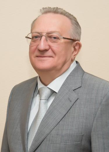
В.В.Гладько

Dear friends, dear colleagues!

The Medical Institute of Continuing Education (MINO) is a structural subdivision of the Federal State Budgetary Educational Institution of Higher Education «Moscow State University of Food Production».

MINO's mission is to create conditions and mechanisms for effective training of medical and pharmaceutical specialists, providing them with high competitiveness and making it possible to integrate into the socio-economic space of the country. The clinical and scientific bases of the departments of the institute are specialized divisions of hospitals of the Ministry of Defense of the Russian Federation, hospitals, sanatoriums and other medical and preventive institutions of the Ministry of Internal Affairs of the Russian Federation and the Russian Guard, as well as specialized departments of research institutes and large medical institutions of the city of Moscow and the Moscow region, in which on the basis of scientific and practical cooperation, practical work of students and research activities are carried out on the most pressing and socially significant health problems.

Dear readers! I am proud to present to your attention the first issue of the journal «Bulletin of the Medical Institute of Continuing Education».

The journal «Bulletin of the Medical Institute of Continuing Education» is a platform where original research papers, reviews, practical recommendations, unique and didactic clinical cases and short messages on the problems of medicine and health care and relevant both in Russia and abroad can be published. The medical community, medical scientists and medical practitioners are interested in a printed organ that would combine the advanced medical thought and modern innovative developments in the medical field. The journal is interdisciplinary in nature, and we hope that it will be of interest to doctors of various specialties.

The priority for the journal is materials with a high level of scientific evidence, designed in accordance with international ethical requirements and capable of arousing the interest of Russian and foreign authors and readers.

Best regards, Editor-in-Chief, MD, PhD, Prof. V.V. Gladko

DERMATOVENEREOLOGY

GENERAL APPROACHES AND CHOICE OF THERAPY FOR ACNE OF VARIOUS DEGREE OF SEVERITY

G.L. Belyakova

9-13

Medical Institute of Continuing Education of the Moscow State University of Food Production

Summary

Acne is the most common skin disease; it occurs in 60–95% of adolescents and young people. The pathological process in acne involves sebaceous glands, keratinocytes, hair follicles, as well as endocrine organs – the adrenal glands and ovaries. Acne therapy should be aimed at the main etiopathogenetic factors, namely, hypersecretion of sebum caused by increased androgenic activity; activation of innate immunity by lipids of sebum and Cutibacterium acnes. Modern drugs for systemic and external therapy have anti-comedogenic, keratolytic, antibacterial, anti-inflammatory, anti-androgenic effects. Eucerin Dermopure cosmetics are used both to reduce the side effects of drug therapy and to treat mild to moderate acne. The drugs are safe to use and can be used as monotherapy and in combination with other external drugs.

Key words: acne, treatment, topical drugs, licochalcon A, 1,2-decanediol, L-carnitine

For citation: Belyakova G.L. General approaches and choice of therapy for acne of various degree of severity. Bulletin of the Medical Institute of Сontinuing Education. 2021; (1): 9–13.
DOI 10.46393/2782-1714_2021_1_9_13

Literature

  1. Акне вульгарные. Клинические рекомендации Российского общества дерматовенерологов и косметологов. 2020. 13 с.
  2. Sharma R.K., Dogra S., Singh A., Kanwar A.J. Epidemiological patterns of acne vulgaris among adolescents in North India: a cross-sectional study and brief review of literature. Indian J. Ped. Dermatol. 2017; 18 (3): 196–201.
  3. Skroza N., Tolino E., Mambrin A. et al. Adult acne versus adolescent acne. J. Clin. Aesthet. Dermatol. 2018; 11 (1): 21–25. 4. Монахов С.А., Иванов О.Л. Акне (методическое пособие для врачей). М., 2010. С. 32.
  4. Чеботарева В.В., Козьмина И.Б., Кошель М.В. и др. Поздние акне у женщин: особенности течения заболевания. Медицинский вестник Северного Кавказа. 2016; 11 (4): 544–547.
  5. Oberemok S.S., Shalita A.R. Acne vulgaris, I: pathogenesis and diagnosis. Cutis. 2002; 70 (2): 101–105.
  6. Bhate K., Williams H.C. Epidemiology of acne vulgaris. Br. J. Dermatol. 2013; 168 (3): 474–485.
  7. Kokandi A. Evaluation of acne quality of life and clinical severity in acne female adults [Электронный ресурс]. Dermatol. Res. Pract. 2010. Режим доступа: doi:10.1155/2010/41080910.
  8. Dreno B. Treatment of adult female acne: a new challenge [Электронный ресурс]. J. Eur. Acad. Dermatol. Venereol. 2015; 29: 14–19. Режим доступа: doi: 10.1111/ jdv.13188. 9.
  9. Волкова Н.В. Комплексная терапия acne vulgaris с учетом коморбидных психических расстройств: автореф. дис. ... канд. мед. наук. Екатеринбург, 2013. 28 с.
  10. Ayer J., Burrows N. Acne: more than skin deep. Postgrad. Med. J. 2006; 970 (82): 500–506.
  11. Юсупова Л.А., Юнусова Е.И., Гареева З.Ш., Мавлютова Г.И. Особенности патогенеза и терапии больных акне. Лечащий врач. 2017; (7): 71.
  12. Dreno B. What is new in the pathophysiology of acne, an overview. J. Eur. Acad. Dermatol. Venereol. 2017; 31 (Suppl 5): 8–12.
  13. Баринова О.А. К вопросу терапии акне топическими антибиотиками: современный взгляд на проблему. Эффективная фармакотерапия. Дерматология и венерология. 2014; (3–4): 16–22.
  14. Kurokawa I., Layton A.M., Ogaw R. Updated treatment for acne: targeted therapy based on pathogenesis. Dermatol. Ther. 2021; 11: 1129–1139.
  15. Ramasamy S., Barnard E., Dawson T.L. Jr., Li H. The role of the skin microbiota in acne pathophysiology. Br. J. Dermatol. 2019; 181 (4): 691–699.
  16. Melnik B.C., Schmitz G., Zouboulis C.C. Anti-acne agents attenuate FGFR2 signal transduction in acne. J. Invest. Dermatol. 2009; 129 (8): 1868–1877.
  17. Zaenglein A.L., Pathy A.L., Schlosser B.J. et al. Guidelines of care for the management of acne vulgaris. J. Am. Acad. Dermatol. 2016; 74 (5): 945–973.
  18. Dreno B., Dagnelie M.A., Khammari A., Corvec S. The skin microbiome: a new actor in inflammatory acne. Am. J. Clin. Dermatol. 2020; 21 (Suppl. 1): 18–24.
  19. Силина Л.В., Овод А.И., Бендрик О.Е., Оганисян К.В. Фармакоэкономическая эффективность антибактериального лечения акне средней степени тяжести. Научные ведомости Белгородского государственного университета. Серия: Медицина. Фармация. 2014; 27 (18): 129–133.
  20. Лукьянов А.М., Артамонова О.В. Эффективность местной терапии угревой болезни средней степени тяжести цинк-эритромициновым комплексом. Поликлиника. 2014; 1 (1): 90–95.
  21. Del Rosso J.Q. Topical and oral antibiotics for acne vulgaris. Semin. Cutan. Med. Surg. 2016; 35 (2): 57–61.
  22. Stein Gold L., Baldwin H.E., Lin T. Management of severe acne vulgaris with topical therapy. J. Drugs Dermatol. 2017; 16 (11): 1134–1138.
  23. Головач Н.А., Корсунская С.А., Шилин Р.Р. Новое в терапии акне. В кн.: Перспективы внедрения инновационных технологий в фармации. Сборник материалов заочной научно-практической конференции с международным участием. 2016. С. 244–251.
  24. Корчевая Т.А. Наружная терапия при акне – Зинерит. Вестник дерматологии и венерологии. 2014; (2): 78–82.
  25. Матушевская Е.В. Актуальные подходы к наружной терапии акне, дерматозов, осложненных вторичной инфекцией, и остроконечных кондилом в практике врача-дерматовенеролога. Эффективная фармакотерапия. 2017; (15): 46–54.
  26. Del Rosso J.Q. Clindamycin phosphate-tretinoin combination gel revisited: status report on a specific formulation used for acne treatment. Cutis. 2017; 99 (3): 179–183.
  27. Gold L.S., Weiss J., Rueda M.J. et al. Moderate and severe inflammatory acne vulgaris effectively treated with single-agent therapy by a new fixed-dose combination adapalene 0.3%/benzoyl peroxide 2.5% gel: a randomized, double-blind, parallel-group, controlled study. Am. J. Clin. Dermatol. 2016; (17): 293–303.
  28. Maiti R., Sirka C.S., Ashique Rahman M.A. et al. Efficacy and safety of tazarotene 0.1% plus clindamycin 1% gel versus adapalene 0.1% plus clindamycin 1% gel in facial acne vulgaris: a randomized, controlled clinical trial. Clin. Drug Investig. 2017; 37 (11): 1083–1091.
  29. Burkhart C.N., Burkhart C.G. Microbiology’s principle of biofilms as a major factor in the pathogenesis of acne vulgaris. Int. J. Dermatol. 2003; 42: 925–927.
  30. Tabara K., Tamura R., Nakamura A. et al. Anti-inflammatory effects of ozenoxacin, a topical quinolone antimicrobial agent. J. Antibiot. (Tokyo). 2020; 73: 247–254.
  31. Maarouf M., Clark A.K., Lee D.E., Shi V.Y. Targeted treatments for hidradenitis suppurativa: a review of the current literature and ongoing clinical trials. J. Dermatolog. Treat. 2018; 29: 441–449.
  32. Dinarello C.A., van der Meer J.W. Treating inflammation by blocking interleukin-1 in humans. Semin. Immunol. 2013; 25: 469–484.
  33. Tan J., Miklas M. A novel topical retinoid for acne: trifarotene 50 lg/g cream. Skin Therapy Lett. 2020; 25: 1–2.
  34. Zuliani T., Khammari A., Chaussy H. et al. Ex vivo demonstration of a synergistic effect of adapalene and benzoyl peroxide on inflammatory acne lesions. Exp. Dermatol. 2011; 20: 850–853.
  35. Dispenza M.C., Wolpert E.B., Gilliland K.L. et al. Systemic isotretinoin therapy normalizes exaggerated TLR-2-mediated innate immune responses in acne patients. J. Invest. Dermatol. 2012; 132: 2198–2205.
  36. Hebert A., Thiboutot D., Stein Gold L. et al. Efficacy and safety of topical clascoterone cream, 1%, for treatment in patients with facial acne: two phase 3 randomized clinical trials. JAMA Dermatol. 2020; 156: 621–630.
  37. Marks D.H., Prasad S., De Souza B. et al. Topical antiandrogen therapies for androgenetic alopecia and acne vulgaris. Am. J. Clin. Dermatol. 2020; 21: 245–254.
  38. Melnik B. Acne vulgaris: the metabolic syndrome of the pilosebaceous follicle. Clin. Dermatol. 2018; 36: 29–40.
  39. Баткаев Э.А., Молодова Ю.С. Акнекутан в терапии акне: новая схема низких доз. Клиническая дерматология и венерология. 2014; (2): 79–85.
  40. Snyder S., Grandell I., Davis S.A., Feldman S.R. Medical adherence to acne therapy: a systematic review. Am. J. Clin. Dermatol. 2014; 15: 87–94.
  41. https://onlinelibrary.wiley.com/doi/10.1111/jdv.13529.
  42. Angelova-Fischer I., Rippke F., Fischer T.W. et al. A double-blind, randomized, vehicle-controlled efficacy assessment study of a skin care formulation for improvement of mild to moderately severe acne. J. Eur. Acad. Dermatol. Venereol. 2013; 27 (Suppl. 2): 6–11.
  43. Dall'Oglio F., Fabbrocini G., Tedeschi A. et al. Licochalcone A in combination with salicylic acid as fluid based and hydroxy-complex 10% cream for the treatment of mild acne: a multicenter prospective trial. Clin. Cosmet. Invest. Dermatol. 2019; 12: 961–967.
  44. Kulthanan K., Trakanwittayarak S., Tuchinda P. et al. A double-blinded, randomized, vehicle-controlled study of the efficacy of moisturizer containing licochalcone A, decanediol, L-carnitine, and salicylic acid for prevention of acne relapse in Asian population. Biomed. Res. Int. 2020; 2020: 2857812.
  45. Chularojanamontri L., Tuchinda P., Kulthanan K. et al. A double-blinded, randomized, vehicle-controlled study to access skin tolerability and efficacy of an anti-inflammatory moisturizer in treatment of acne with 0.1% adapalene gel. J. Dermatol. Treatment. 2016; 27 (2): 140–145.
  46. Yang G., Lee H.E., Yeon S.H. et al. Licochalcone A attenuates acne symptoms mediated by suppression of NLRP3 inflammasome. Phytother. Res. 2018; 32 (12): 2551–2559.
  47. Raoufinejad K., Rajabi M., Sarafian G. Licorice in the treatment of acne vulgaris and postinflammatory hyperpigmentation: a review. J. Pharmaceut. Care. 2021; 8 (4): 186–195.
  48. Hoffmann J., Gendrisch F., Schempp C.M., Wölfle U. New herbal biomedicines for the topical treatment of dermatological disorders. Biomedicines. 2020; 8 (2): 27.
  49. Sulzberger M., Folster H., Sattler M. et al. Inhibition of Propionibacterium acnes associated biofilm formation by decanediol. J. Dermatol. Sci. 2016; 83 (2): 159–161.
  50. Peirano R.I., Hamann T., Düsing H.-J. et al. Topically applied L-carnitine effectively reduces sebum secretion in human skin. J. Cosmetic Dermatol. 2012; 11 (1): 30–36.
  51. Bae B.G., Park C.O., Shin H. et al. Salicylic acid peels versus Jessner’s solution for acne vulgaris: a comparative study. Dermatol. Surg. 2013; 39 (2): 248–253.
  52. Arif T. Salicylic acid as a peeling agent: a comprehensive review. Clin. Cosmet. Investig. Dermatol. 2015; 8: 455–461.

COMORBIDITY OF PATHOLOGY IN ATOPIC DERMATITIS AS A FACTOR OF POLYPRAGMASIA FORMATION

T.V. Sokolova1, V.V. Gladko1, M.S. Davidenko2

14-19

1 Medical Institute of Continuing Education of the Moscow State University of Food Production
2 Clinic "Semeynaya", Moscow

Summary

Concomitant pathology in atopic dermatitis (AtD) aggravates its course. The need of her treatment is one of the reasons of polypharmacy.

Objective – to study the frequency of concomitant pathology in AtD of children and to determine the range of drugs (D) for the treatment of the underlying and concomitant diseases.

Material and methods. Retrospective analysis of outpatient cards: children (7–11 years old, 72), adolescents of the early (12–14 years old, 81) and late (15–17 years old, 98) periods.

The author's version of the individual registration card was used. Statistical processing of the material was carried out using Pearson's test (X2) and Spearman Rank Correlation (Sr).

Results. In outpatient practice of children with AtD till 2 years, pathology of 2.38 ± 0.82 body systems was recorded. An exacer- bation of one or another concomitant disease was observed of 41% of patients, including 27.3% 2-3 times. However, the records of consultations of related specialists were only in 57% of cases. The average number of systemic drugs prescribed to the patient was 17.2 ± 6.3 (9.6 ± 3.5), with the maximum proportion (74.3%) and number (7.3 ± 1.8) for the treatment of comorbidities.

Conclusions. The comorbidity of pathology of patients with AtD is a significant factor that should be taken into account when choosing a rational treatment strategy. To exclude polypharmacy, strict accounting of appointments made by both a dermatologist and related specialists is required.

Key words: atopic dermatitis, children, comorbidity, polypharmacy

For citation: Sokolova T.V., Gladko V.V., Davidenko M.S. Сomorbidity of pathology in atopic dermatitis as a factor of polypragmasia formation. Bulletin of the Medical Institute of Сontinuing Education. 2021; (1): 14–19.
DOI 10.46393/2782-1714_2021_1_14_19

Literature

  1. Тихомиров А.А. Клинико-патогенетическое обоснование дифференцированного подхода к терапии тяжелых форм атопического дерматита у детей: автореф. дис. ... д-ра мед. наук. М., 2014. 48 с.
  2. Соколова Т.В., Давиденко М.С. Статистика распространенности и заболеваемости атопическим дерматитом детей и подростков: за и против. Иммунопатология, аллергология, инфектология. 2019; (1): 80–88.
  3. Sepiashvili R.I., Slavyanskaya Т.А. Spectrum and prevalence of allergic diseases in the Commonwealth of Independent States (CIS) Countries. Internat. J. Immunorehab. 2011; 13 (2): 123–126.
  4. Slavyanskaya T.A., Sepiashvili R.I. Spectrum and features of the course of allergic diseases in the CIS countries «Allergy, Asthma & Immunology: from genes to clinical application». MEDIMOND International Proceedings. 2011. Pp. 33–36.
  5. Mallola J., Craneb E., von Mutiusc J. et al. The ISAAC Phase Three Study Group the International Study of Asthma and Allergies in Childhood (ISAAC): a global synthesiseceived. 2012. 56 p.
  6. Методические рекомендации МЗ и РП РФ, ФГУ УрНИИДВиИ Минздравсоцразвития России «Организация специализированной медицинской помощи детям, больным атопическим дерматитом». Екатеринбург, 2011. 14 с.
  7. Ведерникова С.В. Комплексная оценка эффективности озонотерапии у пациентов с атопическим дерматитом: Дис. ... канд. мед. наук. 2009. 22 с.
  8. Панкратова Е.В. Объективизация диагностики атопического дерматита у мужчин призывного возраста с учетом анализа заболеваемости: автореф. дис. ... канд. мед. наук. М., 2010. 27 с.
  9. Кохан М.М., Самцов А.В., Перламутров Ю.Н., Соколовский Е.В. Анализ методов наружной терапии распространенных дерматозов в оказании специализированной медицинской помощи по профилю «дерматовенерология». Вестник дерматологии и венерологии. 2016; (6): 79–93.
  10. Pepys J., Combs R.R.A., Lachman P.J. et al. In: Gill P.G.H. Clinical aspects of immunology. 3rd edn. Oxford: Blackwell Science, 1975. Рp. 877–902.
  11. Wüthrich B. Clinical aspects, epidemiology and prognosis of atopic dermatitis. Ann. Allergy Asthma Immunol. 1999; 83: 464–470.
  12. Volcheck G.W. Clinical allergy: diagnosis and management. Mayo foundation for medical education and research. Abington: Humana Press, 2009. 340 р.
  13. Silverberg N.B., Silverberg J.I. Inside out or outside in: does atopic dermatitis disrupt barrier function or does disruption of barrier function trigger atopic dermatitis? Cutis. 2015; 96 (11): 359–361.
  14. Смолкин Ю.С., Сердобинцев К.В., Смолкина О.Ю. Десять заблуждений, или распространенные ошибки в лечении атопического дерматита у детей. Consilium Medicum. Педиатрия. 2016; (3): 116–120.
  15. Баранов А.А., Хаитов Р.М. Аллергология и иммунология. М.: Союз педиатров России, 2011. 250 с.
  16. Gao L. Polymorphisms in the myosin light chain kinase gene that confer risk of severe sepsis are associated with a lower risk of asthma. J. Allergy Clin. Immunol. 2007; 119: 111–118.
  17. Ширинский В.С., Ширинский И.В. Коморбидные заболевания – актуальная проблема клинической медицины. Сибирский медицинский журнал. 2014; 12 (1): 7–12.
  18. Caughey G.E., Vitry A.I., Cibert A.L. Prevalence of comorbidity of chronic diseases in Australia. BMC Public Health. 2008; (8): 221–225.
  19. Надаль-Гинард К. Когда одно мешает другому – коморбидность на злобе дня. Новая медицина тысячелетия. 2012; (6): 22–24.
  20. Хазов В.С. Доказательная медицина – ступенька вверх по лестнице познания или движение по кругу? Архив внутренней медицины. 2012; (3): 65–68.
  21. Хархарян В.А., Короткий В.Н., Усачева А.Ю. Дермато-респираторный синдром: новый взгляд на старую проблему. Клиническая дерматология и венерология. 2012; 10 (3): 9–15.
  22. Верткин А.Л., Скотников А.С. Коморбидность. Лечащий врач. 2013; (6): 66–69.
  23. Белялов Ф.И. Лечение внутренних болезней в условиях коморбидности. Иркутск: РИО ИГМАПО, 2013. 297 с.
  24. Fortin M., Bravo G., Huddon C. et al. Prevalence of multimorbidity among adults seen in family practice. Fam. Med. 2005; (3): 223–228.
  25. Кениксфест Ю.В. Состояние специализированной медицинской помощи, оказываемой детям и подросткам, больным атопическим дерматитом. Уральский медицинский журнал. 2011; 86 (8): 7–28.
  26. Суворова К.Н., Варданян К.Л. Трудности и ошибки в диагностике и лечении атопического дерматита у детей. Лечащий врач. Медицинский научно-практический портал. 2005; (9). https://www.lvrach. ru/2005/07/4532769/.
  27. Соколова Т.В., Давиденко М.С., Меньшикова А.И. Атопический дерматит: реальная практика использования антигистаминных препаратов у детей в амбулаторной клинической практике. Клиническая дерматология и венерология. 2020; 19 (5): 701–710.
  28. Eigenmann P.A., Hauser C., Bruggen M.C. Skin barrier defects in atopic dermatitis: new treatments? Rev. Med. Suisse. 2011; 321 (7): 2453–2456.
  29. Ключников С.О. Полипрагмазия: пути решения проблемы. Детские инфекции. 2014; (4): 36–41.
  30. Сычев Д.А., Отделенов В.А., Краснова Н.М., Ильина Е.С. Полипрагмазия: взгляд клинического фармаколога. Терапевтический архив. 2016; (12): 94–102.
  31. Guthrie B., McCowan C., Davey P. et al. High risk prescribing in primary care patients particularly vulnerable to adverse drug events: cross sectional population database analysis in Scottish general practice. BMJ. 2011; 42 (3): 51–54.
  32. Duerden M., Avery T., Payne R. Polypharmacy and medicines optimisation. London: Making it safe. King’s Fund, 2013. 176 р.
  33. Molokhia M., Majeed A. Current and future perspectives on the management of polypharmacy. BMC FAM Pract. 2017; 18 (1): 70–75.
  34. Лазарева Н.Б., Ших Е.В., Реброва Е.В., Рязанова А.Ю. Полипрагмазия в педиатрической практике: современные реалии. Вопросы современной педиатрии. 2019; 18 (3): 212–218.

UROLOGY AND ANDROLOGY

SURGICAL RECOMMENDATIONS FOR INTRAOPERATIVE COMPLICATIONS OF FALLOPROSTHETICS

B.R. Gvasaliya, A.G. Kochetov, K.P. Melnik, A.V. Kasaikin, V.V. Khvorov

20-22

Medical Institute of Continuing Education of the Moscow State University of Food Production

Summary

Penile prosthesis implantation is a surgical procedure that is the method of choice for organic erectile dysfunction.
However, as with any other surgical intervention, there is a risk of complications that are quite specific for this type of surgery. Knowledge of their specificity and correct surgical tactics allows in the overwhelming majority of cases to cope with the situation without serious consequences.
In this article, based on many years of our experience, we analyzed the main intraoperative complications and presented the best ways to resolve them.

Key words: penile prosthesis, corpora cavernosa, cylinders

For citation: Gvasaliya B.R., Kochetov A.G., Melnik K.P. et al. Surgical recommendations for intraoperative complications of falloprosthetics. Bulletin of the Medical Institute of Сontinuing Education. 2021; (1): 20–22.
DOI 10.46393/2782-1714_2021_1_20_22

Literature

  1. Burnett A.L., Nehra A., Breau R.H. et al. Erectile dysfunction: AUA guideline. J. Urol. 2018; 200: 633–641
  2. .
  3. Kohler T., Moore A., Ring J. A contemporary series of penile implant complications occurring at a training institution. J. Sex. Med. 2016; 13: S68.
  4. Eid J.F. Surgery for erectile dysfunction. In: Wein A.J., Kavoussi L.R., Partin A.W. et al. (eds.). Campbell-Walsh Urology. 11th edn. Elsevier. Pp. 709–721.
  5. Henry G., Wilson S. Updates in inflatable penile prostheses. Urol. Clin. North Am. 2007; 34: 535–547.
  6. Kramer A., Goldmark E., Reznicek D. Placement of inflatable penile prosthesis with severe corporal fibrosis from priapism. J. Sex. Med. 2014; 11: 2367–2370.
  7. Levine L., Becher E., Bella A. et al. Penile prosthesis surgery: current recommendations from the international consultation on sexual medicine. J. Sex. Med. 2016; 13: 489–518.
  8. Mulcahy J. The prevention and management of noninfectious complications of penile implants. Sex. Med. Rev. 2015; 3: 203–213.
  9. Montague D.K. Penile prosthesis implantation in the era of medical treatment for erectile dysfunction. Urol. Clin. North Am. 2011; 38: 217–225.
  10. Oberlin D., Matulewicz R., Bachrach L. et al. National practice patterns of treatment of erectile dysfunction with penile prosthesis implantation. J. Urol. 2015; 193: 2040–2044.
  11. Щеплев П.А., Гвасалия Б.Р., Меньщиков К.А., Гарин Н.Н. Коррекция гланулоптоза у пациентов при повторной имплантации протезов полового члена. Тезисы научных трудов Всероссийского конгресса по андроло- гии. Сочи, Дагомыс, 27–29 апреля 2007 г. С. 90.

EVALUATION OF THE EFFICACY AND SAFETY OF THE MODIFIED TECHNIQUE OF ORTHOTOPIC BLADDER PLASTY IN PATIENTS WITH MUSCLE-INVASIVE BLADDER CANCER AFTER RADICAL CYSTECTOMY

P.S. Kozlova1, K.M. Nyushko2, B.Ya. Alekseev2

23-27

1 Pirogov Russian National Medical University
2 National Medical Research Radiological Centre of the Ministry of Health of the Russian Federation

Summary

Bladder cancer is an urgent problem of modern urological oncology due to the persisting high rates of morbidity and mortality from this pathology. Radical cystectomy is the gold standard of treatment in patients with muscle-invasive forms of the disease. However, diverting urine after this surgery remains challenging. The techniques of orthotopic bladder plasty after radical cystectomy allow restoring the act of urination in this contingent of patients, however, such operations may be associated with the risk of urinary incontinence, neocyst-urethroanastomosis strictures, urinary obstruction and ascending urinary infection. The article presents an analysis of the results of modified orthotopic ileocystoplasty using an innovative developed technique for the formation of a zone of neocyst-urethroanastomosis, evaluates the results of using this technique in clinical practice, and also compares the new technique with the standard surgical technique.

Key words: radical cystectomy, orthotopic bladder plasty, neocyst-urethroanastomosis

For citation: Kozlova P.S., Nyushko K.M., Alekseev B.Ya. Evaluation of the efficacy and safety of the modified technique of orthotopic bladder plasty in patients with muscle-invasive bladder cancer after radical cystectomy. Bulletin of the Medical Institute of Сontinuing Education. 2021; (1): 23–27.
DOI 10.46393/2782-1714_2021_1_23_27

Literature

  1. 1. Daneshmand S. Epidemiology and risk factors of urothelial (transitional cell) carcinoma of the bladder. UpToDate. http:// www.uptodate.com/contents/epidemiology-and-risk-factors-of-urothelial-transitional-cell-carcinoma-of-the-bladder (Date of access: 06.07.2017). 2016.
  2. Stein J.P., Lieskovsky G., Cote R. et al. Radical cystectomy in the treatment of invasive bladder cancer: long-term results in 1,054 patients. J. Clin. Oncol. 2001; 19 (3): 666–675.
  3. Palou J., Sylvester R.J., Faba O.R. et al. Female gender and carcinoma in situ in the prostatic urethra are prognostic factors for recurrence, progression, and disease-specific mortality in T1G3 bladder cancer patients treated with bacillus Calmette-Guérin. Eur. Urol. 2012; 62 (1): 118–125.
  4. Fernandez-Gomez J., Madero R., Solsona E. et al. Predicting nonmuscle invasive bladder cancer recurrence and progression in patients treated with bacillus Calmette-Guerin: the CUETO scoring model. J. Urol. 2009; 182 (5): 2195–2203.
  5. Atala A. New methods of bladder augmentation. BJU Int. 2000; 85 (S3): 24–34.
  6. Qu L.G., Lawrentschuk N. Orthotopic neobladder reconstruction: patient selection and perspectives. Res. Rep. Urol. 2019; 11: 333.
  7. Петров С.Б., Левковский Н.С., Король В.Д., Паршин А.Г. Радикальная цистэктомия как основной метод лечения мышечно-инвазивного рака мочевого пузыря (показания, особенности техники, профилактика осложнений). Практическая онкология. 2003; 4 (4): 225–230.
  8. Hautmann R.E., de Petriconi R.C., Volkmer B.G. Lessons learned from 1,000 neobladders: the 90-day complication rate. J. Urol. 2010; 184 (3): 990–994.
  9. Herdiman O., Ong K., Johnson L., Lawrentchuk N. Orthotopic bladder substitution (neobladder). Part II: Post-operative complications, management, and long-term follow-up. J. Wound Ostomy Continence Nurs. 2013; 40 (2): 171–180.
  10. Kim K.H., Yoon H.S., Yoon H. et al. Risk factors for developing metabolic acidosis after radical cystectomy and ileal neobladder. PLoS One. 2016; 11 (7): e0158220.
  11. Thorstenson A., Jacobsson H., Oelöv E. et al. Gastrointestinal function and metabolic control after construction of an orthotopic ileal neobladder in bladder cancer. Scand. J. Urol. Nephrol. 2007; 41 (1): 14–19.
  12. Asgari M.A., Safarinejad M.R., Shakhssalim N. et al. Sexual function after non-nerve-sparing radical cystoprostatectomy: a comparison between ileal conduit urinary diversion and orthotopic ileal neobladder substitution. Int. Braz. J. Urol. 2013; 39 (4): 474–483.
  13. Furrer M.A., Roth B., Kiss B. et al. Patients with an orthotopic low pressure bladder substitute enjoy long-term good function. J. Urol. 2016; 196 (4): 1172–1180.
  14. Kretschmer A., Grimm T., Buchner A. et al. Prognostic features for quality of life after radical cystectomy and orthotopic neobladder. Int. Braz. J. Urol. 2016; 42 (6): 1109–1120.

IMMUNOLOGY

THE ROLE OF IMMUNE SYSTEM IN THE NEW CORONAVIRUS DISEASE SARS-CоV-2

M.I. Varfolomeeva1,2, I.Ts. Kulagina1,2, A.A. Zaicev1,2

28-31

1 Medical Institute of Continuing Education of the Moscow State University of Food Production
2 N.N. Burdenko Main Military Clinical Hospital

Summary

When a person is infected with the SARS-CoV-2 virus, the immune system plays a leading role. This article analyzes up-to-date information on the influence of the immune system on the features of the course of a new coronavirus infection in humans and the possibilities for the development of autoimmune reactions. We analyzed the role and dynamic change of the humoral immune system during SARS-CoV-2 infection causes a humoral immune response: the timing of the appearance and dynamics of changes in the level of specific serum immunoglobulins.

Key words: SARS-CoV-2 virus, new coronavirus infection, immune system, immunoglobulins, cytokine storm

For citation: Varfolomeeva M.I., Kulagina I.Ts., Zaicev A.A. The role of immune system in the new coronavirus disease SARS-CоV-2. Bulletin of the Medical Institute of Сontinuing Education. 2021; (1): 28–31.
DOI 10.46393/2782-1714_2021_1_28_31

Literature

  1. Профилактика и лечение новой коронавирусной инфекции (COVID-19). Временные методические рекомендации. Версия 10 от 08.02.2021.
  2. Fu Y., Cheng Y., Wu Y. Understanding SARS-CoV-2-mediated inflammatory responses: from mechanisms to potential therapeutic tools. Virol. Sin. 2020. URL: https://doi.org/10.1007/s12250-020-00207-4.
  3. Gu J., Korteweg Ch. Pathology and pathogenesis of severe acute respiratory syndrome. Am. J. Pathol. 2007; 170: 1136–1147.
  4. Channappanavar R., Perlman S. Pathogenic human coronavirus infections: causes and consequences of cytokine storm and immunopathology. Semin. Immunopathol. 2017; 39 (5): 529–539.
  5. Wang X., Xu W., Hu G. et al. SARS‐CoV‐2 infects T-lymphocytes through its spike protein‐mediated membrane fusion. Cell. Mol. Immunol. 2020.
  6. Jansen J.M., Gerlach T., Elbahesh H. et al. Influenza virus‐specific CD4+ and CD8+ T cell‐mediated immunity induced by infection and vaccination. J. Clin. Virol. 2019; 119: 44–52.
  7. Tseng C.T., Perrone L.A., Zhu H. et al. Severe acute respiratory syndrome and the innate immune responses: modulation of effector cell function without productive infection. J. Immunol. 2005; 174: 7977–7985.
  8. Siu K.L., Kok K.H., Ng M.H. et al. Severe acute respiratory syndrome coronavirus M protein inhibits type I interferon production by impeding the formation of TRAF3.TANK.TBK1/IKKepsilon complex. J. Biol. Chem. 2009; 284 (24): 16202–16209.
  9. Qu R., Ling Y., Zhang Y.H. et al. Platelet‐to‐lymphocyte ratio is associated with prognosis in patients with Corona Virus Disease‐19. J. Med. Virol. 2020.
  10. Schmidt M.E., Varga S.M. The CD8 T-cell response to respiratory virus infections. Front. Immunol. 2018; 9: 678.
  11. Yang M. Cell pyroptosis, a potential pathogenic mechanism of 2019-nCoV infection. SSRN. 2020. URL: https://doi.org/10.2139/ssrn. 3527420.
  12. Fulop T., Larbi A., Wikby A. et al. Dysregulation of T-cell function in the elderly: scientific basis and clinical implications. Drugs Aging. 2005; 22 (7): 589–603.
  13. Yoneyama M., Kikuchi M., Natsukawa T. et al. The RNA helicase RIG-I has an essential function in double-stranded RNA-induced innate antiviral responses. Nat. Immunol. 2004; 5 (7): 730–737.
  14. Long Q.X., Liu B.Z., Deng H.J. et al. Antibody responses to SARS‐CoV‐2 in patients with COVID‐19. Nat Med. 2020.
  15. Lee Y.L., Liao C.H., Liu P.Y. et al. Dynamics of anti‐SARS‐Cov‐2 IgM and IgG antibodies among COVID‐19 patients. J. Infect. 2020.
  16. Azkur A.K., Akdis M., Azkur D. et al. Immune response to SARS‐CoV‐2 and mechanisms of immunopathological changes in COVID‐19. Allergology. 2020; 75 (7): 1564–1581.
  17. Peiris J.S., Yuen K.Y., Osterhaus A.D., Stohr K. The severe acute respiratory syndrome. N. Engl. J. Med. 2003; 349: 2431–2441.
  18. Xiong Y., Liu Y., Cao L. et al. Transcriptomic characteristics of bronchoalveolar lavage fluid and peripheral blood mononuclear cells in COVID-19 patients. Emerg. Microbes Infect. 2020; 9 (1): 761–770.
  19. Nicholls J.M., Poon L.L., Lee K.C. Lung pathology of fatal severe acute respiratory syndrome. Lancet. 2003; 361: 1773–1778.
  20. Xu Z., Shi L., Wang Y. et al. Pathological findings of COVID-19 associated with acute respiratory distress syndrome. Lancet Respir. Med. 2020 Feb 18 [published online ahead of print].
  21. Lee C.H., Chen R.F., Liu J.W. et al. Altered p38 mitogen-activated protein kinase expression in different leukocytes with increment of immunosuppressive mediators in patients with severe acute respiratory syndrome. J. Immunol. 2004; 172: 7841–7847.
  22. Behrens E.M., Koretzky G.A. Review: cytokine storm syndrome: looking toward the precision medicine era. Arthritis Rheumatol. 2017; 69 (6): 1135–1143.
  23. Zhang L., Zhang F., Yu W. et al. Antibody responses against SARS coronavirus are correlated with disease outcome of infected individuals. J. Med. Virol. 2006; 78 (1): 1–8.
  24. Lin Y.S., Lin C.F., Fang Y.T. et al. Antibody to severe acute respiratory syndrome (SARS)-associated coronavirus spike protein domain 2 cross-reacts with lung epithelial cells and causes cytotoxicity. Clin. Exp. Immunol. 2005; 141: 500–508.
  25. Yang Y.H., Huang Y.H., Chuang Y.H. et al. Autoantibodies against human epithelial cells and endothelial cells after severe acute respiratory syndrome (SARS)-associated coronavirus infection. J. Med. Virol. 2005; 77: 1–7.
  26. Qin С., Zhou L., Hu Z. et al. Dysregulation of immune response in patients with COVID-19 in Wuhan, China. Clin. Infect. Dis. 2020.
  27. Yao X.H., Li T.Y., He Z.C. et al. A pathological report of three COVID-19 cases by minimally invasive autopsies. Chin. J. Pathol. 2020; 49: E009.

NEUROSURGERY

COMBINED ENDOSCOPIC TRANSNASAL AND TRANSCRANIAL ACCESS IN SURGERY OF TRANSETMOIDAL MENINGOENCEPHALOCELE. CLINICAL CASE

N.S. Grachev1,2, A.E. Samarin1, I.N. Vorozhtsov1, A.V. Lezhinskaya1, A.I. Gorozhanina1, I.V. Zyabkin1, I.I. Morozov2

32-35

1 Dmitry Rogachev National Medical Research Center of Pediatric Hematology, Oncology and Immunology
2 Medical Institute of Continuing Education of the Moscow State University of Food Production

Summary

Congenital transethmoidal meningoencephalocele is a rare neural tube malformation with a frequency of 1 in 35,000 live births. Despite the asymptomatic course, the presence of a skull base defect requires surgical treatment by traumatic transcranial access. The search and development of minimally invasive methods of treating this pathology is an urgent problem of head and neck surgery in children. The article presents a clinical case of successful surgical treatment transethmoidal meningoencephalocele in an 8-year-old child using combined endoscopic transnasal and transbasal approach. This clinical case demonstrates the possibilities of minimally invasive endoscopic endonasal skull base surgery and multidisciplinary approach of transethmoidal meningoencephalocele treatment.

Key words: transethmoidal meningoencephalocele, basal cranial hernia, combined approach, skull base surgery

For citation: Grachev N.S., Samarin A.E., Vorozhtsov I.N. et al. Combined endoscopic transnasal and transcranial access in surgery of transetmoidal meningoencephalocele. Clinical case. Bulletin of the Medical Institute of Сontinuing Education. 2021; (1): 32–35.
DOI 10.46393/2782-1714_2021_1_32_35

Literature

  1. Асадов Р.Н., Притыко А.Г., Петраки В.Л. и др. Методические рекомендации «Современное лечение детей с врожденными передними черепно-мозговыми грыжами». 2019 г.
  2. Гофман В.Р., Андроненков В.А., Воронов А.В. Обоснование доступа при эндоскопической хирургии назальной ликвореи. Вестник оториноларингологии. 2012; 77 (4): 16–17.
  3. Кaпитaнов Д.Н. Эндоскопическaя диaгностикa и лечение нaзaльной ликвореи и энцефaлоцеле передних отделов основaния черепa. Вестник отолaрингологии. 2000; (6): 51–54.
  4. Castelnuovo P., Bignami M., Pistochini A. et al. Endoscopic endonasal management of encephaloceles in children: an eight year experience. Int. J. Pediatr. Otorhinolaryngol. 2009; 73: 1132–1136.
  5. Асадов Р.Н., Бельченко В.А., Притыко А.Г., Петров Ю.А. Хирургическое лечение передних черепно-мозговых грыж. Вопросы нейрохирургии. 2007.
  6. Cassano M., Felippu A. Endoscopic treatment of cerebrospinal fluid leaks with the use of lower turbinate grafts: a retrospective review of 125 cases. Rhinology. 2009; 47 (4): 362–368.
  7. Marton E., Billeci D., Schiesari E., Longatti P. Transnasal endoscopic repair of cerebrospinal fluid fistulas and encephaloceles: surgical indications and complications. Minim. Invasive Neurosurg. 2005; 48 (3): 175–181.
  8. Zuckerman J.D., DelGaudio J.M. Utility of preoperative high-resolution CT and intraoperative image guidance in identification of cerebrospinal fluid leaks for endoscopic repair. Am. J. Rhinol. 2008; 22 (2): 151–154.

LEGAL REGULATION

QUALITY OF PREVENTION IN THE STRUCTURE QUALITY OF CARE

O.V. Sharapova1,2, D.I. Kicha3, O.V. Rukodaynyy3, L.I. Gerasimova1,4, N.M. Zakharchenko5, D.F. Моrоgа6, R.V. Fomina7

36-39

1 V.V. Vinogradov City Clinical Hospital, Moscow
2 I.M. Sechenov First Moscow State Medical University
3 Peoples' Friendship University of Russia
4 Medical Institute of Сontinuing Education of the Moscow State University of Food Production
5 Center for Rehabilitation of the Social Insurance Fund of the Russian Federation 'Taraskul', Tyumen
6 Medical Center for the Treatment of Spine and Joints'DEMA', Moscow Region
7 I.N. Ulyanov Chuvash State University, Cheboksary

Summary

The quality of health care (QHC) is not usually considered in terms of the quality of prevention and dispensary work. However, the contribution of dispensary and prevention activities to optimizing health can be considered as a more effective measure of public health.

The aim of the study was to develop objective indicators capable of adequately assessing the quality of preventive care (CPC) based on studies of services provided under the basic program of state guarantees of free medical care for citizens.

Materials and methods. The data of the dispensary and preventive medical examinations of certain groups of the adult population according to the registers – accounts of the region were analyzed. To assess the results of control measures in the preventive direction, the groups of violations and deviations from the ICH in the field of dispensary and preventive examinations, grouped according to the types of violations of the ICH, were applied.

Results. Based on the results of the assessments, a quality assessment of 5,451 cases of preventive interventions (2017) was organized and conducted with the participation of the health insurance organization (HIO). During the examination process, according to the aforementioned criteria, irregularities were identified or not identified in 1,459 cases (27%).

Conclusion. The preventive control measures carried out have led to a significant reduction in the number of defects in prevention and dispensary work in the region. The effectiveness of the designed and applied technological processes of CPT in the structure of CMP is confirmed by the positive indicators of the morbidity reduction in the region. The received results show the actuality and necessity of development and introduction of principles and methods of quality evaluation of preventive, rehabilitative care, as a system of knowledge and practical activity, saving and restoring the potential of public health.

Key words: ВИЧ, абсцесс, селезенка.

For citation: Пучков С.С., Фаллер А.П. Трудности диагностики абсцессов селезенки у ВИЧ-инфицированных пациентов. Вестник Медицинского института непрерывного образования. 2023; 3 (1): 44–51.

Literature

  1. WHA60.29 Health technologies. The Sixtieth World Health Assembly adopted resolution. May 2007.
  2. De Ville K. Assembly resolution, formed the basis for establishing the Global Initiative on Health Technologies (GIHT). Int. J. Technol. Assess. Health Care. 1998; 14 (2): 197–211.
  3. Szlezák N.A., Bloom B.R., Jamison D.T. et al. The global health system: actors, norms, and expectations in transition. PLoS Med. 2010; 7 (1): e1000183.
  4. Health technology assessment of medical devices. World Health Organization. Geneva 27, Switzerland. 2011. 44 p.
  5. Health systems: improving performance. The world health report 2000. Geneva: WHO, 2000. 215 р.
  6. Практикум общественного здоровья и здравоохранения: Учеб. пособие. 2-е изд., перераб. и доп. Под ред. И.Н. Денисова, Д.И. Кичи, А.В. Фоминой, О.С. Сауриной. М.: Медицинское информационное агентство, 2015. 424 с.
  7. Александрова О.Ю., Григорьев И.Ю., Аржанцева О.М. Оценка качества медицинской помощи в свете нового законодательства. Здравоохранение. 2012; (1): 64–71.
  8. Постановление Правительства РФ от 10.12.2018 № 1506 «О программе государственных гарантий бесплатного оказания гражданам медицинской помощи на 2019 год и на плановый период 2020 и 2021 годов».

HISTORY OF DEVELOPMENT OF THE CRIMINAL-LAW ASPECT RELATIONSHIP OF DOCTOR AND PATIENT IN RUSSIA

V.V. Alshevsky, D.S. Kadochnikov

40-44

Medical Institute of Сontinuing Education of the Moscow State University of Food Production

Summary

In the medical community, discussions are periodically resumed on the issue of a doctor's jurisdiction for harm caused to a patient's health in the provision of medical care. The reason for its discussion is the periodic 'stuffing' in the Internet of cases of initiation of criminal cases and convictions of doctors. The reason for the continuing interest in discussing this topic is the discrepancy between the current legal norms and the physician’s worldview, which is based on the historically established tradition of attitudes towards the patient.

Key words: paternalism, morality, social security, medical care, patient

For citation: Alshevsky V.V., Kadochnikov D.S. History of development of the criminal-law aspect relationship of doctor and patient in Russia. Bulletin of the Medical Institute of Сontinuing Education. 2021; (1): 40–44.
DOI 10.46393/2782-1714_2021_1_40_44

Literature

  1. Тураев Б.А. Законы Хаммурапи. [Электронный ресурс]. https://culture/wikireading.ru/3590 (30.03.2020).
  2. Цыпин В.А. Церковное право. Церковный суд в Древней Руси. [Электронный ресурс]. https://religion.wikireading.ru/9802 (26.03.2020).
  3. Исхаков Э.Р., Аксенов С.Г. Государственно-правовое регулирование здравоохранения в период правления Петра I. [Электронный ресурс]. https://cyberleninka.ru/article/n/gosudarstvenno-pravovoe-regulirovaniezdravoohraneniya-v-period-pravleniya-petra-i/viewer (27.03.2020).
  4. Ковригина Г.Д. Правовое регулирование целительской деятельности в России: история вопроса и современные общеюридические начала. Медицинское право. 2008; (4).
  5. РИА Новости. Военные реформы Петра I. Справка. [Электронный ресурс]. https://ria.ru/20090626/175508456 (25.03.2020).
  6. Смирнова Е.М. Частная медицинская практика в России XVIII – начала XX вв. Новый исторический вестник. 2014; (4): 44–63.
  7. Акопов В.И. Медицинское право в вопросах и ответах. М.: ПРИОР, 2001. 208 с.
  8. Казакова Е.Б., Миронова М.Ю. История развития института информированного согласия и современное правовое регулирование данного института. История государства и права. 2012; (21): 35–38.
  9. Основы законодательства Российской Федерации об охране здоровья граждан. Федеральный закон от 22.06.1993 № 5487-1. Ведомости СНД РФ и ВС РФ. 1993; (33): 1318.
  10. Федеральный закон от 21.11.2011 № 323-ФЗ «Об основах охраны здоровья граждан в Российской Федерации». Собрание законодательства РФ. 2011; (48): 6724.

PULMONOLOGY

RADIATION DIAGNOSTICS OF LONG-TERM CONSEQUENCES OF VIRAL PNEUMONIA CAUSED BY COVID-19

E.A. Kukushkina1,2, K.M. Shashkin1, I.S. Obelchak1,2, N.V. Rodina1, K.N. Nikolaev1, E.N. Protsyk1

46-48

1 Main Military Clinical Hospital of the National Guard Troops of the Russian Federation
2 Medical Institute of Continuing Education of the Moscow State University of Food Production

Summary

The article provides an analysis of changes in the pulmonary parenchyma in pneumonia caused by a new coronavirus infection in the acute and separated period of the course of the disease. Based on clinical material (60 patients with viral pneumonia, who underwent classical lung radiography and computed tomography at various times of the disease) changes in radiological signs of lung tissue damage were assessed. The dependence of the course and regression of the disease on the degree of lung tissue damage was revealed according to the results of radiography and computed tomography

Key words: coronavirus infection, viral pneumonia, radiography, computer tomography, «frosted glass», fibrosis

For citation: Kukushkina E.A., Shashkin K.M., Obelchak I.S. et al. Radiation diagnostics of long-term consequences of viral pneumonia caused by COVID-19. Bulletin of the Medical Institute of Сontinuing Education. 2021; (1): 46–48.
DOI 10.46393/2782-1714_2021_1_46_48

Literature

  1. Ye Zh., Zhang Y., Wang Y. et al. Chest CT manifestations of new coronavirus disease 2019 (COVID-19): a pictorial review. Eur. Radiol. 2020; 30 (8): 4381–4389.
  2. Zhang K., Liu X., Shen J. et al. Clinically applicable AI system for accurate diagnosis, quantitative measurements, and prognosis of COVID-19 pneumonia using computed tomography. Cell. 2020; 181 (6): 1423–1433.
  3. Никифоров В.В., Суранова Т.Г., Миронов А.Ю., Забозлаев Ф.Г. Новая коронавирусная инфекция (COVID-19):
  4. Обеспечение COVID-готовности отделений лучевой диагностики: учеб. пособие. Под ред. С.П. Морозова. М., 2020. 72 с.
  5. Синицын В.Е., Тюрин И.Е., Митьков В.В. Временные согласительные методические рекомендации Российского общества рентгенологов и радиологов (РОРР) и Российской ассоциации специалистов ультразвуковой диагностики в медицине (РАСУДМ) «Методы лучевой диагностики пневмонии при новой коронавирусной инфекции COVID-19» (версия 2). Вестник рентгенологии и радиологии. 2020; 101 (2): 72–89.

GENERAL MEDICAL PRACTICE

USING BODY TRACKING TECHNOLOGY ON THE HABILECT APC FOR THE PURPOSE OF EVALUATION OF THE EFFICIENCY OF PHYSICAL REHABILITATION OF PATIENTS WITH VERTEBROBASILAR INSUFFICIENCY

S.M. Starikov1, V.E. Yudin1, A.M. Shchegolkov2, M.A. Ryumshin2

50-52

1 Medical Institute of Сontinuing Education of the Moscow State University of Food Production
2 S.M. Kirov Military Medical Academy

Summary

The article discusses movement disorders in vertebrobasilar insufficiency (VBI) and modern methods of their diagnosis. In particular, this is the technology of non-contact motion capture with the construction of a human model and a three-dimensional projection of the movements of the main body segments in 3 planes: frontal, horizontal, sagittal. The study involved 15 patients with chronic VBI, who were examined on the Habilect APC with an assessment of the position of the general center of gravity, its speed characteristics, and the center of gravity of the head. After a course of physical rehabilitation lasting 8 weeks, a repeated study of these indicators was carried out in order to assess the effectiveness of the correction of motor disorders.

Key words: vertebrobasilar insufficiency, dizziness, stabilography, tracking of body position, biomechanics, physical rehabilitation, functional diagnostics

For citation: Starikov S.M., Yudin V.E., Shchegolkov A.M., Ryumshin M.A. Using body tracking technology on the Habilect APC for the purpose of evaluation of the efficiency of physical rehabilitation of patients with vertebrobasilar insufficiency. Bulletin of the Medical Institute of Сontinuing Education. 2021; (1): 50–52.
DOI 10.46393/2782-1714_2021_1_50_52

Literature

  1. Барыш А.Е., Долуда Я.А. Симптоматика и диагностика повреждений позвоночных артерий при травматических деформациях шейного отдела позвоночника. Ортопедия, травматология и протезирование. 2012; (3): 119–124.
  2. Волков С.К. Вертебрально-базилярная недостаточность: клинические и диагностические аспекты. Неврология, нейропсихиатрия, психосоматика. 2010; (2): 33–39.
  3. Дуданов И.П., Субботина Н.С., Доршакова О.В. Этиологические и клинические особенности недостаточности мозгового кровообращения в вертебрально-базилярной системе. Региональное кровообращение и микроциркуляция. 2006; 5 (4): 6–11.
  4. Ситель А.Б., Кузьминов К.О., Бахтадзе М.А. Влияние дегенеративно-дистрофических процессов в шейном отделе позвоночника на нарушения гемодинамики в вертебрально-базилярном бассейне. Мануальная терапия. 2010; 1 (37): 10–21.
  5. Стариков С.М., Поляев Б.А., Болотов Д.Д. Физическая реабилитация в комплексном лечении больных с дорсопатиями: Монография. 4-е изд., перераб. Под ред. С.М. Старикова. М.: Граница, 2019. 216 с.

DENTISTRY

THE RATIONALE FOR EVALUATING THE RESULTS OF ORTHOPEDIC TREATMENT OF PATIENTS WITH AN UNSTABLE BITE. OVERVIEW

S.V. Tereshchuk1,2, V.V. Kapralova2, L.S. Kolomeicev2, H. Nabahat2

54-56

1 N.N. Burdenko Main Military Clinical Hospital
2 Medical Institute of Continuing Education of the Moscow State University of Food Production

Summary

The article discusses the main problems of orthopedic treatment in patients with an unstable bite. Data are presented on the possibilities of assessing the quality of rehabilitation of patients with an unfocused bite using orthopedic structures based on intraosseous dental implants. Comprehensive rehabilitation of patients with partial and complete adentia consists of the most adapted restoration of the functionality of the dentition by normalizing the impaired discrete occlusal, anatomically static, and dynamically integrated compensation mechanisms. The problem of orthopedic treatment of patients with an unfocused bite lies in the unpredictability of changes in the dentition, which affect the quality of verification of the central ratio, and the lack of algorithms for a straightforward diagnostic process for such a specific category of dental patients.
Particular attention is paid to the specificity and dynamics of changes in bone tissue around titanium dental implants in the immediate and whitened periods, depending on the influence of specific factors. Solving the objectification of quality indicators of orthopedic treatment based on intraosseous titanium dental implants by recording volume changes in bone tissue and stability indicators of dental implants at different stages of iatrogenic intervention is of significant scientific, practical, and public interest and requires other solutions.

Key words: unstable bite, occlusal relationship, central jaw relationships, the defects of dental row, dental implants, prosthetics

For citation: Tereshchuk S.V., Kapralova V.V., Kolomeicev L.S., Nabahat H. The rationale for evaluating the results of orthopedic treatment of patients with an unstable bite. Bulletin of the Medical Institute of Сontinuing Education. 2021; (1): 54–66.
DOI 10.46393/2782-1714_2021_1_54_66

Literature

  1. Абдулаев Б.А., Сабуров С.К. Результаты изучения распространенности дефектов зубных рядов при планировании ортопедической стоматологической помощи. Вестник Авиценны. 2018; 20 (1): 73–76.
  2. Шкарин В.В. Междисциплинарный подход в оказании стоматологической ортопедической помощи при дефектах зубных рядов. Современные проблемы здравоохранения и медицинской статистики. 2019; (1): 105–122.
  3. Ахмедова Н.А. Медико-социальная характеристика пациентов с частичной вторичной адентией, осложненной и не осложненной зубочелюстными аномалиями. Исследования и практика в медицине. 2018; 5 (2): 114–120.
  4. Костенко Е.Я., Мышелов В.Д., Сливка М.М., Гончарук-Хомин М.Ю. Клинико-экспериментальное обоснование судебной классификации стоматологического статуса. Вестник проблем биологии и медицины. 2013; 4 (1): 361–364.
  5. Раздорский В.В., Котенко М.В., Мамытова А.Б. и др. Особенности импластрукции у пациентов со значительными дефектами зубного ряда верхней и нижней челюстей. Сибирский медицинский журнал. 2009; 88 (5): 146–148.
  6. Садыков М.И., Нестеров А.М. Ортопедическое лечение больных с малым количеством зубов на челюстях под контролем электромиографии жевательных мышц. Украинский стоматологический альманах. 2012; (1): 78–81.
  7. Ожоган С.Г., Вдовенко Л.П. Особенности клинической картины дефектов зубных рядов у лиц молодого возраста. Дентальные технологии. 2006; (3/6): 19–21.
  8. Сроковский И.С., Готь И.М. Профилактика атрофии альвеолярных отростков челюстей. Современные методы и их результаты. Acta Medica Leopoliensia. 2014; 20 (2): 62–68.
  9. Rossetti P.H.O., Bonachela W.C., Rossetti L.M.N. Relevant anatomic and biomechanical studies for implant possibilities on the atrophic maxilla: critical appraisal and literature review. J. Prosthodont. 2010; 19 (6): 449–457.
  10. Luraschi J., Schimmel M., Bernard J.P. et al. Mechanosensation and maximum bite force in edentulous patients rehabilitated with bimaxillary implant‐supported fixed dental prostheses. Clin. Oral Implants Res. 2012; 23 (5): 577–583.
  11. Угрин Н.Н., Готь И.М., Смоляр Н.И. и др. Методики дентальной имплантации при стоматологическом лечении и реабилитации у детей с врожденной адентией. Профилактическая и детская стоматология. 2009; (1): 28–35.
  12. Anweigi L., Allen P.F., Ziada H. The use of the Oral Health Impact Profile to measure the impact of mild, moderate and severe hypodontia on oral health‐related quality of life in young adults. J. Oral Rehabil. 2013; 40 (8): 603–608.
  13. Kim Y., Oh T.J., Misch C.E., Wang H.L. Occlusal considerations in implant therapy: clinical guidelines with biomechanical rationale. Clin. Oral Implants Res. 2005; 16 (1): 26–35.
  14. Цимбалистов А.В., Гайворонский И.В., Колтунов А.В., Гайворонская М.Г. Морфометрические и прочностные характеристики капсулы височно-нижнечелюстного сустава при различных состояниях окклюзии. Вестник Санкт-Петербургского университета. Серия 11. Медицина. 2009; (4): 207–212.
  15. Иорданишвили А.К., Толмачев И.А., Бобунов Д.Н. и др. Алгоритм судебно-медицинской экспертизы при оказании стоматологического ортопедического лечения. Институт стоматологии. 2009; (1): 42.
  16. Виноградов А.В., Мазур Л.Г., Мазур К.В., Руднев А.И. Анализ эффективности алгоритма профилактических мероприятий, проводимых пациентам с соматической патологией перед дентальной имплантацией. Здоровье и образование в XXI веке. 2008; 10 (2): 341–342.
  17. Киселева И.В., Стрельников В.Н., Слюсарь Н.Н., Кочкуров О.В. Новые подходы к диагностике состояния костной ткани челюстей у пациентов после реконструктивных операций и проведенной имплантации. Верхневолжский медицинский журнал. 2014; 12 (1): 29–32.
  18. Походенько-Чудакова И.О., Пашкевич Л.А., Шевела Т.В. Сравнительная оценка морфологических изменений остеоинтеграции в динамике применения различных лечебных комплексов в условиях эксперимента. Медицинские новости. 2011; (10): 61–64.
  19. Clementini M., Morlupi A., Agrestini C., Barlattani A. Immediate versus delayed positioning of dental implants in guided bone regeneration or onlay graft regenerated areas: a systematic review. Int. J. Oral Maxillofac. Surg. 2013; 42 (5): 643–650.
  20. Fürhauser R., Mailath‐Pokorny G., Haas R. et al. Esthetics of flapless single‐tooth implants in the anterior maxilla using guided surgery: association of three‐dimensional accuracy and pink esthetic score. Clin. Implant Dent. Relat. Res. 2015; 17 (S2): e427–e433.
  21. Jung R.E., Benic G.I., Scherrer D., Hämmerle C.H. Cone beam computed tomography evaluation of regenerated buccal bone 5 years after simultaneous implant placement and guided bone regeneration procedures – a randomized, controlled clinical trial. Clin. Oral Implants Res. 2015; 26 (1): 28–34.
  22. Vittorini Orgeas G., Clementini M., De Risi V., de Sanctis M. Surgical techniques for alveolar socket preservation: a systematic review. Int. J. Oral Maxillofac. Implants. 2013; 28: 1049–1061.
  23. Походенько-Чудакова И.О., Карсюк Ю.В. Обоснование исследования по разработке системы прогнозирования исходов дентальной имплантации. Аналитический обзор литературы. Вестник Витебского государственного медицинского университета. 2014; 13 (1): 7–11.
  24. Özhayat E.B., Gotfredsen K. Oral health-related quality-of-life in patients to be treated with fixed or removable partial dental prostheses. Acta Odontologica Scandinavica. 2013; 71 (1): 113–119.
  25. Strassburger C., Heydecke G., Kerschbaum T. Influence of prosthetic and implant therapy on satisfaction and quality of life: a systematic literature review. Part 1: Characteristics of the studies. Int. J. Prosthodont. 2004; 17 (1): 83–93.
  26. McKenna G., Allen P.F., O’Mahony D. et al. The impact of rehabilitation using removable partial dentures and functionally orientated treatment on oral health-related quality of life: a randomised controlled clinical trial. J. Dent. 2015; 43 (1): 66–71.
  27. Tsakos G., Allen P.F., Steele J.G., Locker D. Interpreting oral health‐related quality of life data. Community Dent. Oral Epidemiol. 2012; 40 (3): 193–200.
  28. Ascher A., Carlsson G.E., Kronström M. Use of implant-supported prostheses in edentulous mandibles among prosthodontists in Sweden. Swed. Dent. J. 2014; 38 (4): 161–167.
  29. Carlsson G.E. Implant and root supported overdentures – a literature review and some data on bone loss in edentulous jaws. J. Adv. Prosthodont. 2014; 6 (4): 245–252.
  30. Unell L., Johansson A., Ekbäck G. et al. Dental status and self‐assessed chewing ability in 70‐ and 80‐year‐old subjects in Sweden. J. Oral Rehabil. 2015; 42 (9): 693–700.
  31. Jung R.E., Pjetursson B.E., Glauser R. et al. A systematic review of the five years survival and complication rates of implant-supported single crown. Clin. Oral Implants Res. 2013; 19: 188–195.
  32. Belser U.C., Grütter L., Vailati F. et al. Outcome evaluation of early placed maxillary anterior single-tooth implants using objective esthetic criteria: a cross-sectional, retrospective study in 45 patients with a 2-to 4-year follow-up using pink and white esthetic scores. J. Periodontol. 2009; 80 (1): 140–151.
  33. Pjetursson B.E., Zwahlen M., Lang N.P. Quality of reporting of clinical studies to assess and compare performance of implant‐supported restorations. J. Clin. Periodontol. 2012; 39 (12): 139–159.
  34. Zarb G.A., Smith D.E. Criteria for success of osseointegrated endosseous implants. J. Prosthetic Dent. 1989; 62 (5): 567–572.
  35. Albrektsson T., Sennerby L., Wennerberg A. State of the art of oral implants. Periodontol. 2000. 2008; 47 (1): 15–26.
  36. Chrcanovic B.R., Albrektsson T., Wennerberg A. Reasons for failures of oral implants. J. Oral Rehabil. 2014; 41 (6): 443–476.
  37. Qian J., Wennerberg A., Albrektsson T. Reasons for marginal bone loss around oral implants. Clin. Implant Dent. Relat. Res. 2012; 14 (6): 792–807.
  38. Karthik K., Sivaraj S., Thangaswamy V. Evaluation of implant success: a review of past and present concepts. J. Pharm. Bioallied Sci. 2013; 5: 117–119.
  39. Papaspyridakos P., Chen C.J., Singh M. et al. Success criteria in implant dentistry: a systematic review. J. Dent. Res. 2012; 91 (3): 242–248.
  40. Мамчиц Е.В. Системная оценка эффективности приживления и функционирования дентальных имплантатов. Медицинская наука и образование Урала. 2009; 10 (4): 24–26.
  41. Потапчук А.М., Политун А.М. Оценка эффективности результатов ортопедического лечения с использованием дентальных имплантатов. Современная стоматология. 2002; (1): 17.
  42. Prosper L., Redaelli S., Pasi M. et al. A randomized prospective multicenter trial evaluating the platform-switching technique for the prevention of postrestorative crestal bone loss. Int. J. Oral Maxillofac. Implants. 2009; 24 (2): 299–308.
  43. Crespi R., Capparè P., Gherlone E. Radiographic evaluation of marginal bone levels around platform-switched and non-platform-switched implants used in an immediate loading protocol. Int. J. Oral Maxillofac. Implants. 2009; 24 (5): 920–926.
  44. Cappiello M., Luongo R., Di Iorio D. et al. Evaluation of peri-implant bone loss around platform-switched implants. Int. J. Periodontics Restorative Dent. 2008; 28 (4): 347–355.
  45. Rodríguez X., Vela X., Méndez V. et al. The effect of abutment dis/reconnections on peri‐implant bone resorption: a radiologic study of platform‐switched and non‐platform‐switched implants placed in animals. Clin. Oral Implants Res. 2013; 24 (3): 305–311.
  46. Lang N.P., Berglundh T. Periimplant diseases: where are we now? Consensus of the Seventh European Workshop on Periodontology. J. Clin. Periodontol. 2011; 38 (s11): 178–181.
  47. Sanz M., Ivanoff C.J., Weingart D. et al. Clinical and radiologic outcomes after submerged and transmucosal implant placement with two‐piece implants in the anterior maxilla and mandible: 3‐year results of a randomized controlled clinical trial. Clin. Implant Dent. Relat. Res. 2015; 17 (2): 234–246.
  48. Misch C.E., Perel M.L., Wang H.L. et al. Implant success, survival, and failure: the International Congress of Oral Implantologists (ICOI) Pisa consensus conference. Implant Dent. 2008; 17 (1): 5–15.
  49. Gallucci G.O., Doughtie C.B., Hwang J.W. et al. Five‐year results of fixed implant‐supported rehabilitations with distal cantilevers for the edentulous mandible. Clin. Oral Implants Res. 2009; 20 (6): 601–607.
  50. Gallucci G.O., Morton D., Weber H.P. Loading protocols for dental implants in edentulous patients. Int. J. Oral Maxillofac. Implants. 2009; 24: 132–146.
  51. Annibali S., Cristalli M.P., Dell’Aquila D. et al. Short dental implants: a systematic review. J. Dent. Res. 2012; 91 (1): 25–32.
  52. Dewan S., Arora A., Sehgal M., Khullar A. Implant failures: a broader perspective. J. Dent. Implants. 2015; 5 (1): 53–59.
  53. Brennan M., Houston F., O'Sullivan M., O'Connell B. Patient satisfaction and oral health-related quality of life outcomes of implant overdentures and fixed complete dentures. Int. J. Oral Maxillofac. Implants. 2010; 25 (4): 791–800.
  54. Erkapers M., Ekstrand K., Baer R.A. et al. Patient satisfaction following dental implant treatment with immediate loading in the edentulous atrophic maxilla. Int. J. Oral Maxillofac. Implants. 2011; 26 (2): 356–364.
  55. Levi A. Patient self-reported satisfaction with maxillary anterior dental implant treatment. A thesis submitted in partial fulfillment of the requirements for the degree of master of dental science at the University of Connecticut. 2001. 89 p.
  56. Fürhauser R., Florescu D., Benesch T. et al. Evaluation of soft tissue around single‐tooth implant crowns: the pink esthetic score. Clin. Oral Implants Res. 2005; 16 (6): 639–644.
  57. Schmalz G., Ryge G. Reprint of criteria for the clinical evaluation of dental restorative materials. Clin. Oral Investig. 2005; 9 (4): 215–232.
  58. Misch C.E. Dental Implant Prosthetics. 2nd edn. Elsevier Health Sciences, 2014. 1008 p.
  59. Misch C.E. Rationale for dental implants. In: Dental Implant Prosthetics. 1st edn. Elsevier Health Sciences, 2005. Pp. 1–17.
  60. Букаев М.Ф., Суров А.О., Суров О.Н. Дентальная имплантология. Алматы: Раритет, 2004. 103 с.
  61. Ушаков A.И., Серова Н.С., Ушаков А.А. и др. Лучевая диагностика при дентальной имплантации в условиях дефицита костной ткани [Электронный ресурс]. REJR. 2014; 4 (2): 86–96. Режим доступа: http://www.rejr.ru/tom-4/tom-4-2-2014.html.
  62. Базаева И.К., Золоев Р.В., Кастуева Н.З., Аванесян К.И. Динамика микроциркуляторных показателей в опорных тканях при дентальной имплантации. Электронный научно-образовательный вестник «Здоровье и образование в XXI веке». 2009; 11 (5): 227–228.
  63. Горчаков В.Н., Колмогоров Ю.П., Логинов А.Г. и др. Роль микроэлементов во взаимодействии между имплантатом и структурами лимфатического узла. Поверхность. Рентгеновские, синхротронные и нейтронные исследования. 2003; (12): 23–26.
  64. Логинов А.Г. Состояние энергетического метаболизма лимфоцитов регионарного лимфатического узла при имплантации никелида титана. Бюллетень СО РАМН. 2005; (2): 139–142.
  65. Meredith N., Books K., Fribergs B. et al. Resonance frequency measurements of implant stability in vivo. A cross‐sectional and longitudinal study of resonance frequency measurements on implants in the edentulous and partially dentate maxilla. Clin. Oral Implants Res. 1997; 8 (3): 226–233.
  66. Kronström M., Palmqvist S., Söderfeldt B., Vigild M. Subjective need for implant treatment among middle‐aged people in Sweden and Denmark. Clin. Implant Dent. Relat. Res. 2002; 4 (1): 11–15.
  67. Nastych O., Melnychuk I., Pryshlyak V. et al. Differences of the bacterial, biological and immunological aspects of periimplantitis and periodontitis. Literature review. Новини стоматології. 2016; (2): 61–64.
  68. Frost H.M. A 2003 update of bone physiology and Wolff's Law for clinicians. Angle Orthod. 2004; 74 (1): 3–15.
  69. Wolff J. The law of bone remodeling. Berlin: Springer Science & Business Media, 2012. P. 126.
  70. Carr A.B. Evidence and the practice of prosthodontics: 20 years after EBD introduction. J. Prosthodont. 2015; 24 (1): 12–16.
  71. De Smet E., Jaecques S.V., Wevers M. et al. Constant strain rate and peri‐implant bone modeling: an in vivo longitudinal micro‐CT analysis. Clin. Implant Dent. Relat. Res. 2013; 15 (3): 358–366.
  72. Eser A., Tonuk E., Akca K., Cehreli M.C. Predicting time-dependent remodeling of bone around immediately loaded dental implants with different designs. Med. Eng. Phys. 2010; 32 (1): 22–31.
  73. Jilka R.L. Biology of the basic multicellular unit and the pathophysiology of osteoporosis. Med. Pediatr. Oncol. 2003; 41 (3): 182–185.
  74. Consolaro A., Carvalho R.S.D., Francischone C.E. Jr. et al. Saucerization of osseointegrated implants and planning of simultaneous orthodontic clinical cases. Dent. Press J. Orthodont. 2010; 15 (3): 19–30.
  75. Hof M., Pommer B., Zukic N. et al. Influence of prosthetic parameters on peri‐implant bone resorption in the first year of loading: a multi‐factorial analysis. Clin. Implant Dent. Relat. Res. 2015; 17 (S1): 183–191.
  76. Misch C.E. An implant is not a tooth: a comparison of periodontal indices. In: Dental Implant Prosthetics. 2nd edn. Elsevier Health Sciences, 2014. P. 46.
  77. Vervaeke S., Collaert B., Vandeweghe S. et al. The effect of smoking on survival and bone loss of implants with a fluoride‐modified surface: a 2‐year retrospective analysis of 1106 implants placed in daily practice. Clin. Oral Implants Res. 2012; 23: 758–766.
  78. Hermann F., Lerner H., Palti A. Factors influencing the preservation of the periimplant marginal bone. Implant Dent. 2007; 16 (2): 165–175.
  79. Fickl S., Zuhr O., Wachtel H. et al. Dimensional changes of the alveolar ridge contour after different socket preservation techniques. J. Clin. Periodontol. 2008; 35 (10): 906–913.
  80. Al-Qutub M.N. Radiologic evaluation of the marginal bone loss around dental implants with different neck diameters. Pakistan Oral Dent. J. 2011; 31 (1): 150–153.
  81. Schou S., Holmstrup P., Worthington H.V., Esposito M. Outcome of implant therapy in patients with previous tooth loss due to periodontitis. Clin. Oral Implants Res. 2006; 17 (S2): 104–123.
  82. Смирнов Д.А., Ломакин М.В., Лепилин А.В. и др. Биомеханическое изучение напряженно-деформированного состояния в области коротких дентальных имплантатов в системе костная ткань-имплантат-протез. Российская стоматология. 2013; (1): 21–24.
  83. Becker W., Goldstein M., Becker B.E., Sennerby L. Minimally invasive flapless implant surgery: a prospective multicenter study. Clin. Implant Dent. Relat. Res. 2005; 7 (1): S21–S27.
  84. Заблоцкий Я.В. Планирование, клиническое обоснование и оценка эффективности дентальной имплантации в несъемном протезировании: Автореф. дис. … д-ра мед. наук. Львов, 2006. 28 с.