№ 1 (2023)

FROM THE EDITORS

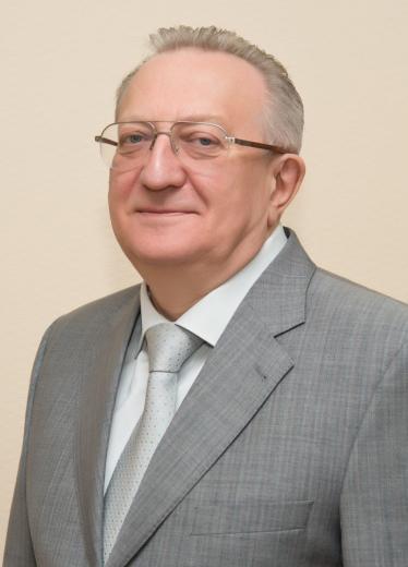
В.В.Гладько

Dear colleagues, Dear friends!

On February 16, 2023, the Academic Council of the Federal State Budgetary Educational Institution of Higher Education "ROSBIOTECH" approved the development concept of the Medical Institute of Continuous Education for the next five years (2023–2027). We have been actively working on it since the beginning of the year.

Since January 2023, the Medical Institute of Continuous Education has been implementing the new areas of training for specialists in higher education programs, such as residency in order to meet the demands for the educational services in the following majors: 31.08.10 Forensic medicine, 31.08.52 Osteopathy, 31.08.53 Endocrinology, 31.08.56 Neurosurgery.

This year the major 31.05.01 "General Medicine" for future specialists with higher medical education will be opened (Specialist degree — 6 years).

The introduction of new areas of training specialists requires significant step-by-step changes in the organizational and staffing structure of the Medical Institute of Continuous Education. From 2023 to 2027, 15 new departments will be created and 3 departments will be reorganized to carry out the educational process at the MINO.

The merger of Russian Biotechnological University with the Pushchino State Institute of Natural Science will increase the scientific and educational potential of ROSBIOTECH, strengthen its positioning as the University of life sciences, where educational, scientific and practical activities are carried out in the field of biology, ecology, food safety, medicine and veterinary medicine. The integration of the Medical Institute of Continuous Education with a number of institutes and departments of ROSBIOTECH will make it possible to create a system and infrastructure of translational medicine as a technological platform for modern medical science. It determines the optimal mechanisms for introducing the most significant achievements of fundamental science into clinical practice for the fastest solving of clinical and preventive medicine problems. The availability of educational technologies in the field of preclinical, experimental and translational studies will allow practicing all types of manual skills on experimental models, developing and introducing into clinical practice new technologies that improve the quality of prevention, diagnostics and treatment of diseases. It will help conduct all types of studies of the safety and efficacy of medications and pharmacological substances, as well as biomedical cell products.

The listed development directions of the Medical Institute of Continuing Education will further expand the range of scientific publications in the journal "Bulletin of the Medical Institute of Continuing Education". The year 2022 has become a fruitful and successful year for the journal. Since December it has been indexed in the RSCI (all articles previously published in our journal received the scientific index RSCI). Congratulations to all of us!

Currently, the editorial board is actively working on including the journal "Bulletin of the Medical Institute of Continuing Education" in the List of peer-reviewed journals of the Higher Attestation Commission. We expect to get the help of our authors in this work.

Best regards, Editor-in-Chief, MD, PhD, Prof. V.V. Gladko

DERMATOVENEREOLOGY

SKIN-LIMITED VASCULITIS

ВV.O. Maksimovich1, S.A. Masyukova2, I.A. Lamotkin2, I.V. Ilyina2, O.V. Gladko2

10-15

1 Aesthetic Medicine Center of Dr. Shumskaya, Moscow, Russia
2 Medical Institute of Continuing Education, Russian Biotechnological University (ROSBIOTECH), Moscow, Russia

Summary

The article considers the classification of cutaneous vasculitis, the main clinical manifestations, differential diagnostics and treatment principles.

Key words: angiitis, dermal vasculitis, viral and bacterial infections, classification, clinical forms.

For citation: Maksimovich V.O., Masyukova S.A., Lamotkin I.A., Ilyina I.V., Gladko O.V. Skin-limited vasculitis. // Bulletin of the Medical Institute of Continuing Education. – 2023. – V. 3. – No 1. – P. 10-15. – DOI 10.36107/2782-1714_2023-3-1-10-15.

The authors declare no funding to study.

The authors declare no apparent or potential conflicts of interest related to the publication of this article.

Literature

  1. Федеральные клинические рекомендации. Дерматовенерология 2015: Болезни кожи. Инфекции, передаваемые половым путем. — 5-е изд., перераб. и доп. — М.: Деловой экспресс, 2016. — 768 с.
  2. Клинические рекомендации. Васкулиты, ограниченные кожей (L95) // Российское общество дерматовенерологов и косметологов. М.:2020 https://www.cnikvi.ru/ klinicheskie-rekomendatsii-2017/КР Васкулиты, ограниченные кожей 2020.docx
  3. Баркаган З.С. Геморрагические заболевания и синдромы. 2-е издание 1988 г.
  4. Насонов Е.Л., Баранов А.А., Шилкина Н.П. Васкулиты и васкулопатии. — Ярославль: Верхняя Волга, 1999. — С. 479–515.
  5. Сорокина Е.В., Масюкова С.А., Ахматова Н.К. Эритемы: руководство для врачей — Москва : КнигИздат, 2019. — 183 с.: цв. ил.
  6. Crowson A.N., Mihm Jr. M.C., Magro C.M. Cutaneous vasculitis: a review. J Cutan Pathol. 2003; 30:161-173.
  7. Клиническая характеристика кожных проявлений при новой коронавирусной инфекции COVID-19, вызванной SARS-CoV-2. Том 21. 3’2020; Москва 2020 г.
  8. Котова И.С., Шишева Р.А. Учебное пособие «Ангииты кожи» Благовещенск 2013 г.
  9. Иванов О.Л. Справочник кожные и венерические болезни. Издательство «Медицина», 2007 г.
  10. Дерматовенерология. Национальное руководство под ред. Ю.С. Бутова, Ю.К. Скрипкина, О.Л. Иванова. — М. : ГЭОТАР-Медиа, 2013. — 896 с.
  11. Millikan L.E., Flynn T.C. Infectious etiologies of cutaneous vasculitis. Clin Dermatol 1999 г.; 17: 509–514
  12. Шапошников О.К., Браиловский А.Я., Разнатовский И.М., Самов В.И. Ошибки в дерматологии. М: Медицина 1987 г.; 88–89.
  13. Клиническая дерматология. Иллюстрированное руководство для врачей / А.Н. Родионов, Д.В. Заславский, А.А. Сыдиков; под ред. А.Н. Родионова. — 2-е изд., перераб. и доп. — М. : ГЭОТАР-Медиа, 2019. — 712 с. : ил.
  14. Елькин В.Д., Митрюковский Л.С., Седова Т.Г. Избранная дерматология. Пермь 2004; 846–847.

TREATMENT OF DEMODEKOSIS: TRADITIONS AND REALITY

Sokolova T.V., Golitsyna M.V., Ilyina I.V., Malyarchuk A.P.

16-23

Medical Institute of Continuing Education, Russian Biotechnological University (ROSBIOTECH), Moscow, Russia

Summary

Demodicosis is a parasitic skin disease caused by mites of the genus Demodex, the most common conditionally pathogenic parasite in the human skin microbiocenosis. An increase in their number aggravates the course of dermatoses of facial localization, which indicates the expediency of determining the degree of tick-borne invasion and the need to use antiparasitic drugs. In clinical practice, for the treatment of demodicosis, not only antiparasitic drugs (benzylbenzoate, permethrin, ivermectine), but also drugs with a different mechanism of action (metronidazole, ornidazole, ketoconazole, isotretinoin) are used. The results of comparing their effectiveness are contradictory. An analysis of the literature data allows us to identify several reasons that confirm this and justify the need for a personalized rational approach to the choice of tactics for the treatment of demodicosis.

Key words: demodicosis, treatment, efficiency, expert opinion.

For citation: Sokolova T.V., Golitsyna M.V., Malyarchuk A.P., Ilyina I.V. Treatment of demodicosis: traditions and reality. // Bulletin of the Medical Institute of Continuous Education. – 2023. – V. 3. – No 1. – P. 16-23. – DOI 10.36107/2782-1714_2023-3-1-16-23.

The authors declare no funding to study.

The authors declare no apparent or potential conflicts of interest related to the publication of this article.

Literature

  1. Akbulatova L.Kh. Morphology of two forms of the demodex folliculorum hominis mite and its role in human skin diseases. Abstract diss ... cand. honey. Sciences. 1968; Tashkent: 27 p.
  2. Kogan B.G. Clinical and immunopathological features, diagnosis and treatment of demodicosis: Ph.D. Dis. ... cand. honey. Sciences. 1995: 23 p.
  3. Sokolov V.O., Morozova N.V., Polovinkina G.V., Khramtsova M.A. Demodicosis of the eyes in outpatients in St. Petersburg (according to the St. Petersburg State Budgetary Healthcare Institution of Healthcare Center No. 7 (ophthalmic). Ophthalmological journals. 2013; VI (3): 83-87.
  4. Kanyukov V.N., Bannikov V.K., Malgina E.K. Demodicosis of the eye: problems and solutions. Ophthalmosurgery. 2015; 1:48-52.
  5. Tretyakova I.P. Diagnosis of a parasitic skin disease. Young scientist. 2018; 191(5): 77-80.
  6. Biernat M.M., Rusiecka-Ziolkowska J., Piatkowska E. et al. Occurrence of Demodex species in patients with blepharitis and inhealthy individuals: a 10-year observational study. Jpn J Ophthalmol. 2018; 62(6): 628-633.
  7. Romanina D.M. study of the market of pharmaceuticals for the treatment of demodicosis. Pharmacy. Series "Medicine". 2014; 175(4): Issue. 25:201-206.
  8. Salem D.A-B., El-shazly A., Nabih N. et al. Evaluation of the efficacy of oral ivermectin in comparison with ivermectin-metronidazole combined therapy in the treatment of ocular and skin lesions of Demodex folliculorum. International Journal of Infectious Diseases. 2013; 17:343-347
  9. Yutskovskaya Ya.A., Kusaya N.V., Klyuchnik S.B. Substantiation of pathogenetic therapy for acneform dermatoses complicated by tick-borne invasion of Demodex folliculorum. Clinical dermatology and venereology. 2010; 3:60-63.
  10. Kubanov A.A., Galyamova Yu.A., Grevtseva A.S. Demodicosis. Tutorial. M. : GBOU DPO RMAPO. 2014; 41 p.
  11. Adaskevich V.P. Demodicosis: etiology, pathogenesis, clinical forms, diagnosis, treatment. "Dermatovenereology Cosmetology". 2015; 1(1):60-71.
  12. Forton F., Seys B., Marchal J.L., Song A.M. Demodex folliculorum and topical treatment: acaricidal action evaluated by standardized skin surface biopsy. Br. J. Dermatol. 1998;138(3): 461–466.
  13. Forton F.M.N., De Maertelaer V. Treatment of rosacea and demodicosis with benzyl benzoate: effects of different doses on Demodex density and clinical symptoms. J Eur Acad Dermatol Venereol. 2020; 34(2): 365–369.
  14. Swenor M.E. Is permetrin 5% cream effective for rosacea? J. Fam. Pract. 2003; 52:183-84.
  15. Dressler C., Rosumeck S., Sunderkötter C. et al. The treatment of scabies. Dtsch Arztebl Int. 2016;113: 757–762.
  16. Hecht I., Melzer-Golik A., Sadi Szyper N., Kaiserman I. Permethrin cream for the treatment of Demodex blepharitis. Cornea. 2019;.38. (12):1513–1518
  17. Sarac G. A comparison of the efficacy and tolerability of topical agents used in facial Demodex treatment. J Cosmetic Dermatol. 2019;18(6):1784–1787.
  18. Verkhoglyad I.V. Modern ideas about demodicosis. Attending doctor. 2011; 5:34-35
  19. Ponic E.S., Savosteeva M.S., Experience with the use of ivermectin in the treatment of rosacea. Health care of Yugra: experience and innovations. 2018; 1:43-45.
  20. Holzchuh F.G., Hida R.Y., Moskovici B.K. et al. Clinical treatment of ocular Demodex folliculorum by systemic ivermectin. Am. J Ophthalmo. 2011;151: 1030-1034.
  21. Gupta G., Daigle D., Gupta A.K., Gold L.S. Ivermectin 1% cream for rosacea. Skin Therapy Lett. 2015:20(4):9–11.
  22. Schaller M., Gonser L., Belge K. et al. Dual anti-inflammatory and anti-parasitic action of topical ivermectin 1% in papulopustular rosacea. J Eur Acad Dermatol Venereol. 2017; 31(11): 1907–1911.
  23. Danilova A.A., Fedorov S.M. Parasitic diseases of the skin. Demodicosis. 2001; 8(6):249–254.
  24. Kogan B.G. Modern aspects of the pathogenesis and clinical course of demodicosis. Ukrainian journal of dermatology, venereology and cosmetology. 2002; 6:13-15.
  25. Sirmais N.S., Ustinov M.V. Clinical efficacy of dermoten gel in the complex treatment and prevention of demodicosis and rosacea. Bulletin of dermatology and venereology. 2011:6:85-90.
  26. Campbell W.C., Fisher M.H., Stapley E.O., et al. Ivermectin: A Potent New Antiparasitic Agent. Science. 1983; 221:823-828.
  27. Scopets B., Wilson R.P., Griffith J.M., Lang C.M. Ivermectin toxicity in young mice. Lab Anim Sci. 1996; 46:111-112.
  28. Kane N.S., Hirschberg B., Qian S et. al. Drug-resistant Drosophila indicate glutamate-gated chloride channels are targets for the antiparasitics nodulisporic acid and ivermectin. Proc Natl Acad Sci USA. 2000; 97: 13949-13954.
  29. Martin R.J., Robertson A.P., C.houdhary S. Ivermectin: an anthelmintic, an insecticide, and much more. Trends Parasitol. 2021; 37(1): 48–64.
  30. Samtsov A.V. The evolution of ideas about rosacea. Dermatology and cosmetology. 2020; 39:52-58.
  31. Stankiewicz M., Cabaj, W., Jonas, W. E. et al. Influence of ivermectin on cellular and humoral immune responses of lambs. Veterinary Immunology and Immunopathology. 1995, 44(3-4): 347–358.
  32. Schaller M., Gonser L., Belge K. et al. Dual anti-inflammatory and anti-parasitic action of topical ivermectin 1% in papulopustular rosacea. J Eur Acad Dermatol Venereol. 2017; 31(11): 1907–1911.
  33. Logger J.G.M., Peppelman M., van Erp P.E.J. et al. Value of reflectance confocal microscopy for the monitoring of rosacea during treatment with topical ivermectin. J Dermatolog Treat. 2022; 33:195–203.
  34. Schmaidel L.K., McEvoy C.K. Topical metronidazole: a new therapy in rosacea. Clinic Pharm. 1990; 9; 94-101.
  35. Werner H., Krasemann C., Kandler R., Wandmacher G. Metronidazole sensitivity of anaerobes. A comparison with other chemotherapeutics (author's transl). MMW Munch Med Wochenschr. 1980; 122:633–636.
  36. Grove D.I., Mahmoud A.A.F., Warren K.S. Suppression of cell mediated Immunity by Metronidazole. Int. Archs Allergy Appl. Immunol. 1997; 54:422–426.
  37. Barnhorst D., Foster J., Chern K. The efficacy of topical metronidazole in the treatment of ocular rosacea. Ophthalmology. 1996;103(11):1880–1883.
  38. Kubanov A.A., Galyamova Yu.A., Kravchenko A.S. Principles of therapy for demodicosis. Pharmateka. 2017; 20:25-30.
  39. Shelley W.B., Shelley E.D., Burmeister V. Unilateral demodectic rosacea. J Am Acad Derma Atol. 1989; 20:915-917.
  40. Pallotta S., Cianchni G., Martelloni E. et al. Unilateral demodicidosis. Eur J Dermatol. 1998; 8:191-192.
  41. Frankenberg A.A., Shevchenko V.A., Krivko S.V., Shlyakhova V.A. Experience with the use of ornidazole in the complex therapy of demodicosis. Worm. 2007; 2:10-12.
  42. Cardwell L.A., Alinia H., Moradi Tuchayi S., Feldman S.R. New developments in the treatment of rosacea - role of once-daily ivermectin cream. ClinCosmet Investig Dermatol. 2016; 9:71-77.
  43. SatRtler E.C., Hoffmann V.S., Ruzicka T. et al. Reflectance confocal microscopy for monitoring the density of Demodex mites in patients with rosacea before and after treatment. Br J Dermatol. 2015; 173(1): 69–75.
  44. Shchetkina M.V. Clinical features, diagnosis and treatment of skin demodicosis. Health care of the Far East. 2018; 1:99-102.
  45. Kravchenko A.N. Clinical and diagnostic features of rosacea in the detection of mites of the genus Demodex. Abstract dis. ... cand. honey. Sciences. 2019. M: 24 p.
  46. Chen W., Plewig G. Human demodicosis: revisit and a proposed classification. British J. Dermatol. 2014; 170:1219–1225.
  47. Belousova N.Yu., Poltanova T.I. Demodex in humans (literature review). Ural Medical Journal, section "Dermatology". 2019; 180 (12:126-132)
  48. Armagan B.Y., Atakan N. The Effect of Different Therapeutic Modalities on Demodex Densities and Clinical Symptoms of Patients with Demodicosis. Acta Medica. 2022; 53(1): 83-89.
  49. Taieb A., J.P. Ortonne J.P., Ruzicka T. et al. Superiority of ivermectin 1% cream over metronidazole 0•75% cream in treating inflammatory lesions of rosacea: a randomized, investigator-blinded trial. Br. J Dermato. 2015; 172(4): 1103-1010.
  50. Grossmann B., Jung K., Linse R. Tubero-pustular demodecosis. Hautarzt. 1999 50 491-494.
  51. Anane S., Mokni M., Beltaief O. Rosacea-like demodicidosis and chronic blepharitis. Ann Dermatol Venerol. 2011; 138:30-30
  52. Batyrshina S.V., Gordeeva A.M., Bogdanova M.A. Efficiency of skinoren gel in external therapy of patients with acne and rosacea. Bulletin of dermatology and venereology. 2005; (4): 25-28.
  53. Polesko I.V., Osipov G.A., Kabaeva T.I. Microecology of the human body in seborrhea and acne. Children's infections. 2006; 5(3):26-33.
  54. Norn M.S. Demodex folliculorum. Incidence, regional distribution, pathogenicity. Danish Med Bulletin. 1971; 18(1):14-17.
  55. Lacey N., Delaney S., Kavanagh K. et al. Mite related bacterial antigens stimulate inflammatory cells in rosacea. Br J Dermatol. 2007; 157:474-481.
  56. Mokronosova M.A., Glushakova A.M., Golysheva E.V., Zheltikova T.M. Mites of the genus Demodex and yeast of the genus Malassezia in patients with seborrheic dermatitis. Bulletin of dermatology and venereology. 2012; 3:92-98.
  57. Tchernev G. Folliculitis et perifolliculitis capitis abscedens et suffodiens controlled with a combination therapy: Systemic antibiosis (Metronidazole Plus Clindamycin), dermatosurgical approach, and high-dose isotretinoin. Indian Journal of Dermatology 2011; 56(3): 318-320.
  58. Anane S., Anane T. R., Malouche N. et al. Which is the role of parasites and yeasts in the genesis of chronic blepharitis? Pathologie Biology. 2007; 55(7): 323-327.
  59. Akilov O.E. Clinical assessment of the relationship between immune system disorders and HLA-histiotype features in patients with skin demodicosis. Abstract dis.... cand. honey. Sciences. M. 2002: 25 p.
  60. Olisova O.Yu., Kochergin N.G., Smirnova E. Modern topical therapy for moderate forms of rosacea. Russian journal of skin and venereal diseases. 2016; 19(6):328-324.
  61. Wolff K., Goldsmith L.A., Katz S.I., Gilchrest B.A., Paller A.S., Leffell D.J. Fitzpatrick's Dermatology in General Medicine. Transl. from English. Moscow: BINOM; 2012; 1; 769–774.
  62. Demina O.M., Potekaev N.N. Modern etiopathogenetic mechanisms of rosacea development and new methods of therapy. Clinical dermatology and venereology. 2017; 16:313-323.

CHERNOGUBOV — EHLERS — DANLOS SYNDROME AT THE INTERSECTION OF CLINICAL SPECIALTIES

V.V. Mordovtseva, V.V. Gladko

24-28

Medical Institute of Continuing Education, Russian Biotechnological University (ROSBIOTECH), Moscow, Russia

Summary

The majority of patients with hereditary connective tissue diseases have cutaneous signs, joint hypermobility and sometimes pathology of other organs and systems. The review of the major symptoms of this syndrome is given in the article.

Key words: hereditary connective tissue diseases, cutis hyperelastica, joint hypermobility.

For citation: Mordovtseva V.V., Gladko V.V. Chernogubov — Ehlers — Danlos syndrome at the intersection of clinical specialties. // Bulletin of the Medical Institute of Continuing Education. – 2023. – V. 3. – No 1. – P. 24-28. – DOI 10.36107/2782-1714_2023-3-1-24-28.

The authors declare no funding to study.

The authors declare no apparent or potential conflicts of interest related to the publication of this article.

Literature

  1. Tschernogobow A. Cutis laxa (Moscow Dermatologic and Venereologic Society, November 1891). Mhft Prakt Derm; 1892:14e76.
  2. Castori M. Ehlers-danlos syndrome, hypermobility type: an underdiagnosed hereditary connective tissue disorder with mucocutaneous, articular, and systemic manifestations. ISRN Dermatol. 2012;2012:751768.
  3. Hamonet C., Ducret L., Baeza-Velasco C.L., Layadi K. Ehlers-Danlos-Tschernogobow: Histoire contrarlée de la maladie. Hist Sci Med. 2016 Jan-Mar; 50 (1): 29–41. French. PMID: 27349123.
  4. Denko C.W. Chernogubov's syndrome: a translation of the first modern case report of the Ehlers-Danlos syndrome. J Rheumatol. 1978 Fall;5(3):347–52.
  5. Islam M., Chang C., Gershwin M.E. Ehlers-Danlos Syndrome: Immunologic contrasts and connective tissue comparisons. J Transl Autoimmun. 2020 Dec 20;4:100077.
  6. Kirk J.A., Ansell B.M. Bywaters EGL. The hypermobility syndrome: musculoskeletal complaints associated with generalized joint hypermobility. Ann Rheum Dis. 1967;26:419–425.
  7. Mishra M.B., Ryan P., Atkinson P. et al. Extra-articular features of benign joint hypermobility syndrome. Br J Rheumatol1996;35:861–6.
  8. Grahame R. Joint hypermobility and genetic collagen disorders: are they related? Arch Dis Child 1999;80:188–91. 9. Tofts LJ., Elliot EJ., Munns C. et al. The differential diagnosis of children with joint hypermobility: a review of the literature. Pediatric Rheumatology 2009, 7:1
  9. F. Malfait, A.J. Hakim, A. De Paepe and R. Grahame. The genetic basis of the joint hypermobility syndromes. Rheumatology 2006;45:502–507.
  10. Tinkle B., Castori M., Berglund B., Cohen H., Grahame R., Kazkaz H., Levy H. Hypermobile Ehlers-Danlos syndrome (a.k.a. Ehlers-Danlos syndrome Type III and Ehlers-Danlos syndrome hypermobility type): Clinical description and natural history. Am J Med Genet C Semin Med Genet. 2017 Mar; 175 (1): 48–69.
  11. Jelsma L.D., Geuze R.H., Klerks M.H., et al. The relationship between joint mobility and motor performance in children with and without the diagnosis of developmental coordination disorder Jelsma et al. BMC Pediatrics 2013, 13:35.
  12. van der Giessen L.J., Liekens D., Rutgers KJ.M., et al. Validation of Beighton score and prevalence of connective tissue signs in 773 Dutch children. J of Rheum 2001, 28:2726–2730.
  13. Bulbena A., Baeza-Velasco C., Bulbena-Cabre A., Pailhez G., Critchley H., et al. (2017) Psychiatric and psychological aspects in the Ehlers-Danlos syndromes. Am J Med Genet Part C Semin Med Genet 175: 237–245.
  14. Hamonet C., Brissot R., Anne Gompel A., Baeza-Velasco C., Guinchat V., et al. (2018) Ehlers-Danlos Syndrome, contribution to clinical diagnosis about 853 cases. EC neurology10: 6.
  15. Malfait F., Francomano C., Byers P., Belmont J., Berglund B., et al. The 2017 international classification of the Ehlers-Danlos syndromes. Am J Med Genet C Semin Med Genet. 2017 Mar; 175 (1): 8–26.
  16. Adib N., Davies K., Grahame R., Woo P., Murray K.J. Joint hypermobility syndrome in childhood. A not so benign multisystem disorder? Rheumatology (Oxford). 2005Jun; 44 (6): 744–50.
  17. Smits-Engelsman B., Klerks M., Kirby A. Beighton score: a valid measure for generalized hyper mobility in children. J Pediatr 2011, 158 (1): 119–123.

RESTORATIVE MEDICINE

THE USE OF CPAP THERAPY IN THE MEDICAL REHABILITATION OF PATIENTS WITH CORONARY HEART DISEASE WITH OBSTRUCTIVE SLEEP APNEA SYNDROME AFTER CORONARY ANGIOPLASTY

V.E. Yudin1, V.V. Klimko1, A.M. Shchegolkov1,3, S.V. Kalinina2,3, T.V. Chursina1

30-33

1 Medical Institute of Continuing Education, Russian Biotechnological University, Moscow, Russia
2 Branch No. 2 of The National Medical Research Center of High Medical Technologies, The Central Military Clinical Hospital named after A.A.Vishnevsky, Russian Ministry of Defense, Moscow, Russia
3 Branch of Military Medical Academy, Moscow, Russia

Summary

The article analyzes the diagnostic results of 245 patients with coronary heart disease after coronary angioplasty. Obstructive sleep apnea was detected in 35.7% of patients. According to the rehabilitation programs, the patients were divided into two groups of 30 people in each one, comparable in age, severity of coronary artery disease, number of surgical interventions, severity of OSA and pharmaseutical treatment. The patients of the main group were prescribed CPAP therapy along with the standard therapy. The effectiveness of CPAP therapy use in the medical rehabilitation of patients with ischemic heart disease with OSA syndrome after coronary angioplasty has been studied. The inclusion of the technique in the comprehensive program of medical rehabilitation in this category leads to a decrease in the number of complaints, improvement of the functional and psychological state, stabilization of the cardiorespiratory system and increased tolerance to physical activity.

Key words: Coronary heart disease, OSA, CPAP therapy, medical rehabilitation, coronary angioplasty.

For citation: Yudin V.E., Klimko V.V., Shchegolkov A.M. [et al.] The use of C-PAP therapy in the medical rehabilitation of patients with coronary heart disease with obstructive sleep apnea after coronary angioplasty. // Bulletin of the Medical Institute of Continuing Education. – 2023. – V. 3. – No 1. – P. 30-33. – DOI 10.36107/2782-1714_2023-3-1-30-33.

The authors declare no funding to study.

The authors declare no apparent or potential conflicts of interest related to the publication of this article.

Literature

  1. Араблинский А.В. Транслюминальная баллонная ангиопластика у больных с многососудистым поражением коронарного русла // Клиническая медицина. — 2001.—NoI.—С.14–18.
  2. Золотухин Н.Н., Коньков А.В. Лечение и медицинская реабилитация при остром коронарном синдроме на стационарном этапе. Вестник Медицинского института непрерывного образования. 2022;(3):8–11. DOI 10.46393/27821714_2022_3_8
  3. Базылев В.В., Тунгусов Д.С., Микуляк А.И. Результаты коронарного шунтирования у пациентов с низкой фракцией выброса. Вестник Медицинского института непрерывного образования.2022; (1): 62–65.DOI 10.46393/27821714_2022_1_62
  4. Мандрыкин С.Ю., Щегольков А.М., Анучкин А.А. Современное состояние проблемы эндоваскулярного лечения и реабилитации больных ишемической болезнью сердца.// Физиотерапия, бальнеология, реабилитация: Научно-практический журнал. — 2004. —No3.—С.38–42.
  5. Калинина С.В. Автореферат. канд. мед. наук 2013. Особенности клинико-функционального состояния и медицинской реабилитации больных ишемической болезнью сердца с синдромом обструктивного апноэ-гипопноэ сна после коронарной ангиопластики. С. 26.
  6. De Scheerder., Chevalier B., Vassanelli C. et al. European freedom stent registry //Eur. Heart J. — 1997. — Vol.l8. — P. 156.
  7. Shamsuzzaman AS, Gersh BJ et al. Obstructive sleep apnea: implications for cardiac and vascular disease. JAMA 2003; 290: 1906–14.
  8. Milleron O, Pilliere R et al. Benefits of obstructive sleep apnoea treatmentin coronary atery disease:a long-term follow-up study. Eur Heart J 2004; 25: 728–34.
  9. Пальман А.Д. Синдром обструктивного апноэ во сне в клинике внутренних болезней / под редакцией А.И. Синопальникова — М., 2007—78. С. 59–60.
  10. Мостовой Л.В. Определение дополнительных критериев тяжести Синдрома обструктивного апноэ сна и прогноза приверженности больных к долгосрочной СИПАП-терапии: Дис. канд. мед. наук, — М., 2022 С. 4.
  11. Бузунов Р.В., Пальман А.Д., Мельников А.Ю., Авербух В.М., Мадаева И.М., Куликов А.Н. Диагностика и лечение синдрома обструктивного апноэ сна у взрослых. Рекомендации Российского общества сомнологов «Клиническая сомнология»// 2018 — С. 35–45.

FEATURES OF THE CORONAVIRUS DISEASE COURSE OF A PATIENT IN THE THIRD TRIMESTER OF PREGNANCY: CLINIC, TREATMENT AND APPROACHES TO REHABILITATION

V.E. Yudin, V.V. Klimko, T.V. Chursina, A.M. Chegolkov, K.A. Soboleva

34-38

Russian Biotechnological University, Moscow, Russia

Summary

The article presents a clinical case of rehabilitation of a young patient in the third trimester of pregnancy who had a severe form of coronavirus disease. The case demonstrates the importance of timely specialty medical rehabilitation care, which may be crucial in preserving health, improving life quality of patients with pneumonia associated with the new coronavirus infection COVID-19.

Key words: new coronavirus infection, the third trimester of pregnancy, medical rehabilitation.

For citation: Yudin V.E., Klimko V.V., Chursina T.V. [et al.] Features of the coronavirus disease course of a patient in the third trimester of pregnancy: clinic, treatment and approaches to rehabilitation. // Bulletin of the Medical Institute of Continuing Education. – 2023. – V. 3. – No 1. – P. 34-38. – DOI 10.36107/2782-1714_2023-3-1-34-38.

The authors declare no funding to study.

The authors declare no apparent or potential conflicts of interest related to the publication of this article.

Literature

  1. Временные методические рекомендации: «Профилактика, Диагностика и лечение новой коронавирусной инфекции Covid-19». Минздрав России. Версия 15 (22.02.2022).
  2. Варфоломеева М.И., Кулагина И.Ц., Зайцев А.А. Роль иммунной системы при новой коронавирусной инфекции SARS-CoV-2. Вестник медицинского института непрерывного образования, 2021; (1): С. 28–31
  3. Макарова М.А., Шакурова Л.Р., Новоселова О.Н., Руденкина О.А., Баймаканова Г.Е., Красовский С.А., Бродская О.Н., Белевский А.С. Новая коронавирусная болезнь 2019 г. в Москве на примере клинических случаев. Практическая пульмонология, 2020, No 1, С. 29–36
  4. Разумов А.Н., Пономаренко Г.Н., Бадтиева В.А. Медицинская реабилитация пациентов с пневмониями, ассоциированными с новой коронавирусной инфекцией COVID-19. Вопросы курортологии, физиотерапии и лечебной физической культуры, 2020, Т. 97, No 3, С. 5–13.
  5. Гетманская Ю.М., Трегер Ю.А. Реабилитация тяжелых больных после коронавирусной инфекции в рамках госпитализации в реабилитационном отделении. Анализ конкретных случаев. Вестник восстановительной медицины, Том 20, No 3 (97). 2020 — С. 14–19.
  6. Ефремова О.Ю., Зайцев А.А., Золотухин О.В. Опыт применения гипербарической оксигенации у кислородзависимых пациентов с тяжелыми формами коронавирусной инфекции. Вестник медицинского института непрерывного образования, 2022; (2): С. 25–27
  7. Малявин А.Г., Бабак С.Л., Горбунова М.В. Респираторная реабилитация постковидных пациентов. The Russian Archives of Internal Medicine, 2021, No 1, С. 22–33.
  8. Методические рекомендации: Организация оказания медицинской помощи беременным, роженицам, родильницам и новорожденным при новой коронавирусной инфекции Сovid-19. Версия 4 (05.07.2021)
  9. Шмонин А.А., Мальцева М.Н., Мельникова Е.В., Мишина И.Е., Иванова Г.Е. Медицинская реабилитация при коронавирусной инфекции: Новые задачи для физической и реабилитационной медицины в России. Вестник восстановительной медицины No 3 (97). 2020 — С. 14–19.
  10. Юдин В.Е., Щегольков А.М., Ярошенко В.П., Будко А.А., Климко В.В., Калинина С.В. Физические методы медицинской реабилитации пациентов, перенесших новую коронавирусную инфекцию COVID-19. Учебное пособие — М. : МГУПП, 2021. — 40 с.

PHANTOM LIMB SYNDROME: CLINIC, DIAGNOSTIC ASSESSMENT AND TREATMENT (LITERATURE REVIEW)

V.E. Yudin, V.P. Yaroshenko, E.S. Kosukhin, A.A. Budko, M.E. Ustinova, V.G. Trubina

39-43

Russian Biotechnological University, Moscow, Russia

Summary

The article analyzes the literature data on the problem of diagnostic assessment, prevention, treatment and medical rehabilitation of the victims with the phantom limb pain. According to the various authors, the phantom limb pain occurs in 35-85% of the cases. There are different views on the pathogenesis of phantom pain syndrome, modern approaches of the treatment choice and rehabilitation methods. The analysis of the Russian and foreign literature data showed that the study of pain syndrome, the development of modern prevention and treatment techniques of patients with phantom limb syndrome is a relevant medical objective nowadays.

Key words: phantom limb syndrome , chronic pain, diagnostic assessment, medical rehabilitation.

For citation: Yudin V.E., Yaroshenko V.P., Kosukhin E.S. [et al.] Phantom limb syndrome: clinic, diagnostic assessment and treatment (literature review). // Bulletin of the Medical Institute of Continuing Education. – 2023. – V. 3. – No 1. – P. 39-43. – DOI 10.36107/2782-1714_2023-3-1-39-43.

The authors declare no funding to study.

The authors declare no apparent or potential conflicts of interest related to the publication of this article.

Literature

  1. 1. Гнездилов A.B. «Диагностика и лечение фантомного и вертеброгенного болевых синдромов»: Дис. д-р мед. наук /Российский научный центр хирургии РАМН (РНЦХ РАМН). М., 1999. 232 с.
  2. Кукушкин М.Л. Системные механизмы развития нейрогенных болевых синдромов и их патогенетическая коррекция: Автореф. дисс. д-ра мед. наук. — М., 1995. — 35 С.
  3. Плохов Д.А. Профилактика выраженных клинических проявлений фантомно-болевого синдрома после ампутации нижней конечности на уровне бедра у пациентов с критической ишемией //Дисс...канд. мед. наук. — М., 2007. — 133 с.
  4. Архангельский А.Е. Фантомно-болевой синдром. Лекция для слушателей факультета подготовки врачей / А.Е. Архангельский. — СПб.: ВМедА, 2006. — С. 1–16.
  5. Давыдов А.Т., Тюкавин А.И., Резванцев М.В., Конончук В.В., Шабанов П.Д. Фантомная боль, роль и место различных видов лечения фантомно-болевого синдрома // Обзоры по клинической фармакологии и лекарственной терапии. — 2014. — С. 35–58.
  6. Чегуров О.К. Фантомно-болевой синдром: патогенез, лечение, профилактика (обзор литературы) / О.К. Чегуров [и др.] // Гений ортопедии. — 2014. — No 1. — С. 89–93.
  7. Архангельский А.Е. Фантомно-болевой синдром. Лекция для слушателей факультета подготовки врачей / А.Е. Архангельский. — СПб.: ВМедА, 2006. — С. 1–16.
  8. Сайко А.В. Современное состояние проблемы фантомной боли // Междунар. неврол. журн. —No 4. — 2018. — С. 84–93. 9. ГанпацуроваО.Б.,ПомогаеваА.Б. Фантомные боли как психологический феномен // Смальта. — No 2. — 2019. - C. 11–15.
  9. Юдин В.Е., Ярошенко В.П., Поправка С.Н. Опыт организации протезирования и медицинской реабилитации военнослужащих с ампутацией конечностей // Военно-медицинский журнал. — No4. — 2021. — С. 18–25.
  10. Давыдов О.С , Яхно Н.Н ,Кукушкин М.Л, Чурюканов М.В, Абузарова Г.Р , Амелин А.В , Балязин В.А и др. Невропатическая боль: клинические рекомендации по диагностике и лечению Российского общества по изучению боли. — 2018. — С. 5–41.
  11. Устинова М.Е., Трубина В.Г., Ишук Ю.Г. Комбинация зеркальной техники и ТКМС в лечении фантомных болей // сборник материала научно-практического семинара СПб ЦПО ФМБА России . — No 10. — 2022.
  12. Albe-Fessard D., Giamberardino M.A., Rampin O. Comparison of animal models of chronic pain. //Adv. Pain Res. and Ther. 1990. — V. 13. — P. 11–27
  13. Wool C., Chong M. Preemptive analgesia treating postoperative pain by preventing the establishment of central sensitization // Anesth.Analg. — 1993. — Vol. 77. — P.1–18.
  14. Woolf C.J. Central mechanisms of acute pain. // Proceeding of the Vl-th World Congress on Pain (eds. M.R. Bond, J.E. Charlton, C.J. Woolf), 1991. — P. 25–34.
  15. Kenneth Maiese MD "neurotransmitters and receptors disorders associated with defects in neurotransmission" // MSD manuals . April 2022 p. 6–7
  16. Burchiel K.J. "Carbamazepine inhibits spontaneous activity in experimental neuromas". // Exp. Neurolog. 1988. V. 102. p. 249–253.
  17. Loeser J.D. Tic douloureux and atypical facial pain. In Wall P.D., Melzack R., eds. Textbook of Pain, 3nd Ed. — London, Churchill Livingstone, 1994. — P. 699–710.
  18. Seltzer Z., Beilin B., Ginzburg R. et al. The role of injury discharge in the induction of neuropathic pain behavior in rats. // Pain. — 1991. V. 46. No. 3 — P. 327–336.
  19. Muhhammed Ernes Gunduz MD, Kevin Pacheco–Barrios MD, Fellipe Fregni MD "Effects of cimbined and alone transcranial motor cortex stimulation and mirrow therapy in phantom limb Pain, randomised factoral trial" // Sage Journal . June 2021 . — P. 10–11.
  20. James C., Watson M.D. "overview of pain" // MSD manual. 2022 p. 3–5.

SURGERY

CHALLENGES IN DIAGNOSING SPLENIC ABSCESSES IN HIV-INFECTED PATIENTS

S.S. Puchkov1, A.P. Faller1,2

44-51

1 Infectious Clinical Hospital No. 2, Moscow, Russia
2 Medical Institute of Continuing Education, Russian Biotechnological University, Moscow, Russia

Summary

Objective: Improve the quality of treatment for HIV-infected patients with heart attacks and splenic abscesses by optimizing the diagnosis of these pathological processes.

Material and Methods: From 2004 to 2019, 14,405 HIV-infected patients, including 34 patients with suspected splenic abscesses, were treated in the surgical department of the Infectious Clinical Hospital State Budgetary Healthcare Institution (ICH SBHI) No.2. The average age was 34.9 years, 22 males and 12 females. Three patients had stage 3, ten people had stage 4A, ten patiens had stage 4B, and eleven people had stage 4B HIV infection. Patient management followed the generally approved algorithms of the Ministry of Health of the Russian Federation for surgical patients (taking into account the HIV-infection co-morbidity). The following was done: complaints and anamnesis were collected, the duration of HIV-infection, adherence to ART, presence of opportunistic and concomitant diseases were carefully determined, an abdominal ultrasound imaging and an abdominal CT scan were performed. Four patients were diagnosed with splenic infarction; they were treated conservatively and discharged in a satisfactory condition. Surgical treatment was performed in 30 out of 34 patients in the period from 1 to 57 days from admission to the hospital, usually on day 9. 26 of them were discharged and 4 patients died. All 30 surgical patients received antibiotic therapy, surgical material was examined histologically, the cause of the splenic process was determined and therapy was adjusted (if tuberculosis had been diagnosed, patients were moved to an anti-TB hospital, if lymphoma had been diagnosed, patients were moved to an oncohematology department).

Results: When examining HIV-infected patients with complaints of abdominal pain and hyperthermia, a detailed history of HIV infection, adherence to ART, viral load and immune status (if the patient knows these data), the presence of concomitant pathological conditions should always be clarified, as wells as abdominal ultrasound and abdominal CT scan should be performed as early as possible. All surgical materials should be examined histologically to verify the cause of the splenic process. Patients in stages 4B-4B, not adhering to ART, with a high viral load and low immune status, suffering from drug addiction and HCV infection, more often get complicated forms of the disease.

Key words: HIV, abscess, spleen.

For citation: Puchkov S.S., Faller A.P.Challenges in diagnosing splenic abscesses in HIV-infected patiens. // Bulletin of the Medical Institute of Continuing Education. – 2023. – V. 3. – No 1. – P. 44-51. – DOI 10.36107/2782-1714_2023-3-1-44-51.

The authors declare no funding to study.

The authors declare no apparent or potential conflicts of interest related to the publication of this article.

Literature

  1. ЮНЭЙДС. Информационный бюллетень. — Switzerland: UNAIDS, 2021. — 1 p., 4 p. URL: https:// www.unaids.org/sites/default/files/media_asset/ UNAIDS_FactSheet_ru.pdf.
  2. Lucien P.J. Ooi, Swan S. Leong. Splenic Abscesses from 1987 to1995. // The American Journal of Surgery. 1997. Vol. 174. P. 87-93. https://doi.org/10.1016/S0002- 9610(97)00030-5
  3. Won-Suk Lee, S.T. Choi, K.K. Kim. Splenic Abscess: A Single Institution Study and Review of the Literature. // Yonsei Medical Journal. 2011. Vol. 52, no. 2. P. 288–292. https://doi.org/10.3349/ymj.2011.52.2.288
  4. Liu K.Y., Shyr Y.M., Su C.H., Wu C.W., Lee L.Y., Lui W.Y. Splenic abscess a changing trend in treatment. // South African Journal of Surgery. 2000. Vol. 38, no. 3. P. 55–57. PMID: 11392197.
  5. B. Davido, A. Dihn, E. Rouveix, P. Crenn, T. Hansik, J.Salomon. Splenic abscesses: From diagnosis to therapy. // La Revue de medicine interne. 2017. Vol. 38, no. 9. P.614–616.https://doi.org/10.1016/j.revmed.2016.12.025
  6. B. Tiri, L.M. Saraca, E. Luciano, F.R. Burkert, S. Cappanera, E. Cenci, D. Francisi. Splenic tuberculosis in a patient with newly diagnosed advanced HIV infection. // IDCases. 2016. Vol. 6. P. 20–22. https://doi.org/10.1016/j. idcr.2016.08.008
  7. Deba Prasad Dhibar, Bharath A Chhabria, Nanili Gupta and Subhash Chander Varma. Isolated Splenic Cold Abscesses in an Immunocompetent Individual. // Oman Medical Journal. .2018. Vol. 33, no. 4. P. 352-355. https:// doi.org/10.5001/omj.2018.64
  8. Sarr M.G., Zuidema G.D. Splenic abscess-presentation, diagnosis, and treatment. // Surgery. 1982. Vol. 92, no. 3. P. 480–485. PMID: 7112399.
  9. Gerzof S.G., Johnson W.C., Robbins A.H., Nabseth D.C. Expanded criteria for percutaneous abscess drainage. // Archives of Surgery-Chicago. 1985. Vol. 120, no. 2. P. 227–232. https://doi.org/10.1001/archsurg.1985.01390260085012
  10. Lerner R.M., Spatario R.F. Splenic abscess: percutaneous drainage. // Radiology. 1984. Vol. 153, no. 3. P. 643-645. https://doi.org/10.1148/radiology.153.3.6494461
  11. Paris S., Weiss S.M., Ayers W.H., Clarke L.E. Splenic abscess. // The American Surgeon. 1994. Vol. 60, no. 5. P. 358–361. PMID: 8161087.
  12. Gadacz T.R. Splenic abscess. // World Journal of Surgery. 1985. Vol. 9, no. 3. P. 410–415. https://doi.org/10.1007/ bf01655275
  13. Alonso Cohen M.A., Galera M.J., Ruiz M., Puig la Calle J. Ruis X., Artigas V. et al. Splenic abscess. // World Journal of Surgery. 1990. Vol. 14. P. 513–516. https://doi.org/10.1007/ bf01658678
  14. Herman P., Oliveiera e Silva A., Chaib E., D’Albuquerque L.C., Pugliese V., Machado M.C. et al. Splenic abscess. // British Journal of Surgery. 1995. Vol. 82, no. 3. P. 335. https://doi.org/10.1002/bjs.1800820323
  15. Phillips G.S, Radosevich M.D., Lipsett P.A. Splenic abscess: another look at an old disease. // Archives of Surgery. 1997. Vol. 132, no. 12. P. 1331–1336. https://doi. org/10.1001/archsurg.1997.01430360077014
  16. Пронина С.И. Дети и ВИЧ. Профилактика вертикальной передачи ВИЧ-инфекции от матери к ребенку. // Информационно-методический сборник, 2014, C. 114.
  17. Таишева Л.А. Оценка поведенческого риска ВИЧ-инфицирования среди молодежи. // Казанский медицинский журнал. 2009. С. 445–448.

GYNECOMASTIA: THE RELATION OF PATIENT COMPLAINTS, TYPE OF CONSTITUTION AND CLINICAL TYPE OF A DISEASE

R. A. Pakhomova1,2, D. I. Sergeev1,2, S. V. Drobiazgo1,2, A. S. Maslovskiy1

52–55

1 Medical Institute of Continuing Education, Russian Biotechnological University (ROSBIOTECH), Moscow, Russia
2 JSC "Family Doctor", Moscow, Russia

Summary

The article provides a brief literature review of the problem of gynecomastia, diagnostics and treatment of this disease, the relation between the frequency of detection of gynecomastia and oncological processes in the breast. It has been shown that gynecomastia is detected in male patients of different age groups, different in BMI, body type. The correlation of complaints and body type in patients with gynecomastia is shown, as well as the connection of clinical types of gynecomastia which depend on the patient's constitution. The article discribes the necessity of further studies of the relation between gynecomastia and body types in order to find the optimal surgical treatment for this problem.

Key words: breast, gynecomastia, diagnosis, treatment.

For citation: Pakhomova R. A., Sergeev D. I., Drobiazgo S. V. [et al.] Gynecomastia: the relation of patient complaints, type of constitution and clinical type of a disease. // Bulletin of the Medical Institute of Continuing Education. – 2023. – V. 3. – No 1. – P. 52-55. – DOI 10.36107/2782-1714_2023-3-1-52-55.

The authors declare no funding to study.

The authors declare no apparent or potential conflicts of interest related to the publication of this article.

Literature

  1. Гинекомастия: клинико-морфологические и молекулярно-биологические особенности / Т.А. Новицкая [и др.] // Медицинский альманах. — 2012. — No 4 (23). — С. 39–41.
  2. Гинекомастия / Д.Г. Бельцевич [и др.] // Эндокринная хирургия. — 2012 — No 1. — С. 18–23.
  3. Манушарова Р.А. Гинекомастия (патофизиология, клиника, диагностика, лечение) / Р.А. Манушарова, Э.И. Черкезова // Медицинский совет. — 2008 — No 7–8 — С. 48–52.
  4. Яшина Ю.Н. Современные представления об эпидемиологии, этиологии и патогенезе гинекомастии / Ю.Н. Яшина, Р.В. Роживанов, Д.Г. Курбатов // Андрология и генитальная хирургия. — 2014. — No 3. — С. 8–15.
  5. Летягин В.П. Рак молочной железы у мужчин / В.П. Летягин // Вестник РОНЦ им. Н.Н. Блохина. — 2000. — Т.11, No 4. — С. 58–62.
  6. Метаболические осложнения андрогенной депривации при раке предстательной железы / А.А. Грицкевич [и др.] // Онкология. — 2014. — No 2. — С. 68–74.
  7. Терновой С.К. Лучевая маммология / С.К. Терновой, А.Б. Абдураимов. — Москва: ГЭОТАР-Медиа, 2007. —128с.
  8. Gynecomastia: Clinical evaluation and management / N. Cuhaci [et al.] // Indian Journal of Endocrinology and Metabolism. — 2014. — No 18, N 2. — P. 150–158.
  9. Горячев, В. В. Некоторые особенности проявления телесного сознания юношей в современной школе / В. В. Горячев // Актуальные проблемы психологического знания. — 2015. — No 1(34). — С. 126–131. — EDN TNRIDT.
  10. Корженкова Г.П. Комплексная рентгено-сонографическая диагностика заболеваний молочной железы: практическое руководство / Г.П. Корженкова / под ред. Н.В. Кочергиной. — Москва: СТРОМ, 2004. — 123 с.
  11. Акимова В.Б. Сравнительный анализ результатов ультразвукового исследования и рентгеновской маммографии у мужчин с объемной патологией в проекции грудной железы / В.Б. Акимова, Д.В. Акимов // Опухоли женской репродуктивной системы. — 2015.—No3—С.35–42.
  12. The modern views of the diagnosis of male breast cancer / Moshurov I.P. [et al.] // Bulletin of experimental and clinical surgery. — 2016. — Vol. 9, No 4. — P. 289–295.
  13. Lapid O. Pathological findings in gynecomastia: analysis of 5113 breasts / O. Lapid, F. Jolink, S.L. Maijer // Annals of Plastic Surgery. — 2015. — Vol. 74, N 2. — P. 163–166.
  14. Акимов Д.В. Ультразвуковое исследование в комплексной диагностике и оценке эффективности лечения больных с гинекомастией : дис. ... канд. мед. наук / Д.В. Акимов. — Москва, 2014. — 124 c.
  15. Akimova V.B. Comparative analysis of ultrasound and X-ray mammography in men with breast pathology / V.B. Akimova, D.V. Akimov // Tumors of Female Reproductive System. — 2015. — N 3. — P. 35–42.
  16. Fine needle aspiration cytology diagnosis of male breast lesions / Y. Davey [et al.] // Nederlandstijdschriftvoorgene eskunde. — 2008. — Vol. 138, N 13. — Р. 276–280.
  17. Carlson H.E. Approach to the patient with gynecomastia / H.E. Carlson // The Journal of Clinical Endocrinology and Metabolism. — 2011. — Vol. 96. — P. 15–21.
  18. Ohguni S. Lactorrea / S. Ohguni, V. Notsu, Y. Kato. // J. Endocr. Jap. — 2007. — Vol. 44, No4. — P. 581–587.
  19. Adverse effects of androgen deprivation therapy and strategies to mitigate them. / P.L. Nguyen [et al.] // European Urology. — 2015. — Vol. 68. — P. 909–910.
  20. Bowman J.D. Drug-Induced Gynecomastia / J.D. Bowman, H. Kim, J.J. Bustamante // Pharmacotherapy. — 2012. — Vol. 32. — P. 1123–1140.
  21. Lapid O. Pathological findings in gynecomastia : analysis of 5113 breasts / O. Lapid, F. Jolink, S.L. Maijer // Annals of Plastic Surgery. — 2015. — Vol. 74, N 2. — P. 163–166.
  22. Olsson H. Male gynecomastia and risk for malignant tumors — a cohort study / H. Olsson // BMC Cancer. — 2002. — N 2. — P. 26.
  23. Современные взгляды на диагностику рака молочной железы у мужчин / И.П. Мошуров [и др.] // Вестник экспериментальной и клинической хирургии. — 2016. — Т. 9, No 4. — С. 289–295.

THE USE OF X-RAY ENDOVACULAR ANGIOGRAPHY FOR THE DIAGNOSIS OF DAMAGED VESSELS WITH LOW-INFORMATIVE VISUALIZATION ON CT ANGIOGRAPHY

R.M. Shabaev1,2, A.V. Ivanov1,2,4, V.A. Ivanov3, A.N. Lishchuk2

56-62

1 Medical Institute of Continuing Education, ROSBIOTECH, Moscow, Russia
2 Central Military Clinical Hospital named after A.V. Vishnevsky, Russian Ministry of Defense, Department of endovascular surgical methods of diagnosis and treatment (with an operating unit and a group of anesthesiology-resuscitation) of the Center of Cardiac Surgery, Krasnogorsk, Novy, Russia
3 Military Medical Academy named after S.M. Kirov, Russian Ministry of Defense (Moscow branch)
4 Peoples' Friendship University of Russia, Moscow, Russia

Summary

Vascular injuries of the extremities have a predominant majority in the structure of combat injuries. In case of suspected damage to the vessel with various injuries, instrumental research methods are used for clarification, such as MRI, MSCT, ultrasound, selective angiography, which all victims with vascular injuries need. Due to contraindications for MRI due to the presence of metal fragments in the tissues and the presence of metal structures for osteosynthesis, as well as the presence of artifacts and "highlights" on CT angiography, which do not allow to accurately determine the size of the affected area of the vessel, selective angiography (SA) was used for subsequent diagnosis of vascular damage, and in some cases, simultaneous closure was performed arterial wall defect with special tools.

Key words: angiography, selective angiography, CT angiography, X-ray endovascular method, a vascular injury, a vascular injury, arteriovenous fistula, a stent graft.

For citation: Shabaev R.M., Ivanov A.V., Ivanov V.A., Lishchuk A.N. The use of X-ray endovascular angiography for the diagnosis of damaged vessels with low-informative visualization on CT angiography. // Bulletin of the Medical Institute of Continuing Education. – 2023. – V. 3. – No 1. – P. 56-62. – DOI 10.36107/2782-1714_2023-3-1-56-62.

The authors declare no funding to study.

The authors declare no apparent or potential conflicts of interest related to the publication of this article.

Literature

  1. Нечаев Э.А., Грицанов А.И., Фомин Н.Ф. Минно-взрывная травма // Рос. НИИ травматол. и ортоп. им. Р.Р. Вредена. — СПб: АОЗТ «Альд», 1994. — С. 487.
  2. Жихарев А.А., Мензул В.А. и др. Результаты комбинированного лечения обширных огнестрельных ран с помощью пленочных окклюзионных повязок и фотодинамической терапии // Современная военная медицина. Актуальные вопросы и перспективы развития: Сборник трудов Общероссийской межведомственной научно-практической конференции с международным участием, посвященной 75-летию ГВКГ войск национальной гвардии. — М.: Редакция журнала «На боевом посту», 2023. — 119–122.
  3. Баумгартнер Р., Ботта П. Ампутация и протезирование нижних конечностей. — М.: Медицина, 2002. — 486 с.
  4. Potter B.K., Scoville C.R. Amputation is not isolated: an overview of the US Army Amputee Patient Care Program and associated amputee injuries. The Journal of the American Academy of Orthopaedic Surgeons. 2006; 14(10 Spec No.): S. 188–190.
  5. Смирнов И.А., Крюков В.И. Характеристика механических факторов поражений в современных войнах и вооруженных конфликтах //Воен. мед. журн. — 2005. — No 5. — С. 38–46.
  6. Штейнле A.B., Дудузинский К.Ю., Ефтеев Л.A. и др. Кровообращение трубчатых костей и возможности его коррекции при огнестрельных переломах // Сибирский мед. журн. 2009. No 1. С. 125–134.
  7. Гуманенко Е.К., Самохвалов И.М., Трусов A.A. Хирургическая помощь раненым в контртеррористических операциях на Северном Кавказе в отдельных медицинских отрядах специального назначения // Воен.-мед.журн. 2006. No 1.С. 12–19.
  8. Тришкин Д.В., Крюков Е.В., Чуприна А.П., Котив Б.Н., Самохвалов И.М. Министерство обороны Российской Федерации Главное военно-медицинское управление методические рекомендации по лечению боевой хирургической травмы. 1 сент. 2022 г.
  9. ЕлоевР.М.Современные подходы к диагностике и лечению огнестрельных ранений конечностей: специальность 14.01.17 «Хирургия», 14.01.15 «Травматология и ортопедия»: автореферат диссертации на соискание ученой степени кандидата медицинских наук / Елоев Руслан Магометович. — Москва, 2010. — 24 с. — EDN QHLQYP.
  10. Мануковский В.А., Тулупов А.Н. Огнестрельные ранения груди, живота, таза и позвоночника: руководство для врачей. Москва: ГЭОТАР-Медиа; 2022.
  11. Muckart D.J., Pillay B., Hardcastle T.C., Skinner D.L. Vascular injuries following blunt polytrauma. Eur J Trauma Emerg Surg. 2014 Jun;40(3):315-22. doi: 10.1007/s00068- 014-0382-y. Epub 2014 Feb 6. PMID: 26816066
  12. Konstantinidis A., Inaba K., Dubose J., Barmparas G., Lam L., Plurad D., Branco B.C., Demetriades D. Vascular trauma in geriatric patients: a national trauma databank review. J Trauma. 2011 Oct; 71(4): 909-16. doi: 10.1097/ TA.0b013e318204104e. PMID: 21399549
  13. Branco B.C., DuBose J.J., Zhan L.X., Hughes J.D., Goshima K.R., Rhee P., Mills J.L. Sr. Trends and outcomes of endovascular therapy in the management of civilian vascular injuries. J Vasc Surg. 2014 Nov; 60(5):1297-1307.e1. doi: 10.1016/j. jvs.2014.05.028. Epub 2014 Jun 26. PMID: 24974784
  14. Patel J.A., White J.M., White P.W., Rich N.M., Rasmussen T.E. A contemporary, 7-year analysis of vascular injury from the war in Afghanistan. J Vasc Surg. 2018 Dec;68(6): 1872-1879. doi: 10.1016/j.jvs.2018.04.038. Epub 2018 Jun 23. PMID: 29945835
  15. Rasmussen T.E., Clouse W.D., Jenkins D.H., Peck M.A., Eliason J.L., Smith D.L. The use of temporary vascular shunts as a damage control adjunct in the management of wartime vascular injury. J Trauma. 2006 Jul; 61(1):8-12; discussion 12- 5. doi: 10.1097/01.ta.0000220668.84405.17. PMID: 16832244
  16. Stannard A., Morrison J.J., Scott D.J., Ivatury R.A., Ross J.D., Rasmussen T.E. The epidemiology of noncompressible torso hemorrhage in the wars in Iraq and Afghanistan. J Trauma Acute Care Surg. 2013 Mar; 74(3):830-4. doi: 10.1097/TA.0b013e31827a3704. PMID: 23425743
  17. Sharrock A.E., Tai N., Perkins Z., White J.M., Remick K.N., Rickard R.F., Rasmussen TE. Management and outcome of 597 wartime penetrating lower extremity arterial injuries from an international military cohort. J Vasc Surg. 2019 Jul; 70(1):224-232. doi: 10.1016/j.jvs.2018.11.024. Epub 2019 Feb 18. PMID: 30786987
  18. Завражнов А.А. Ранения шеи. Диагностика и лечение на этапах медицинской эвакуации и в условиях лечебных учреждений мирного времени: специальность 14.00.27: диссертация на соискание ученой степени доктора медицинских наук / Завражнов Анатолий Анатольевич. — Санкт-Петербург, 2005. — 328 с. — EDN NQCFVH.
  19. De Souza I.S., Benabbas R., McKee S., Zangbar B., Jain A., Paladino L., Boudourakis L., Sinert R. Accuracy of Physical Examination, Ankle-Brachial Index, and Ultrasonography in the Diagnosis of Arterial Injury in Patients With Penetrating Extremity Trauma: A Systematic Review and Meta-analysis. Acad Emerg Med. 2017 Aug; 24(8): 994- 1017. doi: 10.1111/acem.13227. PMID: 28493614
  20. Gifford S.M., Aidinian G., Clouse W.D., Fox C.J., Porras C.A., Jones W.T., Zarzabal L.A., Michalek J.E., Propper B.W., Burkhardt G.E., Rasmussen TE. Effect of temporary shunting on extremity vascular injury: an outcome analysis from the Global War on Terror vascular injury initiative. J Vasc Surg. 2009 Sep;50(3):549-55; discussion 555-6. doi: 10.1016/j.jvs.2009.03.051. PMID: 19595542
  21. Medina O., Arom G.A., Yeranosian M.G., Petrigliano F.A., McAllister DR. Vascular and nerve injury after knee dislocation: a systematic review. Clin Orthop Relat Res. 2014 Sep; 472(9): 2621-9. doi: 10.1007/s11999-014-3511-3. PMID: 24554457; PMCID: PMC4117866
  22. Военно-полевая хирургия локальных войн и вооруженных конфликтов: Руководство для врачей / Е.К. Гуманенко, И.М. Самохвалов, В.И. Бадалов [и др.]. — Москва: Общество с ограниченной ответственностью Издательская группа «ГЭОТАР-Медиа», 2011. — 672 с. — ISBN 978-5-9704-1901-4. — EDN UAOCZD.
  23. Giorgetti A., Giraudo C., Viero A., Bisceglia M., Lupi A., Fais P., Quaia E., Montisci M., Cecchetto G., Viel G. Radiological investigation of gunshot wounds: a systematic review of published evidence. Int J Legal Med. 2019 Jul; 133(4):1149-1158. doi: 10.1007/s00414-019-02071-8. Epub 2019 May 2. PMID: 31049663
  24. Применение современных подходов в лечении раненых с огнестрельными ранениями конечностей / Л.К. Брижань, В.В. Хоминец, Д.В. Давыдов [и др.] // Кафедра травматологии и ортопедии. — 2016. — No Спецвыпуск. — С. 31. — EDN XWFMLB.
  25. Clark K.R. Imaging Assessment of Gunshot Injuries. Radiol Technol. 2016 Jul; 87(6):627-44. PMID: 27390231.
  26. Pinto A., Russo A., Reginelli A., Iacobellis F., Di Serafino M., Giovine S., Romano L. Gunshot Wounds: Ballistics and Imaging Findings. Semin Ultrasound CT MR. 2019 Feb; 40(1):25–35. doi: 10.1053/j.sult.2018.10.018. Epub 2018 Nov 2. PMID: 30686364
  27. Tisherman S.A. Management of Major Vascular Injury: Open. Otolaryngol Clin North Am. 2016 Jun; 49(3):809-17. doi: 10.1016/j.otc.2016.02.004. PMID: 27267027
  28. Kuwahara J.T., Kord A., Ray C.E. Jr. Penetrating Extremity Trauma Endovascular versus Open Repair? Semin Intervent Radiol. 2020 Mar; 37 (1): 55–61. doi: 10.1055/s-0039- 3401840. Epub 2020 Mar 4. PMID: 32139971; PMCID: PMC7056340
  29. Soto J.A., Múnera F., Morales C., Lopera J.E., Holguín D., Guarín O., Castrillón G., Sanabria A., García G. Focal arterial injuries of the proximal extremities: helical CT arteriography as the initial method of diagnosis. Radiology. 2001 Jan;218(1):188-94. doi: 10.1148/ radiology.218.1.r01ja13188. PMID: 11152800
  30. Zerhouni A., Toughrai I. Plaies abdominales par arme à feu: expériences des urgences CHU Hassan II, Fès, Maroc [Abdominal gunshot wounds: experiences in the Emergency Department at the Hassan II University Hospital in Fes, Morocco]. Pan Afr Med J. 2018 Aug 8;30:265. French. doi: 10.11604/pamj.2018.30.265.9133. PMID: 30637050; PMCID: PMC6317401
  31. Mansor S., Bendardaf R., Issa Y., Moftah M. The impact of urgent computed tomography angiography for gunshot wounds in extremities with concomitant vascular injuries on diagnosis and postoperative outcomes. Vascular. 2018 Dec; 26(6):600-607. doi: 10.1177/1708538118777445. Epub 2018 Jul 24. PMID: 30041567
  32. Обельчак, И.С. Лучевая диагностика повреждений магистральных сосудов при огнестрельной боевой травме / И. С. Обельчак // — 2018. — No 5(108). — С. 41. — EDN XTFFRB.
  33. Nasser Eldine R., Dehaini H., Hoballah J.J., Haddad F.F. Management of dual traumatic arterial-venous fistula from a single shotgun injury: a case report and literature review. BMC Surg. 2020 Aug 5;20(1):177. doi: 10.1186/s12893-020- 00833-5. PMID: 32758209; PMCID: PMC7430811
  34. Storm R.K., Sing A.K., de Graaf E.J., et al. Latrogenic arterial trauma associated with hip fracture treatment. J Trauma 2000; 48:957–9.
  35. Chao J., Barnard J., deJong J.L., Prahlow J.A. A Case Series of Anterograde and Retrograde Vascular Projectile Embolization. Acad Forensic Pathol. 2018 Jun;8(2):392-406. doi: 10.1177/1925362118782079. Epub 2018 Jun 6. PMID: 31240049; PMCID: PMC6490119
  36. Иванов А.В., Алияров Р.М., Базанов И.С., Максанов С.Д., Шабаев Р.М. Клинический случай лечения пациента с посттравматической подколенной артериовенозной фистулой. Госпитальная медицина: наука и практика. Т 5. No5. 2022. С 59-64. УДК: 616-089:617.583.5; ISSN: 2658-6681; DOI: 10.34852/GM3CVKG.2022.85.76.010
  37. Franz R.W., Shah K.J., Halaharvi D., Franz E.T., Hartman J.F., Wright M.L. A 5-year review of management of lower extremity arterial injuries at an urban level I trauma center. J Vasc Surg. 2011 Jun; 53(6):1604-10. doi: 10.1016/j. jvs.2011.01.052. Epub 2011 Apr 8. PMID: 21477966
  38. Шабаев Р.М., Иванов А.В., Лищук А.Н. Лечение атериовенозной фистулы рентгенэндоваскулярным методом пациента с политравмой. Описание клинического случая. Вестник медицинского института непрерывного образования. No 4. 2022. С. 41–46.
  39. Иванов А.В., Шабаев Р.М., Алияров Р.А., Максанов С.Д. Опыт применения стент-графтов при лечении травматических повреждений артериальных сосудов. Декабрьские научные чтения посвященные академикам А.В. Вишневскому и А.А. Вишневскому. Материалы научно-практической конференции. Красногорск, 16 декабря 2022 г. С. 44–46. ISBN 978-5-906731-99-9.
  40. White R., Krajcer Z., Johnson M., Williams D., Bacharach M., O'Malley E. Results of a multicenter trial for the treatment of traumatic vascular injury with a covered stent. J Trauma. 2006 Jun; 60(6):1189-95; discussion 1195-6. doi: 10.1097/01.
  41. ta.0000220372.85575.e2. PMID: 16766960

OTORHINOLARYNGOLOGY

EXPERIENCE OF THE SURGICAL TREATMENT OF CHRONIC SUPPURATIVE OTITIS MEDIA WITH MASTOIDOPLASTY

I.I. Morozov1,2, N.S. Grachev1,2 , N.V. Gorbunova2

64–67

1 Medical Institute of Continuing Education, Russian Biotechnological University, Moscow, Russia
2 Clinical Hospital of Ministry of Internal Affairs of the Russian Federation, Moscow, Russia

Summary

Objective: to evaluate the long-term results of mastoidoplasty with bone chips in patients with chronic suppurative otitis media. Materials and methods: Surgical treatment of 38 patients with chronic suppurative otitis media and sclerotic type of mastoid structure was performed. Anatomical and functional results were assessed according to otoscopy, audiometry, CT of the temporal bones.

Results: a satisfactory anatomical result was obtained in 84% (32 patients), unsatisfactory in 16% (6 patients), among them 1 recurrence of cholesteatoma, 3 cases of partial lysis of the osteoplastic material with the formation of retraction pockets, 2 cases of formation of partial fibrous atresia of the external auditory passage. According to audiometry after 1 year, in 16 patients (42%) the air-bone interval was at the level of 0–10 dB, in 14 (36%) patients it was 10–20 dB, in 4 (11%) patients it was 20–30 dB, more than 30 dB in 4 patients (11%). The mean value of the air-bone interval before surgery was 26.7 ± 10.9 dB, after surgery 20.8 ± 10.8 dB (P < 0.05).

Conclusion: the technique of mastoidoplasty using bone chips during primary operations in patients with chronic suppurative otitis media with cholesteatoma and sclerotic structure of the mastoid process, showed high efficiency and allows achieving satisfactory anatomical and functional results in 84%.

Key words: mastoidoplasty, mastoidectomy, chronic suppurative otitis media, cholesteatoma.

For citation: Morozov I.I., Grachev N.S., Gorbunova N.V. Experience of the surgical treatment of chronic suppurative otitis media with mastoidoplasty. // Bulletin of the Medical Institute of Continuing Education. – 2023. – V. 3. – No 1. – P. 64-67. – DOI 10.36107/2782-1714_2023-3-1-64-67.

The authors declare no funding to study.

The authors declare no apparent or potential conflicts of interest related to the publication of this article.

Literature

  1. Brown J.S. A ten-year statistical follow-up of 1142 consecutive cases of cholesteatoma: the closed vs. the open technique. Laryngoscope. 1982 Apr;92(4):390-6. doi: 10.1288/00005537-198204000-00007
  2. Ten Tije F.A., Pauw R.J., Bom SJ.H., Stam M., Kramer S.E., Lissenberg-Witte B.I., Merkus P.; Dutch Cholesteatoma Data consortium. Postoperative Patient Reported Outcomes After Cholesteatoma Surgery. Otol Neurotol. 2022 Jun 1;43(5):e582-e589. doi: 10.1097/ MAO.0000000000003509
  3. Аникин И.А., Бокучава Т.А. Функциональные результаты хирургического лечения пациентов с различными типами приобретенной холестеатомы. Вестник оториноларингологии. 2021;86(5):35–41. https://doi. org/10.17116/otorino20218605135 [Anikin I.A., Bokuchava T.A. Functional results of surgical treatment of patients with various types of acquired cholesteatoma. Bulletin of Otorhinolaryngology = Vestnik otorinolaringologii. 2021;86(5):35-41. (In Russ.). https://doi.org/10.17116/ otorino20218605135]
  4. Астащенко С.В. Причины неудовлетворительных результатов хирургического лечения пациентов с хроническим гнойным средним отитом, перенесших санирующие вмешательства на среднем ухе. Ретроспективный анализ / С.В. Астащенко, И.А. Аникин, Р.В. Карапетян // Российская оториноларингология. — 2011. — No 6(55). — С. 3–11. [Astashchenko, S.V. Prichiny neudovletvoritel'nykh rezul'tatov khirurgicheskogo lecheniya patsiyentov s khronicheskim gnoynym srednim otitom, perenesshikh saniruyushchiye vmeshatel'stva na srednem ukhe. Retrospektivnyy analiz / S.V. Astashchenko, I.A. Anikin, R.V. Karapetyan // Rossiyskaya otorinolaringologiya. — 2011. — No 6(55). — S. 3–11. (in Russ.)]
  5. Грачев Н.С., Полев Г.А., Морозов И.И., Самарин А.Е., Ворожцов И.Н., Щербаков Д.А. Опыт применения эндоскопической техники в отохирургии у детей. Вестник оториноларингологии. 2020;85(1):59-63. https://doi.org/ 10.17116/ otorino20208501159 [Grachev N.S., Polev G.A., Morozov I.I., Samarin A.E., Vorozhtsov I.N., Shcherbakov D.A. Our first experience with endoscopic ear surgery. Vestnik otorinolaringologii. 2020;85(1):59–63. (In Russ.) https://doi.org/10.17116/otorino20208501159]
  6. Gantz B.J., Wilkinson E.P., Hansen M.R. Canal wall reconstruction tympanomastoidectomy with mastoid obliteration. Laryngoscope. 2005 Oct;115(10):1734-40. doi: 10.1097/01.MLG.0000187572.99335.cc
  7. Lee W.S., Choi J.Y., Song M.H., Son E.J., Jung S.H., Kim S.H. Mastoid and epitympanic obliteration in canal wall up mastoidectomy for prevention of retraction pocket. Otol Neurotol. 2005 Nov;26(6):1107-11. doi:10.1097/01. mao.0000184603.32796.6c
  8. Roche P., Coolahan I., Glynn F., Mc Conn W.R. Autologous bone pate in middle ear and mastoid reconstruction. Rev Laryngol Otol Rhinol (Bord). 2011;132(4-5):193-6.
  9. Baek M.K., Choi S.H., Kim D.Y., Cho C.H., Kim Y.W., Moon K.H., Lee J.H. Efficacy of Posterior Canal Wall Reconstruction Using Autologous Auricular Cartilage and Bone Pâté in Chronic Otitis Media with Cholesteatoma. J Int Adv Otol. 2016 Dec;12(3):247–251. doi:10.5152/ iao.2016.2076.
  10. Grote JJ. Results of cavity reconstruction with hydroxyapatite implants after 15 years.Am J Otol. 1998 Sep;19(5):565-8.
  11. Waegeningh H.F., Dinther JJ.S., Vanspauwen R., Zarowski A., Offeciers E. The bony obliteration tympanoplasty in cholesteatoma: safety, hygiene and hearing outcome: allograft versus autograft tympanic membra ne reconstruction. Eur Arch Otorhinolaryngol. 2021 Jun;278(6):1805-1813. doi: 10.1007/s00405-020-06258-3
  12. Фетисов И.С., Грачев Н.С., Наседкин А.Н., Морозов И.И. Импланты в реконструктивной хирургии носа. Head and Neck/Голова и шея. Журнал Федерации специалистов по лечению заболеваний головы и шеи. — 2014. — No 4. — С. 58–63. [I.S. Fetisov, N.S. Grachev A.N. Nasedkin, I.I. Morozov. Implants in reconstructive surgery of nose. Head and neck. Russian Journal. 2014;4:58–63 (in Russ.)]
  13. Ikeda M., Yoshida S., Ikui A., Shigihara S. Canal wall down tympanoplasty with canal reconstruction for middle-ear cholesteatoma: postoperative hearing, cholesteatoma recurrence, and status of reaeration of reconstructed middle-ear cavity. J Laryngol Otol. 2003 Apr; 117(4):249–55.
  14. Aarts M.C., Rovers M.M., van der Veen E.L., Schilder A.G., van der Heijden G.J., Grolman W. The diagnostic value of diffusion-weighted magnetic resonance imaging in detecting a residual cholesteatoma. Otolaryngol Head Neck Surg. 2010 Jul;143(1):12-6. doi:10.1016/j.otohns.2010.03.023
  15. Косяков С.Я., Пчеленок Е.В, Степанова Е.А., Тарасова О.Ю. Совмещение рентгеновской компьютерной и магнитно-резонансной томографии в диагностике холестеатомы среднего уха. Технология Fusion — инструмент точной локализации. Вестник оториноларингологии. — 2021. — Т. 86. — No 5. — С. 90–96. doi:10.17116/ otorino20218605190 [Kosyakov S.Ya., Pchelenok E.V., Stepanova E.A., Tarasova O.Yu. Combination of CT and MRT in the diagnostic of middle ear cholesteatoma. Fusion technology is precise localization tool. Bulletin of Otorhinolaryngology = Vestnik otorinolaringologii. 2021;86(5):90–96. (In Russ.). https://doi.org/10.17116/ otorino20218605190]

ANALYSIS OF THE UNSUCCESSFUL OUTCOME OF OSSICULOPLASTY DUE TO THE PECULIARITIES OF PROSTHESIS FIXATION

I.I. Morozov1,2, N.S. Grachev1,2, N.V. Gorbunova2

68-71

1 Medical Institute of Continuing Education, Russian Biotechnological University, Moscow, Russia
2 Clinical Hospital of Ministry of Internal Affairs of the Russian Federation, Moscow, Russia

Summary

The reasons for unsuccessful functional outcomes of ossiculoplasty are poor aeration of the middle ear, cicatricial deformity, adhesive process in the tympanic cavity, incorrectly selected length of the prosthesis, which ultimately leads to dislocation or extrusion of the implant with deformation of the neotympanic membrane in 1-6% of cases. The article presents a clinical case of dislocation of a partial prosthesis after ossiculoplasty with tympanoplasty, the cause of dislocation of which is not a widespread one. Patient K., 42 years old, previously underwent tympanoplasty with ossiculoplasty with a partial prosthesis for left-sided mesotympanitis with destruction of the ossicular chain. Despite the improvement in hearing in the early postoperative period, the patient began to notice a gradual decrease in hearing in 6 months. The examination revealed left-sided conductive hearing loss of the 3rd degree with a air-bone gap (ABG) of 40 dB, cicatricial deformity of the anterior meatotympanic angle, according to computed tomography of the temporal bones (CT), lateralization of the neotympanic membrane with cicatricial deformity of the anterior meatotympanic angle, diastasis between the head of the stapes and leg of a partial prosthesis. The head plate of the prosthesis is adjacent to the inner surface of the neotympanic membrane together with the cartilaginous plate. During an intraoperative detailed examination of the extracted prosthesis, it was revealed that the head plate of the prosthesis was fixed to the cartilage with three interrupted sutures with a thread of non-absorbable material, the nodes were located on the side of the prosthesis stem. A repeated ossucoloplasty was performed with a longer partial prosthesis, eliminating atresia in the anterior meatotympanic angle. ABG was less than 10 dB, on the control CT of the temporal bones, the tympanic cavity was airy, the partial denture was in the correct position in 10 months. This clinical case is of particular interest, since the unsuccessful outcome of tympanoplasty with ossiculoplasty was due to the peculiarities of fixing the denture plate with suture material combined with the peculiarities of the reparative process.

Key words: tympanoplasty, ossiculoplasty, tympanoplasty complications, prosthesis dislocation in tympanoplasty.

For citation: Morozov I.I., Grachev N.S., Gorbunova N.V. Analysis of the unsuccessful outcome of ossiculoplasty due to the peculiarities of prosthesis fixation. // Bulletin of the Medical Institute of Continuing Education. – 2023. – V. 3. – No 1. – P. 68-71. – DOI 10.36107/2782-1714_2023-3-1-68-71.

The authors declare no funding to study.

The authors declare no apparent or potential conflicts of interest related to the publication of this article.

Literature

  1. Ten Tije F.A., Pauw R.J., Bom SJ.H., Stam M., Kramer S.E., Lissenberg-Witte B.I., Merkus P.; Dutch Cholesteatoma Data consortium. Postoperative Patient Reported Outcomes After Cholesteatoma Surgery. Otol Neurotol. 2022 Jun 1;43(5):e582-e589. doi: 10.1097/MAO.0000000000003509
  2. Iñiguez-Cuadra R., Alobid I., Borés-Domenech A., Menéndez-Colino L.M., Caballero-Borrego M., Bernal-Sprekelsen M. Type III tympanoplasty with titanium total ossicular replacement prosthesis: anatomic and functional results. Otol Neurotol. 2010 Apr;31(3):409-14. doi: 10.1097/ MAO.0b013e3181cc04b5
  3. Meulemans J., Wuyts F.L., Forton G.E. Middle ear reconstruction using the titanium Kurz Variac partial ossicular replacement prosthesis: functional results. JAMA Otolaryngol Head Neck Surg. 2013 Oct;139(10):1017-25. doi: 10.1001/jamaoto.2013.4751
  4. Lahlou G., Sonji G., De Seta D., Mosnier I., Russo F.Y., Sterkers O., Bernardeschi D. Anatomical and functional results of ossiculoplasty using titanium prosthesis. Acta Otorhinolaryngol Ital. 2018 Aug;38(4):377–383. doi: 10.14639/0392-100X-1700
  5. Диаб Х.М.А., Дайхес Н.А., Пащинина О.А., Зухба А.Г., Кондратчиков Д.С., Панина О.С. Анатомические и функциональные результаты оссикулопластики с использованием титановых протезов с регулируемой длиной с гидроксиапатитом и без него. Вестник оториноларингологии. 2021;86(3):14–19. https://doi. org/10.17116/otorino20218603114 [Diab KhMA, Daikhes N.A., Pashchinina O.A., Zukhba A.G., Kondratchikov D.S., Panina O.S. Anatomical and functional results of ossiculoplasty with adjustable length titanium prostheses with and without hydroxyapatite. Bulletin of Otorhinolaryngology = Vestnik otorinolaringologii. 2021;86(3):14–19. (In Russ.). https://doi.org/10.17116/otorino20218603114]
  6. Грачев Н.С., Полев Г.А., Морозов И.И., Самарин А.Е., Ворожцов И.Н., Щербаков Д.А. Опыт применения эндоскопической техники в отохирургии у детей. Вестник оториноларингологии. 2020;85(1):59–63. https:// doi.org/10.17116/otorino20208501159 [Grachev N.S., Polev G.A., Morozov I.I., Samarin A.E., Vorozhtsov I.N., Shcherbakov D.A. Our first experience with endoscopic ear surgery. Vestnik otorinolaringologii. 2020;85(1):59–63. (In Russ.) https://doi.org/10.17116/otorino20208501159]
  7. Mardassi A., Deveze A., Sanjuan M., Mancini J., Parikh B., Elbedeiwy A., Magnan J., Lavieille J.P. Titanium ossicular chain replacement prostheses: prognostic factors and preliminary functional results. Eur Ann Otorhinolaryngol Head Neck Dis. 2011 Apr;128(2):53-8. doi: 10.1016/j.anorl.2010.11.005
  8. Schlemmer K., Qingsong L., Linder T. Tympanoplasty with an Intact Stapes Superstructure in Chronic Otitis Media. J Int Adv Otol. 2021 Jul;17(4):282–287. doi: 10.5152/iao.2021.9267

BALLOON DILATION OF THE EUSTACHIAN TUBE IN PATIENTS WITH ISOLATED EUSTACHIAN TUBE DYSFUNCTION

I.I. Morozov1,2, N.S. Grachev1,2, N.V. Gorbunova2, A.V. Shirokaya2

72–75

1 Medical Institute of Continuing Education, Russian Biotechnological University, Moscow, Russia
2 Clinical Hospital of Ministry of Internal Affairs of the Russian Federation, Moscow, Russia

Summary

Balloon Eustachian tube dilatation (BET) is a promising treatment for Eustachian tube dysfunction (ETD). The incidence of isolated ETD of the Eustachian tube (ET) in adults is 4.6%, therefore, the literature data present a few studies of BET in this pathology.
Objective: Evaluation of the effectiveness of BET in isolated ETD in adult patients.
Materials and methods: 14 adult patients with isolated ETD (total 18 cases of ETD) and clinical manifestations were treated for at least 3 months , in the absence of the effect of conservative therapy. In 7 cases (50%), patients had previously undergone tympanostomy surgery. Pre- and postoperative examination included otoscopy, tympanometry, audiometry and ETDQ-7 questionnaire. The control periods for examining patients were 2, 6 and 12 months after BET. Results: in all 18 cases, a type A tympanogram was recorded and there was no air-bone interval on the audiogram in 2 months after BET. ETDQ-7 results were within the normal range in 14 out of 18 cases (77.7%) in 2 months after BET, 6 months later they were in 16 out of 18 (88.9%), 12 months later they were in 17 out of 18 (94.4%) %). The difference between the results before and after surgery was statistically significant (P < 0.05). Only 1 patient noted no change in their condition. Complications of BET were not noted. Conclusions: BET demonstrated efficiency of 94.4%. Isolated ETD is an indication for BED in the absence of the effect of conservative therapy for more than 3 months, in the presence of objective signs of negative pressure in the middle ear and according to the ETDQ-7 questionnaire, as well as in case of negative results of ETD treatment by tympanostomy.

Key words: Eustachian tube, balloon Eustachian tuboplasty, Eustachian tube dysfunction.

For citation: Morozov I.I., Grachev N.S., Gorbunova N.V. [et al.] Balloon dilatation of the Eustachian tube in patients with isolated Eustachian tube dysfunction. // Bulletin of the Medical Institute of Continuing Education. – 2023. – V. 3. – No 1. – P. 72-75. – DOI 10.36107/2782-1714_2023-3-1-72-75.

The authors declare no funding to study.

The authors declare no apparent or potential conflicts of interest related to the publication of this article.

Literature

  1. Tucci D.L., McCoul E.D., Rosenfeld R.M., Tunkel D.E., Batra P.S., Chandrasekhar S.S., Cordes S.R., Eshraghi A.A., Kaylie D., Lal D., Lee J., Setzen M., Sindwani R., Syms C.A. 3rd, Bishop C., Poe D.S., Corrigan M., Lambie E. Clinical Consensus Statement: Balloon Dilation of the Eustachian Tube. Otolaryngol Head Neck Surg. 2019 Jul;161(1):6–17. doi: 10.1177/0194599819848423
  2. Plaza G., Navarro J.J., Alfaro J., Sandoval M., Marco J. Consensus on treatment of obstructive Eustachian tube dysfunction with balloon Eustachian tuboplasty. Acta Otorrinolaringol Esp (Engl Ed). 2020 May-Jun;71(3):181–189. English, Spanish. doi: 10.1016/j. otorri.2019.01.005
  3. Shan A., Ward B.K., Goman A.M., Betz J.F., Reed N.S., Poe D.S., Nieman C.L. Prevalence of Eustachian Tube Dysfunction in Adults in the United States. JAMA Otolaryngol Head Neck Surg. 2019 Oct 1;145(10):974– 975.
  4. Siow J.K., Tan J.L. Indications for Eustachian tube dilation. Curr Opin Otolaryngol Head Neck Surg. 2020 Feb;28(1):31–35. doi: 10.1097/MOO.0000000000000601
  5. Cheng H., Saxby A., Jufas N., Kong J., Patel N. Balloon dilation eustachian tuboplasty for dilatory dysfunction: Safety and efficacy analysis in an Australian cohort. ANZ J Surg. 2021 Jul;91(7–8):1480–1484. doi: 10.1111/ans.16980
  6. Luukkainen V., Vnencak M., Aarnisalo A.A., Jero J., Sinkkonen S.T. Patient satisfaction in the long-term effects of Eustachian tube balloon dilation is encouraging. Acta Otolaryngol. 2018 Feb;138(2):122-127. doi: 10.1080/00016489.2017.1384568
  7. Грачев Н.С., Полев Г.А., Морозов И.И., Самарин А.Е., Ворожцов И.Н., Щербаков Д.А. Опыт применения эндоскопической техники в отохирургии у детей. Вестник оториноларингологии. 2020;85(1):59-63. https:// doi.org/10.17116/otorino20208501159 [Grachev N.S., Polev G.A., Morozov I.I., Samarin A.E., Vorozhtsov I.N., Shcherbakov DA. Our first experience with endoscopic ear surgery. Vestnik otorinolaringologii. 2020;85(1):59-63. (In Russ.) https://doi.org/10.17116/otorino20208501159]
  8. Meyer T.A., O'Malley E.M., Schlosser R.J., Soler Z.M., Cai J., Hoy M.J., Slater P.W., Cutler J.L., Simpson R.J., Clark M.J., Rizk H.G., McRackan T.R., D'Esposito C.F., Nguyen S.A. A Randomized Controlled Trial of Balloon Dilation as a Treatment for Persistent Eustachian Tube Dysfunction With 1-Year Follow-Up. Otol Neurotol. 2018 Aug;39(7):894-902. doi: 10.1097/MAO.0000000000001853
  9. Kourtidis S., Hempel J.M., Saravakos P., Preyer S. Diagnostic value of computed tomography in Eustachian tube dysfunction. Auris Nasus Larynx. 2022 Jun;49(3):352– 359. doi: 10.1016/j.anl.2021.09.001

HEALTH ORGANIZATION

SOME SAFETY ORGANIZING ISSUES OF MEDICAL AND OTHER WORKERS WHILE PERFORMING EMPLOYMENT DUTIES AT THE SCENE OF A RAILWAY EMERGENCY

A.S. Kataev1,2, A.S. Suvorov1, A.V. Belavin3

76-78

1 Russian Biotechnological University (ROSBIOTECH), Moscow, Russia
2 National Medical and Surgical Center named after N.I. Pirogov, Moscow, Russia
3 International Law Institute, Moscow, Russia

Summary

The article examines the issue of a potential danger to the life and health of all participants of the emergency liquidation in railway transport, and also suggests an algorithm for cooperative actions.

Key words: emergency, video recording, inspection of the scene, graphic fixation method, search and rescue.

For citation: Kataev A.S., Suvorov A.S., Belavin A.V. Some safety organizing issues of medical and other workers while performing employment duties at the scene of a railway emergency. // Bulletin of the Medical Institute of Continuing Education. – 2023. – V. 3. – No 1. – P. 76-78. – DOI 10.36107/2782-1714_2023-3-1-76-78.

The authors declare no funding to study.

The authors declare no apparent or potential conflicts of interest related to the publication of this article.

Literature

  1. Инновационные аспекты и тенденции тактики производства осмотров с использованием беспилотных авиационных средств. Современный ученый. 2019. No 6. С. 269–279;
  2. Ильин Н.Н. Применение видеозвукозаписи в ходе осмотра места происшествия и ее роль при назначении транспортно-технической судебной экспертизы. Юридическая наука и правоохранительная практика. 2020. No 1 (51). С. 82–90.;
  3. Некоторые вопросы криминалистического исследования места чрезвычайного происшествия на железнодорожном транспорте. Шуваева М.С., Белавин А.В. В книге: криминалистика — наука без границ: традиции и новации. материалы всероссийской научно-практической конференции. Санкт-Петербург, 2022. С. 362–365.

ORGANIZING OF MEDICAL WASTE MANAGEMENT IN A PEDIATRIC EMERGENCY MEDICAL ORGANIZATION

V.V. Gladko1, E.Z. Maev1, A.V. Vlasenko1,2, N.Yu. Samokhina2

79-83

1 Medical Institute of Continuing Education, ROSBIOTECH, Moscow, Russia
2 Clinical and Research Institute of Emergency Pediatric Surgery and Trauma (CRIEPST)of the Department of Health of Moscow, Moscow, Russia

Summary

Relevance. All over the world environmental protection issues are taken into great consideration. A serious threat of environmental pollution exists from medical waste in case of improper organization of their collection, movement, disinfection, transportation and disposal. The requirements set out in the sanitary rules and regulations for the management of medical waste are not fully understood by the responsible people and require clarifications and commentary.
Objective. Determination of the organizing features of medical waste in a medical organization of an emergency pediatric profile.
Summary. Strict organizing rules following of medical waste management in a medical organization plays an important role in the prevention of infections associated with medical care provision and allows to fully avoid nosocomial infection for both patients and employees of a medical organization. To carry out the correct collection, accounting, movement, disinfection and transportation of medical waste, a number of administrative, organizational and anti-epidemic measures need to be done in a medical organization.

Key words: medical waste, hazard class, epidemiologically hazardous waste, disinfection of medical waste, temporary storage of medical waste, transportation of medical waste, production control, medical waste management site, CRIEPST.

For citation: Gladko V.V., Maev E.Z., Vlasenko A.V. [et al.] Organizing of medical waste management in a pediatric emergency medical organization. // Bulletin of the Medical Institute of Continuing Education. – 2023. – V. 3. – No 1. – P. 79-83. – DOI 10.36107/2782-1714_2023-3-1-79-83.

The authors declare no funding to study.

The authors declare no apparent or potential conflicts of interest related to the publication of this article.

Literature

  1. Власенко, А. В. Организация работы участков по обеззараживанию медицинских отходов в городских поликлиниках департамента здравоохранения города Москвы / А. В. Власенко, Ю. Е. Вязовиченко, Л. Р. Батыршина // Инфекционные болезни. — 2017. — Т. 15, No S1. — С. 58-59. — EDN ZRHFCN.
  2. Федеральный закон от 21.11.2011 No 323-ФЗ (ред. от 26.03.2022) «Об основах охраны здоровья граждан в Российской Федерации», статья 49. Медицинские отходы.
  3. Постановление Правительства РФ от 04.07.2012 No 681 «Об утверждении критериев разделения медицинских отходов на классы по степени их эпидемиологической, токсикологической, радиационной опасности, а также негативного воздействия на среду обитания».
  4. Постановление Главного государственного санитарного врача РФ от 28.01.2021 No 3 (ред. от 14.02.2022) «Об утверждении санитарных правил и норм СанПиН 2.1.3684-21 "Санитарно-эпидемиологические требования к содержанию территорий городских и сельских поселений, к водным объектам, питьевой воде и питьевому водоснабжению, атмосферному воздуху, почвам, жилым помещениям, эксплуатации производственных, общественных помещений, организации и проведению санитарно-противоэпидемических (профилактических) мероприятий"» (вместе с «СанПиН 2.1.3684-21. Санитарные правила и нормы...») (Зарегистрировано в Минюсте России 29.01.2021 No 62297). Глава X. Требования к обращению с отходами.

MEDICAL INFORMATION SYSTEMS IN MODERN CONDITIONS OF MILITARY MEDICAL ORGANIZATIONS' ACTIVITY (Regulatory framework and literature review)

Sukhorukov A.L.1,2, Abushinov V.V.2, Mayev E.Z.1

84-87

1 Medical Institute of Continuing Education, ROSBIOTECH, Moscow, Russia
2 he National Medical Research Center of High Medical Technologies, The Central Military Clinical Hospital named after A.A.Vishnevsky, Russian Ministry of Defense, Moscow, Russia

Summary

The article considers the problematic issues of ensuring communication of military medical organizations in the unified state medical information system. It is shown that a military medical institution is an integral part of the military organization of the state, which is integrated into the nationalheathcare system. Improving the efficiency of military medical institutions is limited by the possibilities of information support for business processes, the speed of information analysis for any period, in any context, and also the ability to establish full management accounting for making reasonable management decisions.

Key words: medical information system, a military medical organization, information analysis.

For citation: Sukhorukov A.L., Abushinov V.V., Maev E.Z. Medical information systems in modern conditions of military medical organizations' activity (Regulatory framework and literature review). // Bulletin of the Medical Institute of Continuing Education. – 2023. – V. 3. – No 1. – P. 84-87. – DOI 10.36107/2782-1714_2023-3-1-84-87.

The authors declare no funding to study.

The authors declare no apparent or potential conflicts of interest related to the publication of this article.

Literature

  1. Федеральный закон от 21.11.2011 No 323-ФЗ «Об основах охраны здоровья граждан в Российской Федерации».
  2. Федеральный закон от 25 ноября 2013 г. No 317-ФЗ «О внесении изменений в отдельные законодательные акты Российской Федерации и признании утратившими силу отдельных положений законодательных актов Российской Федерации по вопросам охраны здоровья граждан в Российской Федерации» (с изменениями и дополнениями).
  3. Приказ Министерства здравоохранения РФ от 29 декабря 2014 г. No 930н «Об утверждении Порядка организации оказания высокотехнологичной медицинской помощи с применением специализированной информационной системы» (Зарегистрирован в Минюсте РФ 31 декабря 2014 г. Регистрационный No 35499).
  4. Приказ Минздрава России от 30.12.2014 No 956н «Об информации, необходимой для проведения независимой оценки качества оказания услуг медицинскими организациями, и требованиях к содержанию и форме предоставления информации о деятельности медицинских организаций, размещаемой на официальных сайтах Министерства здравоохранения Российской Федерации, органов государственной власти субъектов Российской Федерации, органов местного самоуправления и медицинских организаций в информационно-телекоммуникационной сети Интернет» (вместе с «Информацией, предоставляемой медицинскими организациями, необходимой для проведения независимой оценки качества оказания услуг медицинскими организациями») (Зарегистрирован в Минюсте России 20.02.2015 No 36153).
  5. Методические рекомендации по обеспечению функциональных возможностей медицинских информационных систем медицинских организаций (МИС МО) (утв. Министерством здравоохранения РФ 1 февраля 2016 г.).
  6. Есипов А.В., Алехнович А.В., Мешков А.В., Абушинов В.В. Технологии оптимизации управления многопрофильным стационаром/А.В. Есипов, А.В. Алехнович, А.В. Мешков, В.В. Абушинов. М.: Буки-Веди, 2019. — 244 с.
  7. Абушинов В.В., Есипов А.В., Алехнович А.В. Новые требования к внутреннему контролю качества и безопасности медицинской деятельности: пути и инструменты их реализации в многопрофильном стационаре (сообщение второе)/ В.В. Абушинов, А.В. Есипов, А.В. Алехнович //Госпитальная медицина: наука и практика. 2021. Т. 4. No 2. С. 64–71.
  8. Есипов А.В., Абушинов В.В., Алехнович А.В. Новые требования к внутреннему контролю качества и безопасности медицинской деятельности: пути и инструменты их реализации в многопрофильном стационаре (сообщение первое)/А.В. Есипов, В.В. Абушинов, А.В. Алехнович //Госпитальная медицина: наука и практика. 2021. Т. 4. No 1. С. 45–53.
  9. Есипов А.В., Алехнович А.В., Абушинов В.В. Новые требования к внутреннему контролю качества и безопасности медицинской деятельности: пути и инструменты их реализации в многопрофильном стационаре. Содержание и основные результаты производственных процессов военного госпиталя (сообщение четвертое)/ А.В. Есипов, А.В. Алехнович, В.В. Абушинов, Р.В. Зенин, А.А. Агапитов //Госпитальная медицина: наука и практика. 2021. Т. 4. No 4. С. 71–79.
  10. Интеллектуальный и информационный потенциал системы поддержки принятия решений по противодействию биологическим и химическим угрозам в республике Адыгея/ А.Х. Агиров, С.А. Завгородний, А.П. Мурзин, Б.П. Белашев, В.А. Захаров, И.С. Конышев, В.М. Гукасов, А.В. Алехнович, И.Б. Феткевич, А.А. Ермакова //Медицина и высокие технологии. 2015. No 3. С. 11–18.

UROLOGY & ANDROLOGY

THE PROGNOSTIC ROLE OF THE PROSTATE-SPECIFIC ANTIGEN LEVEL IN HORMONOSENSITIVE PROSTATE CANCER: A CLINICAL CASE

A.A. Gritskevich1,2, A.G. Kochetov3,4, T.P. Baitman1, L.T. Savin1, S.V. Khromova1, V.S. Demidova1, A.N. Lebedeva1

88-92

1 National Medical Research Center of Surgery named after A.V. Vishnevsky, Moscow, Russia
2 Peoples' Friendship University of Russia, Moscow, Russia
3 Central Military Clinical Hospital named after A.V. Vishnevsky, Moscow, Russia
4 4 Medical Institute of Continuing Education, Russian Biotechnological University, Moscow, Russia

Summary

The article describes a rare clinical case of prostate cancer with a high initial level of prostate-specific antigen — 4000.0 ng/ml. In the diagnostics prosess of drug and subsequent surgical treatment, there was a consistent dynamics of a decrease in the PSA level and an improvement in the clinical picture. Currently, the relapse-free follow-up period is 36 months. The presented clinical observation emphasizes the importance of a personalized approach to the treatment of patients with prostate cancer, and also confirms the need to continue fundamental research in the field of the molecular genetic nature of this disease.

Key words: prostate cancer, prostate-specific antigen, prostate-specific antigen level dynamics, combined treatment (hormonal and surgical), prostate cancer chemotherapy

For citation: Gritskevich A.A., Kochetov A.G., Baytman T.P. [et al.] The prognostic role of prostate-specific antigen level in hormonesensitive prostate cancer: a clinical case. // Bulletin of the Medical Institute of Continuing Education. – 2023. – V. 3. – No 1. – P. 88-92. – DOI 10.36107/2782-1714_2023-3-1-88-92.

The authors declare no funding to study.

The authors declare no apparent or potential conflicts of interest related to the publication of this article.

Literature

  1. Каприн А.Д. Состояние онкологической помощи населению России в 2021 году. Под ред. А.Д. Каприна, В.В. Старинского, А.О. Шахзадовой. Москва / МНИОИ им. П.А. Герцена: Изд-во МНИОИ им. П.А. Герцена — филиал ФГБУ «НМИЦ радиологии» Минздрава России, Ответственный за издание проф. В.В. Старинский, 2022. — 239 с. Ил. ISBN 978-5-85502-275-9.
  2. Liu A., Wei L., Gardner W.A., Deng C.X., Man Y.G. Correlated alterations in prostate basal cell layer and basement membrane. Int J Biol Sci. 2009; 5(3): 276–285. https://doi.org/10.7150/ijbs.5.276
  3. Асратов А.Т., Калпинский А.С., Тараки И.А., Самсонов Ю.В., Костин А.А. Рак предстательной железы с высоким исходным уровнем простатспецифического антигена после комбинированного лечения. Research'n Practical Medicine Journal. 2017; 4 (4): 133–142. https://doi.org/10.17709/2409-2231-2017-4-4-14
  4. Грицкевич А.А., Мишугин С.В., Теплов А.А., Медведев В.Л., Русаков И.Г. Роль простатспецифического антигена в прогнозе гормоночувстительного рака предстательной железы. Медицинский совет. 2015; 8: 44–46.
  5. Рак предстательной железы: клинические рекомендации [Электронный ресурс]. URL: https://oncology-association.ru/wp-content/uploads/2022/06/rakpredstatelnoj-zhelezy.pdf (дата обращения 18 февраля 2023)/
  6. Bryant A.K., Lee K.M., Alba P.R., Murphy J.D., Martinez M.E., Natarajan L., et al. Association of Prostate-Specific Antigen Screening Rates With Subsequent Metastatic Prostate Cancer Incidence at US Veterans Health Administration Facilities. JAMA oncology. 2022; 8(12): 1747–1755. https:// doi.org/10.1001/jamaoncol.2022.4319
  7. Chang J., Chaudhuri O. Beyond proteases: Basement membrane mechanics and cancer invasion. J Cell Biol. 2019 Aug 5;218(8):2456-2469. doi: 10.1083/ jcb.201903066
  8. Bonollo F., Thalmann G.N., Kruithof-de Julio M., Karkampouna S. The Role of Cancer-Associated Fibroblasts in Prostate Cancer Tumorigenesis. Cancers (Basel). 2020;12 (7): 2–28. https://doi.org/10.3390/ cancers12071887
  9. Eder T., Weber A., Neuwirt H., Grünbacher G., Ploner C., Klocker H., Sampson N., Eder I.E. Cancer-associated fibroblastsmodify the response of prostate cancer cells to androgen and anti-androgens in three-dimensional spheroid culture. Int. J. Mol. Sci. 2016; 17: 1458. https://doi.org/10.3390/ijms17091458
  10. Kato M., Placencio-Hickok V.R., Madhav A., Haldar S., Tripathi M., Billet S., et al. Heterogeneous cancer-associated fibroblast population potentiates neuroendocrine differentiation and castrate resistance in a CD105-dependent manner. Oncogene. 2019; 38: 716–730. https://doi.org/10.1038/s41388-018-0461-3
  11. Ayala G., Tuxhorn J.A., Wheeler T.M., Frolov A., Scardino P.T., Ohori M., Wheeler M., Spitler J., Rowley D.R. Reactive Stroma as a Predictor of Biochemical-Free Recurrence in Prostate Cancer. Clin. Cancer Res. 2003; 9: 4792-4801.

EFFICACY OF ULTRASOUND-GUIDED TRANSPERINEAL BOTULINUM TOXIN INJECTIONS FOR THE DETRUSOR-SPHINCTER DYSSYNERGIA TREATMENT

R.V. Salyukov1, A.Yu. Pavlov1, P.M. Kotlyarov1, Z.S. Tsallagova1, E.V. Kasatonova2

93-97

1 Russian Scientific Center of Roentgenoradiologyof the Ministry of Healthcare of the Russian Federation, Moscow, Russia
2 Scientific Research Institute of Urology and Interventional Radiology named after N.A. Lopatkin, Branch of the National Medical Research Radiological Centre of the Ministry of Healthcare of the Russian Federation, Moscow, Russia

Summary

Transurethral botulinum toxin type A (BT) injections to the external urethral sphincter are the most effective and safe method of treating detrusor-sphincter dyssynergia (DSD) in patients with a spinal cord injury. The technique requires special logistics and anesthetic support and is difficult to reproduce in the conditions of rehabilitation centers. This article presents comparative data on the treatment of detrusor-sphincter dyssynergy with injections of botulinum toxin type A into the men external urethral sphincter with minimally invasive transperineal administration under ultrasound guidance (main group, N = 36) and transurethral administration (control group, N = 41). Evaluation of the treatment results of patients in both groups showed that the incidence of postoperative complications was significantly lower in patients of the main group (11.1% and 53.7%, respectively, p < 0.05) with a shorter duration of intervention (26 and 42 minutes, respectively, p < 0.05). There were no statistically significant differences in the incidence of autonomic dysreflexia and urodynamic parameters after 3 and 6 months of follow-up. The clinical effect with the transperineal technique was achieved in 55.6% by the 3rd month after injection, and in 66.7% of men by the 6th month after the treatment, with an expected decrease in efficiency to 35.7% by 12 months of observation. Transperineal injections of BT into the zone of the external urethral sphincter in the treatment of DSD in patients with traumatic spinal cord disease are comparable in their effectiveness to transurethral endoscopic administration of BT, however, it is accompanied by a lower incidence of perioperative complications.

Key words: detrusor-sphincter dyssynergia, botulinum toxin, spinal cord injury, neurogenic dysfunction of the lower urinary tract.

For citation: Salyukov R.V., Pavlov A.Yu., Kotlyarov P.M., Tsallagova Z.S., Kasatonova E.V. Efficacy of ultrasound-guided transperineal botulinum toxin injections for the detrusor-sphincter dyssynergia treatment. // Bulletin of the Medical Institute of Continuing Education. – 2023. – V. 3. – No 1. – P. 93-97. – DOI 10.36107/2782-1714_2023-3-1-93-97.

The authors declare no funding to study.

The authors declare no apparent or potential conflicts of interest related to the publication of this article.

Literature

  1. Каприн А.Д., Аполихин О.И., Алексеев Б.Я., Сивков А.В., Ромих В.В., Захарченко А.В., Пантелеев В.В., Ромих Ф.Д. Ботулинотерапия в современной урологии. Медицинский совет. 2016; 10: 130–139.
  2. Dykstra D.D., Sidi A.A., Scott A.B., Pagel J.M., Goldish G.D. Effects of botulinum A toxin on detrusor-sphincter dyssynergia in spinal cord injury patients. J Urol 1988; 139: 919–922.
  3. Мазо Е.Б., Кривобородов Г.Г., Школьников М.Е., Ефремов Н.С. Применение ботулинического токсина в урологии. Фарматека. 2006; 10: 125.
  4. Ефремов Н.С. Ботулинический токсин типа А в лечении больных с нейрогенными нарушениями опорожнения мочевого пузыря: автореф. дис. ... канд. мед. наук. М. 2008. 24 с.
  5. Lee Y.K., Kuo H.C. Therapeutic Effects of Botulinum Toxin A, via Urethral Sphincter Injection on Voiding Dysfunction Due to Different Bladder and Urethral Sphincter Dysfunctions. Toxin. 2019; 11(9): 487.
  6. Kuo H.C. Therapeutic outcome and quality of life between urethral and detrusor botulinum toxin treatment for patients with spinal cord lesions and detrusor sphincter dyssynergia. Int. J. Clin. Pract. 2013; 67: 1044–1049.
  7. Kuo H.C. Comparison of the Therapeutic Effects of Urethral Injections of 50 and 100 Units of Botulinum A Toxin for Voiding Dysfunction. Tzu Chi Med. J. 2007; 19: 134–138.
  8. Gallien P., Reman J.M., Amarenco G., Nicolas. Placebo controlled, randomised, double blind study of the effects of botulinum A toxin on detrusor sphincter dyssynergia in multiple sclerosis patients. J Neurol Neurosurg Psychiatry. 2005; 76(12): 1670-6.
  9. Chen S.L., Bih L.I., Chen G.D., Huang Y.H., You Y.H., Lew H.L. Transrectal ultrasound-guided transperineal botulinum toxin a injection to the external urethral sphincter for treatment of detrusor external sphincter dyssynergia in patients with spinal cord injury. Arch. Phys. Med. Rehabil. 2010; 91: 340–344.
  10. Chen S.L, Bih L.I., Chen G.D., Huang Y.H., You Y.H. Comparing a transrectal ultrasound-guided with a cystoscopy-guided botulinum toxin a injection in treating detrusor external sphincter dyssynergia in spinal cord injury. Am J Phys Med Rehabil 2011; 90(9): 723-30.
  11. Chen S.L., Bih L.I., Huang Y.H., Tsai S.J., Lin T.B., Kao Y.L. Effect of single botulinum toxin A injection to the external urethral sphincter for treating detrusor external sphincter dyssynergia in spinal cord injury. J Rehabil Med 2008; 40(9): 744-8.
  12. Huang Y.H., Chen S.L. Concomitant detrusor and external urethral sphincter botulinum toxin-A injections in male spinal cord injury patients with detrusor overactivity and detrusor sphincter dyssynergia. J. Rehabil. Med. 2022; 54: jrm00264.
  13. Kuo H.C. Clinical Application of Botulinum Neurotoxin in Lower-Urinary-Tract Diseases and Dysfunctions: Where Are We Now and What More Can We Do? Toxins. 2022; 14: Ромих Ф.Д. Ботулинотерапия в современной урологии. Медицинский совет. 2016; 10: 130–139.
  14. Wu S.Y., Jhang J.F., Liu H.H., Chen J.T., Li J.R., Chiu B., Cheng L.C., Kuo H.C. Long-Term Surveillance and Management of Urological Complications in Chronic Spinal Cord-Injured Patients. J Clin Med. 2022; 11(24): 7307.
  15. Салюков Р.В., Кадыров З.А., Комаров А.Н., Самко А.А. Опыт применения ботулинического токсина в коррекции нейрогенной дисфункции мочеиспускания у мужчин с цервикальной посттравматической миелопатией. Вопросы урологии и андрологии. 2013; 2(2): 10–15.

ENDOSCOPIC DIAGNOSIS OF BENIGN TUMORS OF THE BLADDER. THE FEATURES OF CLINICAL MANAGEMENT OF PATIENTS WITH CYSTITIS CYSTICA ET GLANDULARIS (CCEG) AND INTESTINAL METAPLASIA

O.A. Plehanova1, A.G. Kochetov1,2, A.G. Martov3,4, B.R. Gvasalia1, A.A. Gritskevich5,6, N.A. Baykov1,2

98-102

1 Medical Institute of Continuing Education, ROSBIOTECH, Moscow, Russia
2 National Medical Research Center for High Medical Technologies, Central Military Clinical Hospital named after A.V. Vishnevsky, Moscow, Russia
3 State Research Center of the Russian Federation, Federal Medical Biophysical Center named after A.I. Burnazyan, Moscow, Russia
4 Medical Research and Educational Center, Moscow State University named after M.V. Lomonosov, Moscow, Russia
5 National Medical Research Center for Surgery named after A.V. Vishnevsky, Moscow, Russia
6 Peoples' Friendship University of Russia, Moscow, Russia

Summary

Cystitis cystica et glandularis (CCEG) and intestinal metaplasia are benign diseases indistinguishable from a bladder tumor on cystoscopy. Despite the benign nature, these pathological changes can be underlying in adenocarcinoma of the bladder. One more unsolved problem is the recurrent glandular cystitis. The article considers the modern classification of benign bladder diseases and clinical examples of recurrent cases of glandular cystitis.

Key words: Cystitis cystica et glandularis (CCEG), intestinal metaplasia, cystoscopy, transurethral resection of the bladder, benign urothelial tumors.

For citation: Plekhanova O.A., Kochetov A.G., Martov A.G., Gvasalia B.R., Gritskevich A.A., Baikov N.A. Endoscopic diagnosis of benign tumors of the bladder. The Features of clinical management of patients with cystitis cystica et glandularis and intestinal metaplasia. // Bulletin of the Medical Institute of Continuing Education. – 2023. – V. 3. – No 1. – P. 98-102. – DOI 10.36107/2782-1714_2023-3-1-98-102.

The authors declare no funding to study.

The authors declare no apparent or potential conflicts of interest related to the publication of this article.

Literature

  1. Sarma K.P. On the nature of cystitis follicularis. J Urology 1970 Nov; 104(5):709-14.
  2. Itano N.M., Malek R.S. Eosinophilic cystitis in adults. J Urol. 2001 Mar;165(3)
  3. Lamm D.L. Complications of bacillus Calmette-Guérin immunotherapy. Urol Clin North Am. 1992 Aug;19(3):565-72.
  4. Khalaf I., Shokeir A., Shalaby M. Urologic complications of genitourinary schistosomiasis. World J Urol. 2012 Feb;30(1):31-8.
  5. Huan Dong, Sarah Dawes, Joe Philip, Shalini Chaudhri, Kesavapilla Subramonian,Malakoplakia of the Urogenital Tract,Urology Case Reports,Volume 3, Issue 1,2015,Pages 6-8,ISSN 2214-442.
  6. Tirzaman O., Wahner-Roedler D.L., Malek R.S. Localized amyloidosis of the urinary bladder: a case series of 31 patients. Mayo Clin Proc. 2000 Dec;75(12):1264-8.
  7. Smith A.K., Hansel D.E., Jones J.S. Role of cystitis cysticaetglandularis and intestinal metaplasia in development of bladder carcinoma. Urology. 2008 May;71(5):915-8.
  8. Readal N., Epstein J.I. Papillary urothelial hyperplasia: relationship to urothelial neoplasms. Pathology. 2010 Jun;42(4):360-3.
  9. Young R.H. Tumor-like lesions of the urinary bladder. Mod Pathol. 2009 Jun;22Suppl 2:S37-52. doi: 10.1038/ modpathol.2008.201.
  10. Lott S., Lopez-Beltran A., Maclennan G.T., Montironi R., Cheng L. Soft tissue tumors of the urinary bladder, Part I: myofibroblastic proliferations, benign neoplasms, and tumors of uncertain malignant potential. Hum Pathol. 2007 Jun;38(6):807-23.
  11. Goluboff E.T., O'Toole K., Sawczuk I.S. Leiomyoma of bladder: report of case and review of literature. Urology. 1994 Feb;43(2):238-41.
  12. Cheng L., Leibovich B.C., Cheville J.C., Ramnani D.M., Sebo T.J., Neumann R.M., Nascimento A.G., Zincke H., Bostwick D.G. Paraganglioma of the urinary bladder: can biologic potential be predicted? Cancer. 2000 Feb 15;88(4):844-52.
  13. Cheng L., Nascimento A.G., Neumann R.M., Nehra A., Cheville J.C., Ramnani D.M., Leibovich B.C., Bostwick D.G. Hemangioma of the urinary bladder. Cancer. 1999 Aug 1;86(3):498-504.
  14. Ahmad I., Barnetson R.J., Krishna N.S. Keratinizing squamous metaplasia of the bladder: a review. Urol Int. 2008;81(3):247-51.
  15. Ito N., Hirose M., Shirai T. et al. Lesions of the urinary bladder epithelium in 125 autopsy cases. Acta Pathol Jpn 1981; 31: 545–57.
  16. Son Y., Madison I., Scali J., Chialastri P., Brown G. Cystitis Cystica Et Glandularis Causing Lower Urinary Tract Symptoms in a 29-Year-Old Male. Cureus. 2021;13(8):e17144. Published 2021 Aug 13. doi:10.7759/cureus.17144
  17. Li A., Zhou J., Lu H., et al. Pathological feature and immunoprofile of cystitis glandularis accompanied with upper urinary tract obstruction. Biomed Res Int. 2014;2014:872170. doi:10.1155/2014/872170
  18. Agrawal A., Kumar D., Jha A.A., Aggarwal P. Incidence of adenocarcinoma bladder in patients with cystitis cystica et glandularis: A retrospective study. Indian J Urol. 2020;36(4):297-302. doi:10.4103/iju.IJU_261_20
  19. Li A., Liu S., Lu H., et al. Clinical character of cystitis glandularis accompanied with upper urinary tract obstruction. Can Urol Assoc J. 2013;7(11-12):E708-E710. doi:10.5489/cuaj.359
  20. Takizawa N., Matsuzaki T., Yamamoto T., et al. Novel strategy for cystitis glandularis: Oral treatment with cyclooxygenase-2 inhibitor. Int J Urol. 2016;23(8):706-708. doi:10.1111/iju.13121
  21. Yuksel O.H., Urkmez A., Erdogru T., Verit A. (2015) The role of steroid treatment in intractable cystitis glandularis: a case report and literature review. Can Urol Assoc J9(5–6):E306–E309.

FORENSIC MEDICINE

SOME ISSUES OF FORENSIC AND CRIMINALISTIC RESEARCH OF GENERAL AND SPECIAL SIGNS IN TRACES OF A SHOT FROM A 12-GAUGE HUNTING WEAPON USING A CARTRIDGE EQUIPPED WITH A WAD-CONTAINER AT A DISTANCE OF 100 CM

A.S. Kataev1, A.S. Suvorov1, A.V. Belavin2, V.K. Einstein3

103-106

1 "Russian Biotechnological University", Moscow, Russia
2 International Law Institute, Moscow, Russia
3 National Medical and Surgical Center named after N.I. Pirogov, Moscow, Russia

Summary

The article deals with the issues of forensic and criminalistic research of the traces of a bullet and additional factors of the shot, formed on the obstacle from firing with a 12-gauge hunting weapon equipped with a polymer wad-container at a distance of 100 cm.

Key words: firearms, muzzle attachment, a polymeric wad-container, ammunition, traces of a shot, damage, forensic science, criminalistics

For citation: Kataev A.S., Suvorov A.S., Belavin A.V., Einstein V.K. Some issues of forensic and criminalistic research of general and special signs in the traces of a shot from a 12-gauge hunting weapon using a cartridge equipped with a wad-container at a distance of 100 cm. // Bulletin of the Medical Institute of Continuing Education. – 2023. – V. 3. – No 1. – P. 103-106. – DOI 10.36107/2782-1714_2023-3-1-103-106.

The authors declare no funding to study.

The authors declare no apparent or potential conflicts of interest related to the publication of this article.

Literature

  1. Лисицин А.Ф. Судебно-медицинская экспертиза при повреждениях из охотничьего гладкоствольного оружия. М., 1968. 236 с.; Смусин Я.С. Судебно-медицинская экспертиза повреждений выстрелами из охотничьего ружья. Л.: Медицина, 1971. 101 с.
  2. Агафонов В.В., Белавин А.В., Шаров А.В. Криминалистическое исследование оружия и следов его применения (криминалистическое оружиеведение): учебно-методическое пособие. М.: Московский университет МВД России, 2012. 63 с.; Устинов А.И., Блюм М.М. Патроны ручного огнестрельного оружия и их криминалистическое исследование. М, 1982. 296 с.
  3. Методика установления факта и расстояния выстрела из охотничьего ружья 12-го калибра по особенностям входных повреждений одежды Суворов А.С., Белавин А.В., Макаров И.Ю., Страгис В.Б. Методические рекомендации / Москва, 2018. с. 32.

FOOD SAFETY

PLANT-BASED MILK ALTERNATIVES

T.V. Tulyakova, N.А. Buravova, A.A. Kolecnikova

107-112

International Institute of Management and Business, Russian Biotechnological University (ROSBIOTECH). Moscow, Russia

Summary

One of the promising and developing areas of modern technology is the production of plant foods. Many people, due to medical reasons or because of their lifestyle, refuse to consume cow's milk and replace it with plant-based drinks. In this regard, the demand for alternatives of dairy products, which are presented in a wide range, is growing. The nutritional properties of such drinks depend on the source of plant raw materials and fortification. The purpose of this article was to study plant-pased beverages and the possibility for its production in our country. A systematic research was conducted on the basis of data from scientific articles. This review includes the materials from the database of Google Scholar dated from 2018 to 2023. The article examines the raw materials used in the production of drinks and the technology of their prosessing. The chemical composition of oats was studied, and also a SWOT-analysis was compiled. The data of this review show the beneficial properties of plant-based drinks, their difference from cow's milk and the trends of alternative production in the world.

Key words: milk, dairy products, plant-based drink, oatmeal drink, production technology, allergens, SWOT-analysis.

For citation: Tulyakova T.V., Buravova N.A., Kolesnikova A.A. Plant-based milk alternatives. // Bulletin of the Medical Institute of Continuing Education. – 2023. – V. 3. – No 1. – P. 107-112. – DOI 10.36107/2782-1714_2023-3-1-107-112.

The authors declare no funding to study.

The authors declare no apparent or potential conflicts of interest related to the publication of this article.

Literature

  1. 1. Медведев, О.С. Растительные заменители молока: особенности, преимущества, использование в питании / О.С. Медведев, Н.А. Медведева // Вопросы диетологии. — 2018. — Т. 8. — No 1. — С. 52-58. — DOI 10.20953/2224- 5448-2018-1-52-58. — EDN XNOCQH.
  2. Чуракова, А.С. «Не молоко» — экологичная альтернатива молоку: преимущества, виды, технология производства / А.С. Чуракова, В.А. Лазарев // Экологическая безопасность в техносферном пространстве : сборник материалов Четвертой Международной научно-практической конференции преподавателей, молодых ученых и студентов, Екатеринбург, 20 мая 2021 года. — Екатеринбург: Российский государственный профессионально-педагогический университет, 2021. — С. 204–209. — EDN FFCRRD.
  3. Silva A.R. A., Silva M.M. N., Ribeiro B.D. Plant-based milk products //Future Foods. — Academic Press, 2022. — С. 233-249.
  4. Технический регламент Таможенного союза 033/2013 «О безопасности молока и молочной продукции». Принят Решением Совета Евразийской экономической комиссии от 9 октября 2013 года No 67.
  5. Корзюкова В.В., Фалынскова Н.П. Роль молока и молочных продуктов в организме. Польза и вред //Технологические инновации и научные открытия. Сборник трудов по материалам VII Международного конкурса научно-исследовательских работ. — 2021.
  6. Akram M. et al. Health benefits of milk and milk products // Functional Foods and Nutraceuticals: Bioactive Components, Formulations and Innovations. — 2020. — С. 211–217.
  7. Корзюкова В.В., Фалынскова Н.П. Роль молока и молочных продуктов в организме. Польза и вред //Технологические инновации и научные открытия. Сборник трудов по материалам VII Международного конкурса научно-исследовательских работ. — 2021.
  8. Bharti B.K., Badshah J., Beniwal B.S. A review on comparison between bovine milk and plant based coconut milk //Journal of Pharmaceutical Innovation. — 2021. — Т. 10. — No 3. — С. 374–378.
  9. Adenuga A.H. et al. Environmental technical efficiency and phosphorus pollution abatement cost in dairy farms: A parametric hyperbolic distance function approach. — 2019. — No 2229-2019-1878.
  10. Сафиуллина, Н.М. Проблемы отходов животноводства и устойчивое развитие природы и общества / Н.М. Сафиуллина, Л.З. Тельцова // Рациональное природополь- зование — основа устойчивого развития : Материалы Всероссийской научно-практической конференции с международным участием, Грозный, 22–23 сентября 2020 года. — Грозный: Чеченский государственный педагогический университет, ИП Овчинников Михаил Артурович (Типография Алеф), 2020. — С. 358–361. — EDN JKXSJF.
  11. Гуща, В.Н. Вредное воздействие животноводства на состояние окружающей среды / В.Н. Гуща, Л.А. Овчинникова // Теория и практика современной аграрной науки : Сборник V национальной (всероссийской) научной конференции с международным участием, Новосибирск, 28 февраля 2022 года. — Новосибирск: Издательский центр Новосибирского государственного аграрного университета «Золотой колос», 2022. — С. 284-288. — EDN CGRSKZ.
  12. ACHAM I.O., EKE M.O., EDAH J. Physicochemical, microbiological and sensory quality of juice mix produced from watermelon fruit pulp and baobab fruit pulp powder //Croatian journal of food science and technology. — 2020. — Т. 12. — No 1. — С. 48-55.
  13. Hamadou B. et al. Innovation in tigernut (Cyperus esculentus L.) milk production: in situ hydrolysis of starch //Polymers. — 2020. — Т. 12. — No. 6. — С. 1404.
  14. Dimitry M. Y. et al. Hypolipidemic and antioxidant effects of vegetal milk produced with Mucuna pruriens L. seed in rats fed a high-fat diet //Heliyon. — 2022. — Т. 8. — No 11. — С. e11835.
  15. BEŞİR A. et al. A Plant-Based Milk Type: Hemp Seed Milk //Akademik Gıda. — 2019. — Т. 20. — No 2. — С. 170–181. 16. Angelino D. et al. Nutritional quality of plant-based drinks sold in Italy: the Food Labelling of Italian Products (FLIP) study //Foods. — 2020. — Т. 9. — No 5. — С. 682.
  16. da Silva L. R., Velasco J. I., Fakhouri F. M. Use of rice on the development of plant-based milk with antioxidant properties: From raw material to residue //LWT. — 2023. — Т. 173. — С. 114271.
  17. Ntukidem V.E. et al. Influence of different pretreatments on the nutritional and organoleptic properties of vegetable milk produced locally from tiger-nut (Cyperus esculentus) tubers //IOSR-JESTFT. — 2019. — Т. 13. — No 6. — С. 55–61.
  18. Kehinde B. A. et al. Vegetable milk as probiotic and prebiotic foods //Advances in food and nutrition research. — Academic Press, 2020. — Т. 94. — С. 115–160.
  19. Медведев, О. С. Растительные заменители молока: особенности, преимущества, использование в питании / О. С. Медведев, Н. А. Медведева // Вопросы диетологии. — 2018. — Т. 8. — No 1. — С. 52–58. — DOI 10.20953/2224- 5448-2018-1-52-58. — EDN XNOCQH.
  20. Рыбалка, А. А. Растительное молоко и его влияние на организм. Перспектива использования растительного молока в пищевой промышленности / А. А. Рыбалка, О. С. Кустова // Академическая публицистика. — 2021. — No 11-2. — С. 43–47. — EDN AXBYMD.
  21. Кобелев, К. В. Овес — перспективный материал для производства растительного молока / К. В. Кобелев, Л. Н. Харламова, И. В. Лазарева [и др.] // Пищевая промышленность. — 2022. — No 7. — С. 89–92. — DOI 10.52653/PPI.2022.7.7.016. — EDN KVRCHW.
  22. Baerle A. Microencapsulation of functional components in the food technology: partially optimistic view //Journal of Engineering Sciences. — 2021. — No 3. — С. 139–157
  23. Froiio F. et al. Vegetable-milk-based yogurt-like structure: Rheological properties influenced by gluten-free carob seed flour //Applied Sciences. — 2020. — Т. 10. — No 19. — С. 6963.