№ 3 (2022)

FROM THE EDITORS

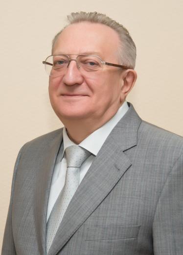
В.В.Гладько

Dear friends, dear colleagues!

I am proud to present to your attention the new issue of the Journal of the Medical Institute of Continuing Education. The journal is a platform where original research papers, reviews, practical recommendations, unique and didactic clinical cases, short reports on the problems of medicine and healthcare and relevant both in Russia and abroad can be published. This issue of our journal is dedicated to modern scientific research in medical rehabilitation and restorative treatment.

Under the term "medical rehabilitation" in the domestic scientific literature understand the restoration (rehabilitation) of the physical and psycholog- ical status of people who have lost this ability due to illness or injury.

Currently, medical rehabilitation as a branch of health care, within the framework of the concept of modern medicine, is considered as a differentiated staged system of therapeutic and preventive measures that ensure the integrity of the functioning of the body and, as a result, the complete restoration of the patient's health to the optimal level of performance through the combined, sequential and successive application of methods pharmacological, surgical, physical and psychophysiological effects on functionally or pathologically altered organs and systems of the body.

The methodological support of rehabilitation based on the achievements of science (development and scientific justification of the concept, approaches and methods) is based on the position that human health is a reflection of the state of adaptation of the organism to various influences, which determines the approaches to rehabilitation based on the principle of optimality.

There are many unresolved scientific problems in rehabilitation – yesterday, today and tomorrow – and those already solved yesterday require a more subtle interpretation today.

Considering the new concepts of rehabilitation and restorative treatment, first of all, it must be indicated that the use of any type of treatment should be based on the evidence of their effectiveness by evidence-based medicine, that is, benign studies. Until now, there are no reliable founda- tions of the theory of physiotherapy, which would lead us out of the field of empiricism at the turn of an accurate calculation of indications, prescriptions and effectiveness. Experimental work (primarily in the field of biophysics, bionics, medical cybernetics) does not sufficiently replenish the theoretical base, leaving, in turn, engineering thought without specific goals.

There is no doubt that there is another problem – the combined use of drugs with non-drug technologies. In this case, many natural or preformed physical factors can significantly reduce the risk of developing drug complications, allow the patient's body to mobilize natural protective resources, and provide them with the necessary metabolic and energy background. In addition, promising areas of scientific research in this area are physiogenetics – the phenomenon of genetic determination of the therapeutic effect of therapeutic physical factors (that is, optimization of adaptation mechanisms), as well as the study and correction of metabolic determinations.

Best regards, Editor-in-Chief, MD, PhD, Prof. V.V. Gladko

MEDICAL REHABILITATION

TREATMENT AND MEDICAL REHABILITATION FOR ACUTE CORONARY SYNDROME AT THE INPATIENT STAGE

N.N. Zolotukhin1,2, A.V. Konkov1

8-11

1 Medical Institute of Continuing Education, FGBOU VO MGUPP
2 Main Clinical Hospital of the Ministry of Internal Affairs of Russian Federation

Summary

Despite the qualitative improvement of medical care for acute coronary pathology, the level of disability and mortality among the most able-bodied population remains sufficiently significant. The term acute coronary syndrome (ACS), which combined acute myocardial infarction (AMI) and unstable angina (NS), was manifested by the introduction of new approaches to the treatment of patients with AMI and NS, increased interest in the problems of the peculiarities of the occurrence, course of ACS, the effectiveness of invasive and noninvasive methods of treatment, and the reassessment of the medical rehabilitation system (MR). The most important component of the treatment of ACS, especially in employees of the internal affairs bodies, whose activities are associated not only with constant stress factors, but also with a direct risk to life, is MR. The analysis of the ratio of clinical cases of AMI and NS, stages of MR in the Main Clinical Hospital of the Ministry of Internal Affairs (GKG MIA) for a period of 6 years was carried out. There are a total of 805 patients with ACS – 227 employees and 578 pensioners of the Ministry of Internal Affairs. With the development of ACS, employees of the Ministry of Internal Affairs are more often diagnosed with AMI, pensioners are dominated by NS. Despite the timely provision of specialized medical care and MR with AMI by the employees of the Ministry of Internal Affairs, 38% of patients were transferred to a specialized rehabilitation center for the second stage of MR. The presence of a rehabilitation center in the structure of the GKG of the Ministry of Internal Affairs makes it possible to carry out the I and II stages of MR within the framework of one hospitalization.

Key words: acute coronary syndrome, acute myocardial infarction, unstable angina, Interior Ministry employee, Interior Ministry pensioner, medical rehabilitation

For citation: Zolotukhin N.N., Konkov A.V. Treatment and medical rehabilitation for acute coronary syndrome at the inpatient stage. Bulletin of the Medical Institute of Сontinuing Education. 2022; (3): 8–11.
DOI 10.46393/27821714_2022_3_8

Literature

  1. Аронов Д.М., Бубнова М.Г., Бабараш О.Л. и др. Острый инфаркт миокарда с подъемом сегмента ST электрокардиограммы: реабилитация и вторичная профилактика. CardioСоматика. 2014; (Suppl. 1): 5–41.
  2. Аронов Д.М., Бубнова М.Г., Краницкий В.Б. Новые подходы к реабилитации и вторичной профилактике у больных, перенесших острый инфаркт миокарда с подъемом сегмента ST электрокардиограммы. Кардиология. 2015; 55 (12): 93–100.
  3. Золотухин Н.Н., Погонченкова И.В., Рассулова М.А., Фесюн А.Д. Вопросы лечения острого коронарного синдрома. Актуальные проблемы медицины. Сборник научных трудов. М., 2018: 11–15.
  4. Острый коронарный синдром. Под ред. И.С. Явелова, С.М. Хохлунова, Д.В. Дуплякова. М.: ГЭОТАР-Медиа, 2017. 384 с.
  5. Franklin K., Goldberg R.J., Spenser F. et al Implications of diabetes in patients with acute coronary syndromes: a multinational observational study. Arch. Intern. Med. 2004; 164 (13): 1457–1463.
  6. Himbert D., Klutman M., Steg G. et al. Cigarette smoking and acute coronary syndromes: a multinacional observation study. Int. J. Cardiol. 2005; 100 (1): 109–117.
  7. Бобровницкий И.П., Фесюн А.Д., Яковлев М.Ю. Восстановительная медицина как научное направление системы санаторно-курортного лечения. Вестник Медицинского института непрерывного образования. 2022; (2): 28–33.
  8. Lablance J.M., Amouel P., Hoden S. The OSCAR registry. Registry of acute coronary syndromes. Ann. Cardiol. Angiol. 2003; 52 (4): 205–211.
  9. Pop C., Pop L., Diсu D. Epidimiology of acute myocardial infarction in Romanian country hospitals: a population-based study in the Baiai Mare District. Rom. J. Intern. Med. 2004; 93 (12): 607–623.
  10. Золотухин Н.Н., Погонченкова И.В., Рассулова М.А., Фесюн А.Д. Оценка показателей состояния организма у лиц опасных профессий. Вопросы восстановительной и спортивной медицины. Сборник научных трудов. М., 2018: 16–20.
  11. Золотухин Н.Н. Острый коронарный синдром: лечение и медицинская реабилитация в главном клиническом госпитале МВД России. Медицинский вестник МВД. 2022; 116 (1): 46–48.

A COMPREHENSIVE PROGRAM OF MEDICAL REHABILITATION OF PATIENTS WITH RHEUMATOID ARTHRITIS USING HYDROGEN SULFIDE BATHS

V.V. Klimko1, R.A. Dzhalilova2, V.E. Yudin1, A.M. Shchegolkov1, T.V. Arseniy1

12-17

1 Medical Institute of Continuing Education, FGBOU VO MGUPP
2 Dagestan State Medical University

Summary

The paper presents the results of the use of hydrogen sulfide baths of the Talgi resort (Republic of Dagestan) in a comprehensive program of medical rehabilitation of patients with rheumatoid arthritis with impaired lipid metabolism. The study included 80 patients with rheumatoid arthritis and hypertension. The average age of the subjects was 62.4 ± 7.5 years (from 53 to 70 years). All the subjects had a low and moderate degree of disease activity according to DAS28 (2.64 ± 1.21). The patients were divided into 2 groups of 40 people each: the main group received statin therapy and hydrogen sulfide baths. The control group received statin therapy only. The course of balneotherapy consisted of 12 baths, the procedures were carried out every other day or two days in a row with a break on the third day, lasting 2 minutes and gradually increasing by 2 minutes, bringing to a maximum duration of 6 minutes. At the same time, a statin group drug, pitavastatin 2–4 mg/day, was used for a year.

The inclusion of balneotherapy in the treatment program of this category made it possible to obtain a more pronounced and persistent anti-inflammatory effect, improve microcirculation, and improve the clinical and functional condition of patients. All patients had favorable dynamics of subjective symptoms of the disease: irritability, headaches and dizziness decreased, the state of the emotional and mental sphere stabilized.

Statin therapy, in addition to the main lipid-lowering effect, contributed to a decrease in the number of joints involved, had an activating effect on

Key words: medical rehabilitation, hydrogen sulfide baths, rheumatoid arthritis, statins

For citation: Klimko V.V., Dzhalilova R.A., Yudin V.E. et al. A comprehensive program of medical rehabilitation of patients with rheumatoid arthritis using hydrogen sulfide baths. Bulletin of the Medical Institute of Сontinuing Education. 2022; (3): 12–17.
DOI 10.46393/27821714_2022_3_12

Literature

  1. Arts E.E.A., Fransen J., den Broeder A.A. et al. The effect of disease duration and disease activity on the risk of cardiovascular disease in rheumatoid arthritis patients. Ann. Rheum. Dis. 2015; 74 (6): 998–1003.
  2. Arts E.E.A., Popa C.D., den Broeder A.A. et al. Prediction of cardiovascular risk in rheumatoid arthritis: performance of original and adapted SCORE algorithms. Ann. Rheum. Dis. 2016; 75 (4): 674–680.
  3. Babaeva A.R., Tarasov A.A., Davidov S.I., Reznikova E.A. The prognostic value of anti-connective tissue antibodies in coronary heart disease and asymptomatic atherosclerosis. Cardiovasc. Res. 2016; 111 (Suppl. 1): S56–S81. Abstract 314.
  4. Земляничкина Н.В., Храмцова Н.А., Дзизинский А.А. и др. Частота артериальной гипертензии при ревматоидном артрите. Сибирский медицинский журнал. 2009; 7: 43–46.
  5. Насонов Е.Л., Попкова Т.В. Кардиоваскулярные проблемы ревматологии. Научно-практическая ревматология. 2004; 42 (4): 4–9.
  6. Попкова Т.В., Новикова Д.С., Насонов Л.Е. Интерлейкин 6 и сердечно-сосудистая патология при ревматоидном артрите. Научно-практическая ревматология. 2011; 49 (4): 64–72.
  7. Ревматология: клинические рекомендации. Под ред. Е.Л. Насонова. М.: ГЭОТАР-Медиа, 2010.
  8. Насонов Е.Л. Фармакотерапия ревматоидного артрита: российские и международные рекомендации. Научно-практическая ревматология. 2016; 54 (5): 557–571.
  9. Никитина Н.М., Ребров А.П. Активность ревматоидного артрита и факторы кардиоваскулярного риска на фоне базисной противовоспалительной терапии. Клиницист. 2010; 1: 25–29.
  10. Тортов И.Н., Улубиева Е.А., Амбалова С.А. и др. Риск кардиоваскулярных осложнений у больных ревматоидным артритом. Sciences оf Europe. 2016; 7–1 (7): 78–83.
  11. Тутунов В.С., Зубарева М.Ю., Малышев П.П., Кухарчук В.В. Cтатины в ревматологии. Атеросклероз и дислипидемии. 2011; 2 (3): 43–48.
  12. Насонов Е.Л. Проблема атеротромбоза в ревматологии. Вестник Российской академии медицинских наук. 2003; 7: 6–10.
  13. Del Rincon I., Williams K., Stern M.P. et al. Association between carotid atherosclerosis and markers of inflammation in rheumatoid arthritis patients and healthy subjects. Arthritis Rheum. 2003; 48: 1833–1840.
  14. Юдин В.Е., Щегольков А.М., Ярошенко В.П. и др. Современные подходы к комплексной медицинской реабилитации артрологических больных, перенесших артроскопические операции на коленном суставе. Вестник Медицинского института непрерывного образования. 2022; 1: 44–49.
  15. Khan M.A. Ankylosing spondylitis. Arthritis Rheum. 2004; 50 (9): 3047–3048.
  16. Jin L., Zhang G., Joshua M. et al. Lack of linkage of IL1RN genotypes with ankylosing spondylitis susceptibility. Arthritis Rheum. 2004; 50 (9): 3047–3048.
  17. Shen Y., Shen Z., Luo S. et al. The cardioprotective effects of hydrogen sulfide in heart diseases: from molecular mechanisms to therapeutic potential. Oxid. Med. Cell. Longev. 2015; 2015: 925167.
  18. Аничкова Д.А., Шостак Н.А. Ревматоидный артрит и сердечно-сосудистый риск: перспективы применения статинов. Рациональная фармакотерапия в кардиологии. 2005; 3: 47–53.
  19. Сероводородные ванны в лечебно-реабилитационных и профилактических программах: клинические рекомендации. М., 2015. 31 с.
  20. Тихомирова И.А., Петроченко Е.П., Петроченко А.С. Сероводород как сигнальная молекула в сердечно-сосудистой системе. Регионарное кровообращение и микроциркуляция. 2021; 20 (1): 5–16.
  21. Бобровницкий И.П., Фесюн А.Д., Яковлев М.Ю. Восстановительная медицина как научное направление системы санаторно-курортного лечения. Вестник Медицинского института непрерывного образования. 2022; 2: 28–33.

UROLOGY & ANDROLOGY

THE POSSIBILITIES OF MODERN FLEXIBLE URETEROSCOPES IN THE TREATMENT OF COMPLEX FORMS OF UROLITHIASIS AND THE ACHIEVEMENT OF "STONE FREE"

A.G. Kochetov1,2, A.G. Martov3,4,5, O.V. Sidorov1, B.R. Gvasalia1,2, N.A. Baykov1,2, A.A. Gritskevich6,7, A.V. Alekhnovich1, A.V. Kasaikin2, O.A. Plekhanova2

18-22

1 National Medical Research Center for High Medical Technologies – A.A. Vishnevsky Central Military Clinical Hospital of the Ministry of Defense of Russia
2 Medical Institute of Continuing Education, FGBOU VO MGUPP
3 Biomedical University of Innovations and Continuing Education, A.I. Burnazyan Federal Medical and Biological Center
4 Medical Research and Education Center, M.V. Lomonosov Moscow State University
5 D.D. Pletnev City Clinical Hospital, Moscow
6 A.V. Vishnevsky National Medical Research Center for Surgery
7 RUDN University

Summary

The article represents three complex clinical cases describing the use of flexible ureteroscopes in patients with urolithiasis. The features of the use of flexible single-use ureteroscopes for percutaneous nephrolithotripsy, retrograde intrarenal surgery and antegrade ureterolithotripsy are demonstrated. The treatment of urolithiasis using flexible ureteroscopes is feasible and effective method. The need of further research and development of this technique is warranted.

Key words: urolithiasis, minimally invasive endourological technologies, percutaneous nephrolithotripsy, retrograde intrarenal surgery, patients with pathologies of the bones of the skeleton, joints of the lower extremities, contact ureterolithotripsy

For citation: Kochetov A.G., Martov A.G., Sidorov O.V. et al. The possibilities of modern flexible ureteroscopes in the treatment of complex forms of urolithiasis and the achievement of "stone free". Bulletin of the Medical Institute of Сontinuing Education. 2022; (3): 18–22.
DOI 10.46393/27821714_2022_3_18

Literature

  1. Мартов А.Г., Ергаков Д.В. Достижения современной эндоурологии: материалы XII Съезда Российского общества урологов. М., 2012: 417–426.
  2. Kupajski M., Tkocz M., Ziala D. Modern menagment of ston disease in patients with a solitary kidney. Wideochir. Inne. Tech. Maloinwazyjne. 2012; 7 (1): 1–7.
  3. Дутов В.В., Уренков С.Б., Паршенкова И.Г., Мамедов Э.А. Особенности чрескожной нефролитотрипсии у пациентов с мочекаменной болезнью единственной почки. Урология. 2015; 2: 52–55.
  4. Кочетов А.Г. Становление, итоги, основные направления и перспективы развития урологического центра. Актовая речь в честь 48-й годовщины со дня основания госпиталя и 20-летия урологического центра. Красногорск, 2016.
  5. Подойницын А.А., Трапезникова М.Ф., Уренков С.Б. и др. Ретроградная нефролитотрипсия в лечении коралловидного нефролитиаза. Урология. 2013; 1: 82–85.
  6. Каприн А.Д., Кочетов А.Г., Сидоров О.В. и др. Сравнительный анализ перкутанной и ретроградной нефролитотрипсии крупных и коралловидных камней почек. Вопросы урологии и андрологии. 2013; 2 (3): 62–66.
  7. Davol P.E., Wood C., Fulmer B. Success in treating renal calculi with singleaccess, single-event percutaneous nephrolithotomy: is a routine "second look" necessary? J. Endourol. 2006; 20: 289–292.
  8. Wang Y., Zhong B., Yang X. et al. Comparison of the efficacy and safety of URSL, RPLU, and MPCNL for treatment of large upper impacted ureteral stones: a randomized controlled trial. BMC Urol. 2017; 17 (1): 50.
  9. Aravantinos E., Anagnostou T., Samarinas M. et al. Two-step minipercutaneous ureterolithotripsy under multimodal analgesia for complicated impacted calculi in proximal ureter. Urology. 2013; 81 (6): 1147–1152.
  10. Drake T., Grivas N., Dabestani S. et al. What are the benefits and harms of ureteroscopy compared with shock-wave lithotripsy in the treatment of upper ureteral stones? A systematic review. Eur. Urol. 2017; 72 (5): 772–786.
  11. Geavlete P., Mulţescu R., Georgescu D. Flexible nephroscopy for upper urinary tract pathology. Chirurgia (Bucur). 2007; 102 (2): 191–196.
  12. Raman J.D., Bagrodia A., Gupta A. et al. Natural history of residual fragments following percutaneous nephrostolithotomy. J. Urol. 2009; 181 (3): 1163–1168.

OTORHINOLARYNGOlOGY

ПРИМЕНЕНИЕ СИПАП-ТЕРАПИИ В МЕДИЦИНСКОЙ РЕАБИЛИТАЦИИ БОЛЬНЫХ ИШЕМИЧЕСКОЙ БОЛЕЗНЬЮ СЕРДЦА С СИНДРОМОМ ОБСТРУКТИНОГО АПНОЭ СНА ПОСЛЕ КОРОНАРНОЙ АНГИОПЛАСТИКИ

I.I. Morozov1,2, N.S. Grachev1, N.V. Gorbunova2, A.V. Shirokaya2, M.Yu. Pogorelov2

24-27

1 Medical Institute of Continuing Education, FGBOU VO MGUPP
2 Main Clinical Hospital of the Ministry of Internal Affairs of Russian Federation

Summary

The literature provides many ways to close perforations of the nasal septum (NS), there is no differential approach to the choice of technique depending on the etiological factor.

Objective: to study the clinical and morphological features of tissues at the edges of postoperative nasal septum perforations (PNSP) to substantiate the choice of the optimal surgical technique.

Material and methods. 52 patients are divided into 3 groups in accordance with the area (S) of PNSP: group 1 – S < 1 cm2 (10%); group 2 – S = 1–2 cm2 (75%), group 3 – S > 2 cm2 (15%). In group 2, the subgroups are: 2A – PNSP without curvature of the NS (CNS) (52%); 2B – PNSP with CNS (48%). The state of the mucous membrane (MM) and supporting tissues at the edges of the PNSP, the results of a cytological study from the edges of the PNSP, and specific complaints of the patients were evaluated.

Results. In group 1 and 2 at the edges of the PNSP, a significant decrease in tissue elasticity compared with group 3, indicating a deficit in supporting tissues (p < 0.05). The ratio of the area of the defect of supporting tissues and PNSP in group 1 – 1 to 3.5, in group 2 – 1 to 1.5, in group 3 – 1 to 1.1. A high degree of elasticity of the posterior margin of PNSP is accompanied by pronounced clinical manifestations, regardless of the size of PNSP, and is confirmed by the cytological picture of inflammation (p < 0.01). The presence of CNS enhances the clinical manifestations of PNSP (p < 0.05).

Conclusion. The structure of the edges of PNSP forms a clinical picture of the disease. When choosing a closure method for PNSP, the surgeon must solve the following problems: the size of the NS defect required for replacement is determined not so much by the dimensions of the PNSP as the area of the defect in the supporting tissues of the NS; with a deficit of local tissues, it is advisable to additionally use grafts; MM in the area of the posterior and lower edges has pronounced signs of inflammation, these tissues should be removed during the operation.

Key words: nasal septum perforation, complication of nasal septum surgery

For citation: Morozov I.I., Grachev N.S., Gorbunova N.V. et al. Study of clinical and morphological features of nasal septum perforations. Bulletin of the Medical Institute of Сontinuing Education. 2022; (3): 24–27.
DOI 10.46393/27821714_2022_3_24

Literature

  1. Bateman N.D., Woolford T.J. Informed consent for septal surgery: the evidence-base. J. Laryngol. Otol. 2003; 117 (3): 186–189.
  2. Quinn J.G., Bonaparte J.P., Kilty S.J. Postoperative management in the prevention of complications after septoplasty: a systematic review. Laryngoscope. 2013; 123 (6): 1328–1333.
  3. Virkkula P., Mäkitie A.A., Vento S.I. Surgical outcome and complications of nasal septal perforation repair with temporal fascia and periosteal grafts. Clin. Med. Insights Ear Nose Throat. 2015; 8: 7–11.
  4. Pedroza F., Patrocinio L.G., Arevalo O. A review of 25-year experience of nasal septal perforation repair. Arch. Facial Plast. Surg. 2007; 9 (1): 12–18.
  5. Молоков К.В. Перфорация перегородки носа - причины возникновения и способы лечения. Автореф. дис. ... канд. мед. наук. М., 2007.
  6. Lindemann J., Scheithauer M., Hoffmann T.K. et al. Long-term results after surgical closures of septal perforations. Laryngorhinootologie. 2014; 93 (11): 751–755.
  7. Базаркина К.П., Козлов В.С. Изучение качества жизни у пациентов с болезнью оперированного носа. Российская оториноларингология. 2015; 5: 20–23.
  8. Протасевич Г.С. Клиническая классификация деформаций перегородки носа. Здравоохранение Туркменистана. 1979; 7: 14–16.

RADIATION DIAGNOSIS

RADIATION DIAGNOSIS OF HEMORRHAGIC COMPLICATIONS IN PATIENTS TREATED FOR A NEW CORONAVIRUS INFECTION: CLINICAL OBSERVATIONS (MSCT, MRI)

I.S. Obelchak1,2, E.A. Kukushkina1,2, A.I. Zherebtsov2, M.V. Bolyavin2, D.A. Taldonov2, A.S. Kovalev2

28-31

1 Medical Institute of Continuing Education, FGBOU VO MGUPP
2 Main Military Clinical Hospital of the National Guard Troops of the Russian Federation

Summary

The new coronavirus infection COVID-19 is a potentially dangerous respiratory infection caused by the SARS-CoV-2 coronavirus, which has several variants of the course with a high risk of death. In addition to the main damage to the respiratory tract, lungs, other organs and systems also suffer, most often the heart, liver, kidneys, nervous system.
The main complication of coronavirus infection, which causes the development of a lethal outcome, is respiratory and multiple organ failure, the development of distress syndrome. Currently, antiviral and anti-inflammatory drugs are used to treat COVID-19. Given the possibility of developing hypercoagulable syndrome, hypocoagulant therapy is widely prescribed. In this case, it is necessary to take into account the risk of developing hemorrhagic complications.

Key words: new coronavirus infection, hemorrhagic complications, bleeding, hematoma

For citation: Obelchak I.S., Kukushkina E.A., Zherebtsov A.I. et al. Radiation diagnosis of hemorrhagic complications in patients treated for a new coronavirus infection: clinical observations (MSCT, MRI). Bulletin of the Medical Institute of Сontinuing Education. 2022; (3): 28–31.
DOI 10.46393/27821714_2022_3_28

Literature

  1. Thachil J., Tang N., Gando S. et al. ISTH interim guidance on recognition and management of coagulopathy in COVID-19. J. Thromb. Hemost. 2020; 18 (5): 1023–1026.
  2. Никифоров В.В., Суранова Т.Г., Миронов А.Ю., Забозлаев Ф.Г. Новая коронавирусная инфекция (COVID-19): этиология, эпидемиология, клиника, диагностика, лечение и профилактика. М., 2020. 48 c.
  3. Временные методические рекомендации «Диагностика и лечение новой коронавирусной инфекции (COVID-19)». Версия 7. Министерство здравоохранения Российской Федерации, 2020. 166 с.
  4. Koupenova M., Corkrey H.A., Vitseva O. et al. SARS-CoV-2 initiates programmed cell death in platelets. Circ. Res. 2021; 129 (6): 631–646.
  5. Буланов И.Б., Симарова Е.Л., Буланова Д.О. и др. Новая коронавирусная инфекция COVID-19: клиническая и прогностическая значимость оценки фибриногена плазмы. Вестник интенсивной терапии им. А.И. Салтанова. 2020; 4: 42–47.
  6. Zhou F., Yu T., Du R. et al. Clinical course and risk factors for mortality of adult inpatients with COVID-19 in Wuhan, China: a retrospective cohort study. Lancet. 2020; 395 (10229): 1054–1062.
  7. Huang C., Wang Y., Li X. et al. Clinical features of patients infected with 2019 novel corona-virus in Wuhan, China. Lancet. 2020; 395 (10223): 497–506.
  8. Wu C., Chen X., Cai Y. et al. Risk factors associated with acute respiratory distress syndrome and death in patients with coronavirus disease 2019 pneumonia in Wuhan, China. JAMA Intern. Med. 2020; 180 (7): 934–943.
  9. Кукушкина Е.А., Шашкин К.М., Обельчак И.С. и др. Лучевая диагностика отдаленных последствий перенесенных вирусных пневмоний, вызванных COVID-19. Вестник Медицинского института непрерывного образования. 2021; 1: 46–49.

SURGERY

CHONDROBLASTOMA OF THE BASE OF THE SKULL: A CLINICAL OBSERVATION

S.V. Tereshchuk1,2,3, V.A. Sukharev1,2, E.A. Vasilyev1

32-36

1 Burdenko Main Military Clinical Hospital
2 RUDN University
3 Medical Institute of Continuing Education, FGBOU VO MGUPP

Summary

Chondroblastoma of the bones of the base of the skull is extremely rare, with the most typical site of its occurrence being the temporal bone. In cases where the base of the skull and adjacent structures are involved in the pathological process, the use of hightech techniques is required to fully restore function and aesthetics. The article describes a clinical observation of chondroblastoma of the temporal bone at the base of the skull, the surgical treatment of which required the use of virtual surgical planning and 3D printing.

Key words: chondroblastoma, skull base, reconstructive surgery, individual implant, temporomandibular joint

For citation: Tereshchuk S.V., Sukharev V.A., Vasilyev E.A. Chondroblastoma of the base of the skull: a clinical observation. Bulletin of the Medical Institute of Сontinuing Education. 2022; (2): 32–36.
DOI 10.46393/27821714_2022_3_32

Literature

  1. Codman E.A. Epiphyseal chondromatous giant cell tumour of upper end of humerus. Surg. Gynaecol. Obstet. 1931; 52: 543–548.
  2. Vandenberg H.J. Jr., Coley B.L. Primary tumour of cranial bones. Surg. Gynaecol. Obstet. 1950; 90: 602–612.
  3. Mirra J.M. Chondroblastoma. In: Bone tumours: clinical, radiologic and pathologic correlations. Vol. 2. Ed. by J.M. Mirra. Lea and Febeger Philadelphia London, 1989: 589–623.
  4. Denko J.V., Krauel C.H. Benign chondroblastoma of bone: an unusual location in temporal bone. Arch. Pathol. (Chicago). 1955; 59: 710–711.
  5. Bian L., Sun Q., Zhao W. et al. Temporal bone chondroblastoma: a review. Neuropathology. 2005; 25: 159–164.
  6. Horn K.L., Hankinson H., Nagel В. et al. Surgical management of temporal bone chondroblastoma. Otolaryngol. Head Neck Surg. 1990; 102: 264–269.
  7. Watanabe N., Kazuhide Y., Shigemi H. et al. Temporal bone chondroblastoma. Otolaryngol. Head Neck Surg. 1999; 121: 327–330.
  8. Brien E.W., Mirra J.M., Kerr R. Benign and malignant cartilage tumours of the bone and joint. Their anatomic and theoretical basis with an emphasis on radiology, pathology and clinical biology. I. The intramedullary cartilage tumours. Skeletal Radiol. 1997; 26: 325–353.
  9. Dahlin D.C., Irvin J.C. Benign chondroblastoma. A study of 125 cases. Cancer. 1972; 30: 401–413.
  10. Narita Y., Morimoto T., Neshihana R. et al. Chondroblastoma of the temporal bone: report of a case and re- view of literature of 54 cases. No To Shinkei. 1992; 44: 143–148.
  11. Cares H.L., Terplan K. Chondroblastoma of the skull. J. Neurosurg. 1971; 35: 614–618.
  12. Blaaw G., Prick J.J.W., Versteege C. Chondroblastoma of temporal bone. Neurosurgery. 1998; 22: 1102–1107.
  13. Bertoni F., Unni K.K., Beabout J.W. et al. Chondroblastoma of the skull and facial bones. Am. J. Pathol. 1987; 88: 19.
  14. Feely M., Keokane C. Chondroblastoma of the skull. J. Neurol. Neurosurg. Psychiatry. 1984; 47: 1348–1350.
  15. Jaffe H.L., Lichtenstein L. Benign chondroblastoma of bone: reinterpretation of the so-called calcifying or chondromatous giant cell tumour. Am. J. Pathol. 1942; 18: 969–991.
  16. Kyriakos M., Land V.J., Pennng L.H. et al. Metastatic chondroblastoma: report of a fatal case with a review of literature on atypical, aggressive and malignant chondroblastoma. Cancer. 1985; 55: 1770–1789.

SURGICAL APPROACHES TO ORGAN-PRESERVING OPERATIONS IN PATIENTS WITH KIDNEY CANCER

B.Ya. Alekseev1,2, K.M. Nyushko1,2, I.M. Shevchuk1, V.M. Perepukhov1, V.P. Aleshin1, S.A. Sergienko1, A.S. Kalpinsky1, V.A. Polyakov1

38-43

1 National Medical Research Radiological Center
2 Medical Institute of Continuing Education, FGBOU VO MGUPP

Summary

Kidney cancer is one of the most significant problems in oncourology. Over the past 10 years, there has been an increase in the incidence of kidney cancer, while the introduction of modern methods of radiation diagnostics makes it possible to more often detect the disease at an early stage. The method of choice for this pathology at stage T1a is kidney resection. It should be noted that this surgical intervention requires careful hemostasis, since it is performed on an organ with intensive blood circulation. To ensure adequate hemostasis during kidney resection, mechanical, physical and chemical methods are used. This review highlights the various methods and techniques used to stop bleeding when performing a kidney resection. It presents the results of large studies that have evaluated various methods of hemostasis and reduce the risk of complications associated with bleeding.

Key words: renal cell carcinoma, kidney resection, organ-preserving operations, hemostasis, bleeding, hemostatics, glue, ischemia, anoxia

For citation: Alekseev B.Ya., Nyushko K.M., Shevchuk I.M. et al. Surgical approaches to organ-preserving operations in patients with kidney cancer. Bulletin of the Medical Institute of Сontinuing Education. 2022; (3): 38–43.
DOI 10.46393/27821714_2022_3_38

Literature

  1. Состояние онкологической помощи населению России в 2019 году. Под ред. А.Д. Каприна, В.В. Старинского, А.О. Шахзадовой. М., 2020.
  2. Sabino L., Andreoni C., Faria E.F. et al. Evaluation of renal defect healing, hemostasis, and urinary fistula after laparoscopic partial nephrectomy with oxidized cellulose. J. Endourol. 2007; 21 (5): 551–556.
  3. Thompson T., Ng C.G., Tolley D. Renal parenchymal hemostatic aids: glues and things. Curr. Opin. Urol. 2003; 13 (3): 209–214.
  4. Van Dijk J.H., Pes P.L. Haemostasis in laparoscopic partial nephrectomy: current status. Minim. Invasive Ther. Allied Technol. 2007; 16 (1): 31–44.
  5. Rane A., Rimington P.D., Heyns C.F. et al. Evaluation of a hemostatic sponge (TachoSil) for sealing of the renal collecting system in a porcine laparoscopic partial nephrectomy survival model. J. Endourol. 2010; 24 (4): 599–603.
  6. Msezane L.P., Katz M.H., Gofrit O.N. et al. Hemostatic agents and instruments in laparoscopic renal surgery. J. Endourol. 2008; 22 (3): 403–408.
  7. Bernie J.E., Ng J., Barman V. et al. Evaluation of hydrogel tissue sealant in porcine laparoscopic partial-nephrectomy model. J. Endourol. 2005; 19 (9): 312–317.
  8. Nadler R.B., Loeb S., Rubenstein R.A. et al. Use of BioGlue in laparoscopic partial nephrectomy. Urology. 2006; 68 (2): 416–418.
  9. Hidas G., Kastin A., Mullerad M. et al. Sutureless nephron‐sparing surgery: use of albumin glutaraldehyde tissue adhesive (BioGlue). Urology. 2006; 67 (4): 697–700.
  10. Schips L., Dalpiaz O., Cestari A. et al. Autologous fibrin glue using the Vivostat system for hemostasis in laparoscopic partial nephrectomy. Eur. Urol. 2006; 50 (4): 801–805.
  11. Bak J.B., Singh A., Shekarriz B. Use of gelatin matrix thrombin tissue sealant as an effective hemostatic agent during laparoscopic partial nephrectomy. J. Urol. 2004; 171 (2 Pt. 1): 780–782.
  12. Shekarriz B., Stoller M.L. The use of fibrin sealant in urology. J. Urol. 2002; 167 (3): 1218–1225.
  13. Oz M.C., Rondinone J.F., Shargill N.S. FloSeal matrix: new generation topical hemostatic sealant. J. Card. Surg. 2003; 18 (6): 486–493.
  14. Rosales A., Salvador J., De Graeve N. et al. Clamping of the renal artery in laparoscopic partial nephrectomy: an old device for a new technique. Eur. Urol. 2005; 47 (1): 98–101.
  15. Porpiglia F., Fiori C., Bertolo R. et al. The effects of warm ischaemia time on renal function after laparoscopic partial nephrectomy in patients with normal contralateral kidney. World J. Urol. 2012; 30 (2): 257–263.
  16. Gill I.S., Abreu S.C., Desai M.M. et al. Laparoscopic ice slush renal hypothermia for partial nephrectomy: the initial experience. J. Urol. 2003; 170 (1): 52–56.
  17. Saitz T.R., Dorsey P.J., Colli J. et al. Induction of cold ischemia in patients with solitary kidney using retrograde intrarenal cooling: 2-year functional outcomes. Int. Urol. Nephrol. 2013; 45 (2): 313–320.
  18. Guillonneau B., Bermudez H., Gholami S. et al. Laparoscopic partial nephrectomy for renal tumor: single center experience comparing clamping and no
  19. Verhoest G., Manunta A., Bensalah K. et al. Laparoscopic partial nephrectomy with clamping of the renal parenchyma: initial experience. Eur. Urol. 2007; 52 (5): 1340–1346.
  20. Rodriguez‐Covarrubias F., Gabilondo B., Borgen J.L., Gabilondo F. Partial nephrectomy for renal tumors using selective parenchymal clamping. Int. Urol. Nephrol. 2007; 39 (1): 43–46.
  21. Beck S.D., Lifshitz D.A., Cheng L. et al. Endoloop assisted laparoscopic partial nephrectomy. J. Endourol. 2002; 16 (3): 175–177.
  22. Vitagliano G., Villasante N. Laparoscopic partial nephrectomy with selective polar clamping using the Simon clamp: initial experience. Arch. Esp. Urol. 2013; 66 (3): 308–312.
  23. Selikowitz S.M., Curtis M.R. Hemostatic control with flexible compression tape used during partial nephrectomy and organ salvage. J. Urol. 1999; 162 (2): 458–459.
  24. Айвазян А.В. Гемостаз при операциях на почке. 2‐е изд., доп. М.: Наука, 1982. 280 с.
  25. Cozar J.M., Tallada M. Open partial nephrectomy in renal cancer: a feasible gold standard technique in all hospitals. Adv. Urol. 2008; 2008: 916463.
  26. Taneja S., Dakwar G., Godoy G. Simplified reconstruction after laparoscopic partial nephrectomy using a single‐pass suturing technique. J. Endourol. 2009; 23 (4): 589–592.
  27. Shikanov S., Wille M., Large M. et al. Knotless closure of the collecting system and renal parenchyma with a novel barbed suture during laparoscopic porcine partial nephrectomy. J. Endourol. 2009; 23 (7): 1157–1160.
  28. Понукалин Н.М. Хирургическое лечение больных с коралловидными камнями почек: дис. ... канд. мед. наук. Саратов, 1969.
  29. Simforoosh N., Noor‐Alizadeh A., Tabibi A. et al. Bolsterless laparoscopic partial nephrectomy: a simplification of the technique. J. Endourol. 2009; 23 (6): 965–969.
  30. Семичев Е.В., Байков А.Н., Бушланов П.С., Дамбаев Г.Ц. Сравнительный анализ методов гемостаза при операциях на селезенке. Бюллетень сибирской медицины. 2015; 4 (2): 91–100.
  31. Sengupta S., Webb D.R. Use of a computer‐controlled bipolar diathermy system in radical prostatectomies and other open urological surgery. ANZ J. Surg. 2001; 71 (9): 538–540.
  32. Terai A., Ito N., Yoshimura K. et al. Laparoscopic partial nephrectomy using microwave tissue coagulator for small renal tumors: usefulness and complications. Eur. Urol. 2004; 45 (6): 744–748.
  33. Nanri M., Udo K., Kawasaki M. et al. Microwave tissue coagulator induces renal apoptotic damage to preserved nor- mal renal tissue following partial nephrectomy. Clin. Exp. Nephrol. 2009; 13 (5): 424–429.
  34. Сoleman J., Singh A., Pinto P. et al. Radiofrequency‐assisted laparoscopic partial nephrectomy: clinical and histologic results. J. Endourol. 2007; 21 (6): 600–605.
  35. Hilal Z., Mavrommati G., Foerster C. et al. Spray versus forced coagulation in large loop excision of the transformation zone: a randomized trial. J. Low Genit. Tract Dis. 2016; 20 (2): 169–173.
  36. Yao P., Gunasegaram A., Ladd L.A., Morris D.L. InLine bipolar radiofrequency ablation device‐assisted partial nephrectomy in a porcine model. ANZ J. Surg. 2008; 78 (7): 564–567.
  37. Sprunger J., Herrell S.D. Partial laparoscopic nephrectomy using monopolar saline‐coupled radiofrequency device: animal model and tissue effect characterization. J. Endourol. 2005; 19 (4): 513–519.
  38. Mattioli S., Muсoz R., Recasens R. et al. What does Revolix laser contribute to partial nephrectomy? Arch. Esp. Urol. 2008; 61 (9): 1126–1129.
  39. Anderson J.K., Baker M.R., Lindberg G. et al. Large volume laparoscopic partial nephrectomy using the potassium titanyl-phosphate (KTP) laser in a survival porcine model. Eur. Urol. 2007; 51 (3): 749–754.
  40. Guzzo T.J. Small renal masses: the promise of thulium laser enucleation partial nephrectomy. Nat. Rev. Urol. 2013; 10 (5): 259–260.
  41. Ramakumar S., Roberts W.W., Fugita O.E. et al. Local hemostasis during laparoscopic partial nephrectomy using biodegradable hydrogels: initial porcine results. J. Endourol. 2002; 16 (7): 489–494.
  42. Xie H., Khajanchee Y.S., Teach J.S., Shaffer B.S. Use of a chitosan‐based hemostatic dressing in laparoscopic partial nephrectomy. J. Biomed. Mater. Res. B. Appl. Biomater. 2008; 85 (1): 267–271.
  43. Аляев Ю.Г., Крапивин А.А. Резекция почки при раке. М.: Медицина, 2001. 224 с.
  44. Зубрицкий В.Ф., Лебедева Ю.Н. Межклеточные и клеточно-матриксные взаимодействия при репарации длительно незаживающих ран. Вестник Медицинского института непрерывного образования. 2022; 1: 66–70.

DERMATOLOGY

PATHOPHYSIOLOGY OF INFLAMMATION AND DIRECTIONS OF THERAPY IN ROSACEA

E.V. Gorbakova, S.A. Masyukova, I.V. Ilyina, I.A. Lamotkin, E.G. Sanakoeva

44-47

Medical Institute of Continuing Education, FGBOU VO MGUPP

Summary

The article discusses the pathogenetic relationship of neurogenic and immune inflammation in rosacea and the most likely causes of dermatosis.

Key words: rosacea, etiology, pathogenesis, patient profile, melatonin, neurogenic inflammation, immune inflammation

For citation: Gorbakova E.V., Masyukova S.A., Ilyina I.V. et al. Pathophysiology of inflammation and directions of therapy in rosacea. Bulletin of the Medical Institute of Сontinuing Education. 2022; (3): 44–47.
DOI 10.46393/27821714_2022_3_44

Literature

  1. Демина О.М., Потекаев Н.Н. Современные этиопатогенетические механизмы развития розацеа и новые методы терапии. Клиническая дерматология и венерология. 2017; 16 (3): 13–23.
  2. Демина О.М., Бутов Ю.С. Клинико-иммунологические показатели у больных розацеа и оценка результатов применения высокотехнологического метода лечения – ФДТ. Вестник эстетической медицины. 2010; 4 (9): 59–64.
  3. Потекаев Н.Н., Новожилова О.Л., Потапова С.В., Хамаганова И.В. Асимметричная розацеа. Клиническая дерматология и венерология. 2019; 18 (2): 150–153.
  4. Самцов А.В. Акне и акнеформные дерматозы. М., 2014. 352 с.
  5. Олисова О.Ю., Кочергин Н.Г., Смирнова Е.А. Современная наружная терапия среднетяжелых форм розацеа. Российский журнал кожных и венерических болезней. 2016; 19 (6): 328–334.
  6. Подымов В.К. Красная волчанка: общая схема патогенеза и принципы патогенетической терапии. Глава II. Порфирин секрета сальных желез кожи человека. Его роль в норме и патологии. Под ред. Л.А. Пирузяна. Ереван: Айастан, 1981: 35–59.
  7. Zhang H., Zhang Y., Li Y. et al. Bioinformatics and network pharmacology identify the therapeutic role and potential mechanism of melatonin in AD and rosacea. Front. Immunol. 2021; 12: 756550.
  8. Алексеев Ю.В., Ткаченко С.Б., Анфимова Н.А. и др. Изменение содержания эндогенных порфиринов в сальных железах при заболеваниях кожи. Экспериментальная и клиническая дерматокосметология. 2004; 1: 8.
  9. Steinhoff M., Buddenkotte J., Aubert J. et al. Clinical, cellular, and molecular aspects in the pathophysiology of rosacea. J. Investig. Dermatol. Symp. Proc. 2011; 15 (1): 2–11.
  10. Anzengruber F., Czernielewski J., Conrad C. et al. Swiss S1 guideline for the treatment of rosacea. J. Eur. Acad. Dermatol. Venereol. 2017; 31 (11): 1775–1791.
  11. Buddenkotte J., Steinhoff M. Recent advances in understanding and managing rosacea. F1000Res. 2018; 7: F1000 Faculty Rev-1885.
  12. Del Rosso J.Q. Medical management of rosacea with topical agents: a thorough appraisal of available treatment options and recent advances. Book of Abstracts of 62st AAD Annu- al Meeting. 2004: 69.
  13. Del Rosso J.Q., Thiboutot D., Gallo R.L. et al. Consensus recommendations from the American Acne & Rosacea Society on the management of rosacea, part 1: a status report on the disease state, general measures, and adjunctive skin care. Cutis. 2013; 92 (5): 234–240.

CARDIAC SURGERY

PREDICTORS OF PACEMAKER IMPLANTATION AFTER THE COX-MAZE IV PROCEDURE IN OPEN HEART SURGERY

V.V. Bazylev1,2, V.A. Karnakhin2, A.A. Kuznetsova2

48-53

1 Medical Institute of Continuing Education, FGBOU VO MGUPP
2 Federal Center for Cardiovascular Surgery, Penza

Summary

The purpose of the study: to determine the rate of pacemaker implantation after Cox-Maze IV surgery and to identify predictors that affect the neсessity of a pacemaker.

Results. In the group of patients with implanted pacemakers, perioperative complications were similar to those in the group without pacemakers. In the group with pacing, the aorta was clamped for a longer time. Patients with implanted pacemakers spent a longer time in the hospital. According to multiple regression data, the only predictor of pacemaker implantation is the age of the patient. For every year of the patient's age, the chance that a pacemaker will need to be implanted after the Cox-Maze procedure increases by 4%.

Conclusions

  1. The rate of pacemaker implantation after simultaneous heart surgery is 4%, but increases with the age of the patient.
  2. Long aortic cross-clamp time and cardiopulmonary bypass time increase the risk of pacemaker implantation.
  3. Artificial neural networks have an advantage in finding leading predictors compared to logistic regression.

Key words: Cox-Maze IV procedure, pacemaker implantation, atrial fibrillation

For citation: Bazylev V.V., Karnakhin V.A., Kuznetsova A.A. Predictors of pacemaker implantation after the Cox-Maze IV procedure in open heart surgery. Bulletin of the Medical Institute of Сontinuing Education. 2022; (3): 48–53.
DOI 10.46393/27821714_2022_3_48

Literature

  1. Cox J.L., Schuessler R.B., Boineau J.P. The development of the Maze procedure for the treatment of atrial fibrillation. Semin. Thorac. Cardiovasc. Surg. 2000; 12 (1): 2–14.
  2. Kim H., Park P.W., Sung K. et al. Mid-term results of the Cox Maze III procedure combined with open mitral commissurotomy for the treatment of rheumatic mitral stenosis. Circ. J. 2010; 74 (7): 1332–1328.
  3. Базылев В.В., Немченко Е.В., Карнахин В.А. Влияние основных факторов риска рецидива фибрилляции предсердий на эффективность операции Cox-Maze 4 при сочетанных открытых вмешательствах на сердце. Кардиология и сердечно-сосудистая хирургия. 2013; 6 (6): 48–52.
  4. Tinetti M., Costello R., Cardenas C. et al. Persistent atrial fibrillation is associated with inability to recover atrial contractility after Maze IV surgery in rheumatic disease. Pacing. Clin. Electrophysiol. 2012; 35 (8): 999–1004.
  5. Yuda S., Nakatani S., Kosakai Y. et al. Long-term follow-up of atrial contraction after the Maze procedure in patients with mitral valve disease. J. Am. Coll. Cardiol. 2001; 37 (6): 1622–1627.
  6. Jessurun E.R., van Hemel N.M., Kelder J.C. et al. The effect of Maze operations on atrial volume. Ann. Thorac. Surg. 2003; 75 (1): 51–56.
  7. Reyes G., Benedicto A., Bustamante J. et al. Restoration of atrial contractility after surgical cryoablation: clinical, electrical and mechanical results. Interact. Cardiovasc. Thorac. Surg. 2009; 9 (4): 609–612.
  8. Lonnerholm S., Blomstrom P., Nilsson L., Blomstrom-Lundqvist C. Long-term effects of the Maze procedure on atrial size and mechanical function. Ann. Thorac. Surg. 2008; 85 (3): 916–920.
  9. Soni L.K., Cedola S.R., Cogan J. et al. Right atrial lesions do not improve the efficacy of a complete left atrial lesion set in the surgical treatment of atrial fibrillation, but they do increase procedural morbidity. J. Thorac. Cardiovasc. Surg. 2013; 145 (2): 356–361; discussion 361–363.
  10. Ad N., Henry L., Hunt S. et al. Results of the Cox-Maze III/ IV procedure in patients over 75 years old who present for cardiac surgery with a history of atrial fibrillation. J. Cardiovasc. Surg. (Torino). 2013; 54 (2): 281–288.
  11. Weimar T., Bailey M.S., Watanabe Y. et al. The Cox-Maze IV procedure for lone atrial fibrillation: a single center experience in 100 consecutive patients. J. Interv. Card. Electrophysiol. 2011; 31 (1): 47–54.
  12. Ad N., Barnett S., Lefrak E.A. et al. Impact of follow-up on the success rate of the cryosurgical maze procedure in patients with rheumatic heart disease and enlarged atria. J. Thorac. Cardiovasc. Surg. 2006; 131 (5): 1073–1079.
  13. Ad N., Cox J.L. The Maze procedure for the treatment of atrial fibrillation: a minimally invasive approach. J. Card. Surg. 2004; 19 (3): 196–200.
  14. Gammie J.S., Haddad M., Milford-Beland S. et al. Atrial fibrillation correction surgery: lessons from the Society of Thoracic Surgeons national cardiac database. Ann. Thorac. Surg. 2008; 85 (3): 909–914.
  15. Worku B., Pak S.W., Cheema F. et al. Incidence and predictors of pacemaker placement after surgical ablation for atrial fibrillation. Ann. Thorac. Surg. 2011; 92 (6): 2085–2089.
  16. Pet M., Robertson J.O., Bailey M. et al. The impact of CHADS(2) score on late stroke after the Cox Maze procedure. J. Thorac. Cardiovasc. Surg. 2013; 146 (1): 85–89.
  17. Lall S.C., Melby S.J., Voeller R.K. et al. The effect of ablation technology on surgical outcomes after the Cox-Maze procedure: a propensity analysis. J. Thorac. Cardiovasc. Surg. 2007; 133 (2): 389–396.
  18. Gillinov A.M., Gelijns A.C., Parides M.K. et al. Surgical ablation of atrial fibrillation during mitral-valve surgery. N. Engl. J. Med. 2015; 372 (15): 1399–1409.
  19. Gillinov A.M., Bhavani S., Blackstone E.H. et al. Surgery for permanent atrial fibrillation: impact of patient factors and lesion set. Ann. Thorac. Surg. 2006; 82 (2): 502–513; discussion 513–514.
  20. Prasad S.M., Maniar H.S., Camillo C.J. et al. The Cox Maze III procedure for atrial fibrillation: long-term efficacy in patients undergoing lone versus concomitant procedures. J. Thorac. Cardiovasc. Surg. 2003; 126 (6): 1822–1828.
  21. Stulak J.M., Dearani J.A., Sundt T.M. 3rd et al. Ablation of atrial fibrillation: comparison of catheter-based techniques and the Cox-Maze III operation. Ann. Thorac. Surg. 2011; 91 (6): 1882–1888; discussion 1888–1889.
  22. Базылев В.В., Тунгусов Д.С., Микуляк А.И. и др. Результаты коронарного шунтирования у пациентов с крайне низкой фракцией выброса. Вестник Медицинского института непрерывного образования. 2022; 1: 62–65.
  23. Byrd G.D., Prasad S.M., Ripplinger C.M. et al. Importance of geometry and refractory period in sustaining atrial fibrillation: testing the critical mass hypothesis. Circulation. 2005; 112 (9 Suppl.): I7–I13.
  24. Gaynor S.L., Ishii Y., Diodato M.D. et al. Successful performance of Cox-Maze procedure on beating heart using bipolar radiofrequency ablation: a feasibility study in animals. Ann. Thorac. Surg. 2004; 78 (5): 1671–1677.
  25. Абдульянов И.В., Вагизов И.И., Мухарямов М.Н. Эффективность лечения фибрилляции предсердий методом радиочастотной аблации у пациентов с хирургическим вмешательством на митральном клапане. Медицинский альманах. 2015; 3 (38): 54–56.
  26. Robertson J.O., Cuculich P.S., Saint L.L. et al. Predictors and risk of pacemaker implantation after the Cox-Maze IV procedure. Ann. Thorac. Surg. 2013; 95 (6): 2015–2020; disussion 2020–2021.
  27. Ad N., Holmes S.D., Ali R. et al. A single center's experience with pacemaker implantation after the Cox Maze procedure for atrial fibrillation. Thorac. Cardiovasc. Surg. 2017; 154 (1): 139–146.e1.
  28. Bussink B.E., Holst A.G., Jespersen L. et al. Right bundle branch block: prevalence, risk factors, and outcome in the general population: results from the Copenhagen city heart study. Eur. Heart. J. 2013; 34 (2): 138–146.
  29. Roger V.L., Go A.S., Lloyd-Jones D.M. et al. Heart disease and stroke statistics–2012 update: a report from the American Heart Association. Circulation. 2012; 125 (1): e2–e220.
  30. Cox J.L., Ad N., Churyla A. et al. The Maze procedure and postoperative pacemakers. Ann. Thorac. Surg. 2018; 106 (5): 1561–1569.
  31. Cox J.L., Gallagher J.J., Cain M.E. Experience with 118 consecutive patients undergoing surgery for the Wolff-Parkinson-White syndrome. J. Thorac. Cardiovasc. Surg. 1985; 90 (4): 490–501.

NEW CORONAVIRUS INFECTION

ACUTE SURGICAL PATHOLOGY IN GYNECOLOGICAL PATIENTS WITH NEW CORONAVIRUS INFECTION

Yu.T. Gafurov1, A.P. Faller1,3, A.V. Sundukov2

54-57

1 Infectious Clinical Hospital No. 2, Moscow
2 Evdokimov Moscow State Medical University of Medicine аnd Dentistry
3 Medical Institute of Continuing Education, FGBOU VO MGUPP

Summary

The article is devoted to the study of the features of surgical interventions in emergency gynecological diseases in women with COVID-19. The study included 218 women aged 36 to 53 years. Most often, the reason for the treatment of patients is abnormal uterine bleeding. Approaches to conservative and surgical methods of treatment have been studied and formulated. Surgical interventions do not adversely affect the course and outcome of coronavirus infection.

Key words: gynecology, COVID-19, abnormal uterine bleeding

For citation: Gafurov Yu.T., Faller A.P., Sundukov A.V. Acute surgical pathology in gynecological patients with new coronavirus infection. Bulletin of the Medical Institute of Сontinuing Education. 2022; (3): 54–57.
DOI 10.46393/27821714_2022_3_54

Literature

  1. Umakanthan S., Sahu P., Ranade A.V. et al. Origin, transmission, diagnosis and management of coronavirus disease 2019 (COVID-19). Postgrad. Med. J. 2020; 96 (1142): 753–758.
  2. Alabi O.C., Okohue J.E., Adewole A.A., Ikechebelu J.I. Association of gynecological endoscopy surgeons of Nigeria (AGES) advisory on laparoscopic and hysteroscopic procedures during the COVID-19 pandemic. Niger. J. Clin. Pract. 2020; 23 (5): 747–749.
  3. Zizzo M., Bollino R., Ruiz C.C. et al. Surgical management of suspected or confirmed SARS-CoV-2 (COVID-19)-positive patients: a model stemming from the experience at level III hospital in Emilia-Romagna, Italy. Eur. J. Trauma Emerg. Surg. 2020; 46 (3): 513–517.
  4. De Simone В., Chouillard E., Di Saverio S. et al. Emergency surgery during the COVID-19 pandemic: what you need to know for practice. Ann. R. Coll. Surg. Engl. 2020; 102 (5): 323–332.
  5. Gupta N., Agrawal H. COVID-19 and laparoscopic surgeons, the Indian scenario – perspective. Int. J. Surg. 2020; 79: 165–167.
  6. Chadi S.A., Keegan G., Caycedo-Marulanda A. et al. More current evidence for minimally invasive surgery during the COVID-19 pandemic and risk mitigation strategies: a narrative review. Ann. Surg. 2020; 272 (2): e118–e124.
  7. Thomas V., Maillard C., Barnard A. et al. International Society for Gynecologic Endoscopy (ISGE) guidelines and recommendations on gynecological endoscopy during the evolutionary phases of the SARS-CoV-2 pandemic. Eur. J. Obstet. Gynecol. Reprod. Biol. 2020; 253: 133–140.

SPECIFICS OF PERSONAL CHARACTERISTICS OF MEDICAL WORKERS WORKING WITH COVID-PATIENTS

A.M. Abdullaev, O.A. Mosolov

58-60

Medical University "Reaviz"

Summary

In a pandemic, medical workers were faced with a choice between working as usual, or working in the COVID zone. What is the specificity of the personality of a medical worker who is ready to be exposed to the risk of a disease on a daily basis, to work in a situation of uncertainty in treatment methods, patient routing, and control difficulties. The aim of the study was to study the specifics of personal characteristics in health workers working with COVID patients by comparing with health workers working with ordinary patients.

Key words: personality traits, pandemic, COVID patients, healthcare workers

For citation: Abdullaev A.M., Mosolov O.A. Specifics of personal characteristics of medical workers working with COVID-patients. Bulletin of the Medical Institute of Сontinuing Education. 2022; (3): 58–60.
DOI 10.46393/27821714_2022_3_58

Literature

  1. Безносов С.П. Профессиональная деформация личности. СПб.: Речь, 2010. 272 с.
  2. Пшеничная Н.Ю., Веселова Е.И., Семенова Д.А. и др. COVID-19 – новая глобальная угроза человечеству. Эпидемиология и инфекционные болезни. Aктуальные вопросы. 2020; 10 (1): 6–13.
  3. Царанов К.Н., Жильцов В.А., Климова Е.М., Тарбастаев А.Г. Восприятие угрозы личной безопасности специалистов-медиков в условиях пандемии COVID-19. Менеджер здравоохранения. 2020; 4: 15–19.
  4. Говорин Н.В., Бодагова Е.А. Психическое здоровье и качество жизни врачей. Томск, Чита: Иван Федоров, 2013.

FOOD SAFETY

MICROBIOLOGICAL SAFETY IN FOOD PRODUCTION

M.B. Moiseyak, N.G. Ilyashenko, A.G. Grishin

64-67

Institute of Food Systems and Health Technologies, FGBOU VO MGUPP

Summary

The main issues of ensuring the quality and safety of food products by microbiological indicators are considered. Various groups of microorganisms (saprophytic, pathogenic and conditionally pathogenic), the ways of their entry into production and the influence of technological factors on their survival are described. The main microbiological criteria for the quality and safety of food products, established in regulatory documents, are given.

Key words: microbiological safety, quality, microorganisms-contaminants, sources of entry of microorganisms-contaminants into production

For citation: Moiseyak M.B., Ilyashenko N.G., Grishin A.G. Microbiological safety in food production. Bulletin of the Medical Institute of Сontinuing Education. 2022; (3): 64–67.
DOI 10.46393/27821714_2022_3_64

Literature

  1. Галынкин Н.А., Заикина В.В., Карцев С.А. и др. Микробиологические основы НАССП при производстве пищевых продуктов: учебное пособие. СПб.: Проспект Науки, 2007. 288 с.
  2. Джей Дж.М., Лесснер М.Дж., Гольден Д.А. Современная пищевая микробиология. 7-е изд. Пер. с англ. М.: БИ- НОМ. Лаборатория знаний, 2014. 886 с.
  3. Микробиологическая порча пищевых продуктов. Под ред. К. де В. Блекберн. Пер. с англ. СПб.: Профессия, 2008. 784 с.
  4. СанПиН 2.3.2.1078–01 «Гигиенические требования безопасности и пищевой ценности пищевых продуктов: санитарно-эпидемиологические правила и нормативы». Федеральный центр Госсанэпиднадзора Минздрава России, 2004. 34 с.
  5. ТР ТС 021 2011 «Технический регламент Таможенного союза о безопасности пищевой продукции».
  6. ГОСТ Р 51705.1–2001 «Системы качества. Управление качеством пищевых продуктов на основе принципов ХАССП. Общие требования» от 23 января 2001 г.
  7. Ильяшенко Н.Г., Шабурова Л.Н. Микроорганизмы и окружающая среда: учебное пособие. 2-е изд., перераб. и доп. М.: ИНФРА-М, 2017. 195 с.
  8. Михеева Е.Н., Сероштан М.В. Управление качеством: учебник для студентов высших учебных заведений. М.: Издательско-торговая корпорация «Дашков и Кo», 2008. 708 с.