We are glad to welcome you on the pages of our journal! We are proud to present to your attention a new issue of the scientific journal "Bulletin of the Medical Institute of Continuing Education".
Russia celebrated Combat Medic's Day on August 26th. Emperor Alexander I established the central agency of military authority responsible for soldiers and officers' health of the Russian Army.
Military medicine has always been the flagship of healthcare. During the harsh war years and in peacetime, specialists of the military medical service demonstrate high professional skills, selflessness, courage, and readiness to help at any moment. And now the combat medics, the staff of our institute and authors of our journal, are on the front line among the participants of the special operation. Many of them have been awarded state and departmental honors.
Today, our country has created a modern system of medical support for the Armed Forces of the Russian Federation. Measures to improve the medical support system are being consistently implemented, and the quality of care provided to soldiers, members of their families and veterans of military service is improving. The capacities of the air ambulance evacuation system are being expanded, military medical organisations are being supplied with high-tech medical equipment, advanced and unique treatment methods are being introduced.
I warmly congratulate you on the Day of Military Medical Workers! You are the hope for those who find themselves in trouble, thanks to your knowledge and experience many lives have been saved and people have been able to return to normal life after severe injuries. I wish you good health, good luck in your work and personal life, happiness and prosperity!
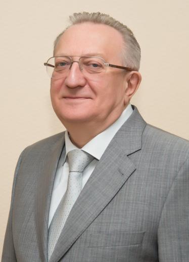
Best regards, Chief Editor, Director of the Medical Institute of Continuing Education of the Russian Biotechnological University (ROSBIOTECH), Academician of the Russian Academy of Medical and Technological Sciences, Member of the European Academy of Dermatology and Venereology, Honored Doctor of the Russian Federation, V.V. Gladko
1 Medical Institute of Continuing Education of the Russian Biotechnological University (ROSBIOTECH), Moscow, Russia 2 Research Center for Immunology and Allergology, Moscow, Russia 3 Scientific and Educational Center "Maruga", Moscow, Russia
Abstract
Background. The analysis of traditional, primarily Chinese, medicine is the subject of close attention of modern medicinal substances developers. One of the important, frequently used plant substrates is Tripterygium Wilfordii, which is used as an antiinflammatory agent, including for the treatment of skin diseases. However, the mechanisms of action of this agent, particularly in such a widespread disease as atopic dermatitis, still need to be investigated. Aim. To study of the effect of Tripterygium Wilfordii extract on in vitro cytokine production in normal and atopic dermatitis patients. Materials and Methods. In a culture of the effect of Tripterygium Wilfordii extract on spontaneous and phytohemagglutinininduced synthesis of IL-2, IL-4, IL-6, IL-10, IL-17, TNFα, IFNγ was evaluated in mononuclear cells culture obtained from healthy donors and patients with atopic dermatitis using enzyme immunoassay test systems Cloud-Clone Corp. (CCC, Wuhan) Results. It was found that the level of spontaneous cytokine synthesis in patients with atopic dermatitis was significantly increased, while the response to the mitogenic stimulus was sharply reduced, absent or inverted. Tripterygium Wilfordii extract in all samples, except for intact cells of healthy donors, showed a pronounced inhibitory effect. Conclusions. Under in vitro conditions, cultured mononuclear cells of patients with atopic dermatitis produce a greater amount of IL-2, IL-4, IL-6, IL-10, IL-17, TNFα, IFNγ, which indicates the activation of the cytokine function. Tripterygium Wilfordii extract has a pronounced inhibitory effect, especially on activated cells. The use of Tripterygium Wilfordii extracts seems reasonable for promising pharmaceutical substances.
For citations: Volchek I. A., Teryaev A. S., Gladko O. V., Rebrova O. M., Michurina A. P., Makarova A. G. Tripterygium Wilfordii extract as a regulator of spontaneous and in vitro stimulated cytokine production with atopic dermatitis. Bulletin of the Medical Institute of Continuing Education. — 2023. — V. 3, № 3. — P. 8–13. — DOI 10.36107/2782-1714_2023-3-3-8-13.
Literature
Severity scoring of atopic dermatitis: the SCORAD index. Consensus Report of the European Task Force on Atopic Dermatitis. Dermatology. 1993;186(1):23-31. doi: 10.1159/000247298. PMID: 8435513.
Практикум по иммунологии: учеб. пособие / Под ред. И. А. Кондратьевой, В. Д. Самуилова. — М. — Изд-во МГУ. — 2001. — 224 с.
Sugaya M. Pe Role of P17-Related Cytokines in Atopic Dermatitis. Int J Mol Sci. 2020 Feb 15;21(4):1314. doi: 10.3390/ijms21041314. PMID: 32075269; PMCID: PMC7072946.
Schuler C.F. 4th, Billi A.C., Maverakis E., Tsoi L.C., Gudjonsson J.E. Novel insights into atopic dermatitis. J Allergy Clin Immunol. 2023 May;151(5):1145-1154. doi: 10.1016/j.jaci.2022.10.023. Epub 2022 Nov 22. PMID: 36428114; PMCID: PMC10164702.
Dutta K., Friscic J., HoRmann M.H. Targeting the tissue-complosome for curbing inQammatory disease. Semin Immunol. 2022 Mar;60:101644. doi: 10.1016/j.smim.2022.101644. Epub 2022 Jul 26. PMID: 35902311.
Chen S.R., Dai Y., Zhao J., Lin L., Wang Y., Wang Y. A Mechanistic Overview of Triptolide and Celastrol, Natural Products from Tripterygium wilfordii Hook F. Front Pharmacol. 2018 Feb 14;9:104. doi: 10.3389/fphar.2018.00104. PMID: 29491837; PMCID: PMC5817256.
Song J., He G.N., Dai L. A comprehensive review on celastrol, triptolide and triptonide: Insights on their pharmacological activity, toxicity, combination therapy, new dosage form and novel drug delivery routes. Biomed Pharmacother. 2023 Jun;162:114705. doi: 10.1016/j.biopha.2023.114705. Epub 2023 Apr 14. PMID: 37062220.
Luo Y.M., Yang S.D., Wen M.Y., Wang B., Liu J.H., Li S.T., Li Y.Y., Cheng H., Zhao L.L., Li S.M., Jiang J.J. Insights into the mechanisms of triptolide nephrotoxicity through network pharmacology-based analysis and RNA-seq. Front Plant Sci. 2023 Mar 7;14:1144583. doi: 10.3389/fpls.2023.1144583. PMID: 36959927; PMCID: PMC10027700.
Волчек И.А., Теряев А.С. Влияние экстрактов дудника амурского (Angelica amurensis) на синтез цитокинов in vitro // Вестник медицинского института непрерывного образования. — 2023 — T. 3. — № 2. — С. 8–3. — EDN AKJOTJ. DOI: 10.36107/2782-1714_2023-3-2-8-13
1 Department of Plastic Surgery Bedford Hospital, Bedford, UK 2 Department of Skin and Venereal Diseases with Cosmetology course Medical Institute of Continuing Education, Russian Biotechnological University (ROSBIOTECH), Moscow, Russia 3 Dermatology Department Bedford Hospital, Bedford, UK
Abstract
Background. Rhinophyma is a chronic and progressive condition, considered a form of severe rosacea, which classically affects the tip of the nose. Some people have rhinophyma without having rosacea. The disease also carries a risk of 3-10% of malignant transformation. Different therapeutic methods have been described over time, with different grades of efficiency, and each of them with its associated risks. At present, the gold standard treatment for the "established" rhinophyma is the ablative laser Aim. To propose a new therapeutic protocol, combining ablative and non-ablative lasers, with an early approach, that results in a better cosmetic outcome and a longer free disease interval. Materials and Methods. The four stages of the new proposed protocol involve early treatment of rosacea with vascular lasers (PDL, NdYAG), then the use of an ablative laser (CO2) and further revision of the post-ablative result (CO2, ErYAG, PDL/NdYAG). Results. The results of the treatment are presented with "before" and "after" photos of the patients. Conclusions. The proposed approach, combining ablative and non-ablative lasers, leads to a better cosmetic result and a longer remission.
For citations: Chiru R. Rhinophyma — from the genetics of the disease to a new therapeutic protocol / Chiru Roxana, Margaj Vishrabdha, Maheshwari Kavish, V.V. Gladko, S.A. Masyukova, I.V. Ilyina, E.P. Burova // Bulletin of the Medical Institute of Continuing Education. — 2023. — V. 3, No. 3. — P. 14–20. — DOI 10.36107/2782-1714_2023-3-3-14-20.
Literature
Madan V., Ferguson J.E., August P.J. Carbon dioxide laser treatment of rhinophyma: a review of 124 patients. Br J Dermatol. 2009 Oct;161(4):814-8. doi: 10.1111/j.1365-2133.2009.09317.x. Epub 2009 Jul 14. PMID: 19624541.
Laun J., Gopman J., Elston J.B., Harrington M.A. Rhinophyma. Eplasty. 2015 May 1;15:ic25. PMID: 25987948; PMCID: PMC4426765.
Fink C., Lackey J., Grande D.J. Rhinophyma: A Treatment Review. Dermatol Surg. 2018 Feb;44(2):275-282. doi: 10.1097/DSS.0000000000001406. PMID: 29140869.
H. Sadick, B. Goepel, C. Bersch, U. Goessler, K. Hoermann, and F. Riedel, "Rhinophyma: diagnosis and treatment options for a disfiguring tumor of the nose," Annals of Plastic Surgery, vol. 61, no. 1, pp. 114-120, 2008
Pandrangi, V., Z. Johnson, C. & A. Krane, N. Rhinophyma: Taking Care of the "WC Fields" Nose. Curr Otorhinolaryngol Rep 10, 262-270 (2022). https://doi.org/10.1007/s40136-022-00409-2
Dick M.K., Patel B.C. Rhinophyma. In: StatPearls. StatPearls Publishing, Treasure Island (FL); 2022. PMID: 31335093.
van Zuuren E.J. Rosacea. N. Engl J. Med. 2017;377(18):1754-1764. doi:10.1056/NEJMcp150663029091565
Curnier A, Choudhary S. Rhinophyma: dispelling the myths. Plast Reconstr Surg. 2004;114(2):351-354. doi:10.1097/01. PRS.0000131875.67987.6915277798
Payne G., Ko F., Anspaugh S., Wheeler C.K., Wright T.E., Robson M.C. Down-regulating causes of fibrosis with tamoxifen: a possible cellular/molecular approach to treat rhinophyma. Ann Plast Surg. 2006;56(3):301-305. doi:10.1097/01. sap.0000199155.73000.2f16508362
Jansen T., Plewig G. Clinical and histological variants of rhinophyma, including nonsurgical treatment modalities. Facial Plast Surg. 1998;14(4):241-253. doi:10.1055/s-2008-106445611816064
Elliot R.A., Ruff L.E. (1980) Rhinophyma and its treatment. Clin Plast Surg 7:277-288
Tuzun Y., Wolf R., Kutlubay Z., Karakus O., Engin B. Rosacea and rhinophyma. Clin Dermatol. 2014;32(1):35-46. doi:10.1016/j. clindermatol.2013.05.02424314376
ReboraA. The management of rosacea.Am J Clin Dermatol. 2002;3(7):489–496. doi:10.2165/00128071-200203070-0000512180896
Stefania Tenna, Pierluigi Gigliofiorito, Marika Langella, Carlo Carusi, Paolo Persichetti, Treatment of rhinophyma with ultrasonic scalpel: case report, Journal of Plastic, Reconstructive & Aesthetic Surgery, Volume 62, Issue 6, 2009, Pages e164-e165, ISSN 1748- 6815, https://doi.org/10.1016/j.bjps.2008.11.006.
Metternich F.U., Wenzel S., Sagowski C., Jäkel K., Koch U. Die operative Therapie des Rhinophyms mit dem ultraschallaktivierten Skalpell (Ultracision Harmonic Scalpel) [Surgical treatment of rhinophyma with the ultrasonic scalpel (Ultracision Harmonic Scalpel)]. Laryngorhinootologie. 2003 Feb;82(2):132-7. German. doi: 10.1055/s-2003-37731. PMID: 12624844.
Lazzeri D., Larcher L., Huemer G.M., et al. Surgical correction of rhinophyma: comparison of two methods in a 15-year-long experience. J Cranio-Maxillofacial Surgery. 2013;41(5):429-436. doi:10.1016/j.jcms.2012.11.009
Goon P.K.Y., Dalal M., Peart F.C. The gold standard for decortication of rhinophyma: combined erbium-YAG/CO2 laser. Aesthetic Plast Surg. 2004;28(6):456-460. doi:10.1007/s00266-004-0012-x15625593
Wenig B.L., Weingarten R.T. Excision of rhinophyma with Nd: yAGlaser: a new technique. Laryngoscope. 1993;103(1 Pt 1):101-103. doi:10.1288/00005537-199301000-000208421411
FEATURES OF THE COURSE OF LICHEN PLANOPILARIS IN MEN
L.A. Safonova
21-26
Medical Institute of Continuing Education of the Russian Biotechnological University (ROSBIOTECH), Moscow, Russia
Abstract
Background. Lichen Planopilaris (LPP) is a variant of primary cicatricial alopecia with a lymphocytic inflammatory infiltrate that occurs at the level of the hair follicle funnel and destroys stem cells of the bulge zone. Timely diagnosis and determination of the correct treatment tactics allows to achieve stabilization of the process and minimize irreversible hair loss. The article presents clinical cases of Lichen Planopilaris in men, initially accompanied by misdiagnoses and lack of adequate therapy, which led to significant irreversible hair loss.
For citations: Safonova L. A. Features of the course of Lichen Planopilaris in men. // Bulletin of the Medical Institute of Continuing Education. — 2023. — V. 3, No. 3. — P. 21–26. — DOI 10.36107/2782-1714_2023-3-3-21-26.
Literature
Summary of North American Hair Research Society (NAHRS) — Sponsored workshop on cicatricial alopecia, Duke University Medical Center, February 10 and 11, 2001. Journal of the American Academy of Dermatology, 48(1), 103-110. https://doi. org/10.1067/mjd.2003.68
Chantal B., Leonard C. Sperling and Jerry Shapiro Lymphocytic primary cicatricial alopecias, including chronic cutaneous lupus erythematosus, lichen planopilaris, frontal fibrosing alopecia, and Graham-Little syndrome. J. Am. Acad. Dermatol 2016;75:1081- 99. http://dx.doi.org/10.1016/j.jaad.2014.09.058
Errichetti E, Figini M, Croatto M, Stinco G. Therapeutic management of classic lichen planopilaris: a systematic review. Clin Cosmet Investig Dermatol. 2018;11:91-102. Published 2018 Feb 27. doi:10.2147/CCID.S137870
Lepe K, Nassereddin A., Salazar F. J. Lichen Planopilaris https://www.ncbi.nlm.nih.gov/books/NBK470325/
Babahosseini H., Tavakolpour S., Mahmoudi H., Balighi K., Teimourpour A., Ghodsi S.Z., Abedini R., Ghandi N., Lajevardi V., Kiani A., Kamyab K., Mohammadi M., Daneshpazhooh M.J. Lichen planopilaris: retrospective study on the characteristics and treatment of 291 patients. Dermatolog Treat. 2019 Sep;30(6):598-604. doi: 10.1080/09546634.2018.1542480. Epub 2019 Jan 4.PMID: 30411987
Rosalynn R Z Conic, Melissa Piliang, Wilma Bergfeld, Natasha Atanaskova-Mesinkovska Association of Lichen Planopilaris With Dyslipidemia DOI: 10.1001/jamadermatol.2018.1749
Kenia Lepe, Nassereddin Ali, Francisco J. Salazar Lichen Planopilaris StatPearls Publishing; 2022 Jan; https://www.ncbi.nlm.nih.gov/books/NBK470325/
Lyakhovitsky A. Amichai B., C Sizopoulou, A Barzilai A case series of 46 patients with lichen planopilaris: Demographics, clinical evaluation, and treatment experience J. Dermatolog treat; 2015 Jun;26(3):275-9. doi: 10.3109/09546634.2014.933165. Epub 2014 Jul 1.
Errichetti E., Figini M., Croatto M., Stinco G. Therapeutic management of classic lichen planopilaris: a systematic review. Clin Cosmet Investig Dermatol. 2018;11:91-102. Published 2018 Feb 27. doi:10.2147/CCID.S137870
Carolina Oliveira Costa Fechine, Neusa Yuriko Sakai Valente, Ricardo Romiti Lichen planopilaris and frontal fibrosing alopecia: review and update of diagnostic and therapeutic feature; Anais Brasileiros de Dermatologia Volume 97, Issue 3, May–June 2022, Pages 348-357
Yang C.C., Khanna T., Sallee B., Christiano A.M, Bordone L.A. Tofacitinib for the treatment of lichen planopilaris: A case series.. Dermatol Ther. 2018 Nov;31(6):e12656. doi: 10.1111/dth.12656. Epub 2018 Sep 27.
Errichetti E., Figini M., Croatto M., Stinco G. Therapeutic management of classic lichen planopilaris: a systematic review. Clin Cosmet Investig Dermatol. 2018;11:91-102. Published 2018 Feb 27. doi:10.2147/CCID.S137870
Charles Chiang, Deborah Sah, Bryan K. Cho, Blanca E. Ochoa, Vera H. Price Hydroxychloroquine and lichen planopilaris: Efficacy and introduction of Lichen Planopilaris Activity Index scoring system; Journal of the American Academy of Dermatology Volume Лечение волос и кожи головы: практ руководство / под ред. А.Тости, Д.Аз-Сигала, Р. Пирмеза; пер. с англ. под ред. А.Г. Гаджигороевой // М. — Геотар-Медиа. — 2021. 176 с. doi:10.33029/9704-5873-0-HST-2021-1-408
LINEAR CLOSURE: A STRAIGHTFORWARD APPROACH TO LARGE DEFECTS ON THE NASOLABIAL CHEEK FOLLOWING MOHS MICROGRAPHIC SURGERY FOR MORPHEAFORM BASAL CELL CARCINOMA
Justin W. Marson, MD1, Rebecca M. Chen, MD1, Michelle Schwartz, MD1, V.V. Gladko2, I.V. Ilyina2 and Daniel M. Siegel, MD, MS1
27-30
1 Department of Dermatology, SUNY Downstate Health Sciences University, Brooklyn, NY 2 Medical Institute of Continuing Education of Russian Biotechnological University (ROSBIOTECH), Moscow, Russia
Abstract
Mohs micrographic surgery (MMS) utilizes intra-operative histological assessment to maximize clearance of cutaneous malignancy while minimizing surgical defect size. [1–4] On cosmetically sensitive areas, most notably the face, transposition, advancement, and interpolation flaps and occasionally skin grafts are often utilized as repair techniques, despite potential for post-operative complications including bleeding, infection, impaired wound healing, and graft/flap loss.[1–5] Here we present a case of a large (6.5 cm by 3.5 cm) surgical defect on the cheek following MMS for the management of a morpheaform basal cell carcinoma and briefly review considerations regarding closure repairs, emphasizing linear closure as a reliable and successful option.
For citations: Marson, Justin W. Linear Closure: a straightforward approach to large defects on the nasolabial cheek following Mohs micrographic surgery for morpheaform basal cell carcinoma./Justin W. Marson, Rebecca M. Chen, Michelle Schwartz, V.V. Gladko, I.V. Ilyina and Daniel M. Siegel, // Bulletin of the Medical Institute of Continuous Education. — 2023. — V. 3, No. 3. — P. 27–30. — DOI 10.36107/2782-1714_2023-3-3-27-30.
Literature
Alam M., Ibrahim O., Nodzenski M., et al. Adverse Events Associated With Mohs Micrographic Surgery. JAMA Dermatology. 2013-12-01 2013;149(12):1378. doi:10.1001/jamadermatol.2013.6255
Johnson A.R., Egeler S.A., Wu W.W., Bucknor A., Ibrahim A.M.S., Lin S.J. Facial Reconstruction After Mohs Surgery: A Critical Review of Defects Involving the Cheek, Forehead, and Perioral Region. J Craniofac Surg. 2019;30(2):400-407. doi:10.1097/SCS.0000000000005074
Brandão C.M., Weimann E.T.S., Terzian L.R., Machado Filho C.D.S., Paschoal F.M., Criado PR. Keep it simple. A ten-year experience in reconstructions after Mohs micrographic surgery. An Bras Dermatol. 2020;95(6):714-720. doi:10.1016/j.abd.2020.05.004
Soliman S., Hatef D.A., Hollier L.H., Thornton JF. The rationale for direct linear closure of facial Mohs' defects. Plast Reconstr Surg. Jan 2011;127(1):142-149. doi:10.1097/PRS.0b013e3181f95978
Cook J.L., Perone J.B. A prospective evaluation of the incidence of complications associated with Mohs micrographic surgery. Arch Dermatol. Feb 2003;139(2):143-52. doi:10.1001/archderm.139.2.143
Kimyai-Asadi A., Goldberg L.H., Peterson S.R., Silapint S., Jih M.H. The incidence of major complications from Mohs micrographic surgery performed in office-based and hospital-based settings. J Am Acad Dermatol. Oct 2005;53(4):628-34. doi:10.1016/j.jaad.2005.03.023
RESULTS OF TYMPANOPLASTY IN TOTAL AND SUBTOTAL TYMPANIC MEMBRANE DEFECTS
I.I. Morozov1,2, N.V. Gorbunova1, N.S. Grachev2,3
31-35
1 Main Clinical Hospital of the Ministry of Internal Affairs of the Russian Federation, Moscow, Russia 2 Medical Institute of Continuing Education of the Federal State Budgetary Educational Institution of Higher Education "Russian Biotechnological University (ROSBIOTECH)". Moscow, Russia 3 Institute of Oncology, Radiology and Nuclear Medicine, Dmitry Rogachev National Medical Research Center of Pediatric Hematology, Oncology and Immunology of the Ministry of Health of the Russian Federation, Moscow, Russia
Abstract
Purpose. To carry out a comparative analysis of the results of endoscopic tympanoplasty with auricle cartilage and microscopic tympanoplasty with temporal autofascia in the surgical treatment of total and subtotal perforations of the tympanic membrane. Materials and Methods. 56 patients were devided into 2 groups, the first group had 28 patients after endoscopic type I tympanoplasty with auricle cartilage; the second group had 28 patients after type I tympanoplasty with temporal autofascia. The patients were examined after the surgery in 2, 3, 6, 12 months, anatomical and functional results were assessed, audiometry, and the number of recurrences of tympanic membrane perforations were extimated. Results. The average operation time in the first group (60.2±11.5 min) was significantly less than in the second group (88.3±15.2min; p<0.5). Complete closure of the tympanic membrane perforation and graft engraftment was observed in group 1 in 92.8 %, in group 2 in 89.3 %. The average preoperative air conduction according to audiometry data in group 1 was 42.1±15.4 dB, in group 2 it was 38.2±12.4 dB. The differences of bone-air conduction before the surgery were 18.8±11.6 dB and 19.6±13.3 dB respectively. The bone-air interval before the surgery was 23.3±6.4 dB in group 1, — it was 19.2±7.8 dB in group 2. After the surgery in 1-year follow-up, the average improvement in air conduction in group 1 was 18.1±7.7 dB, in group 2 it was 19.5±7.5 dB. Postoperative bone conduction was 17.8±10.7 dB and 18.8±14.9 dB respectively. The bone-air interval 1 year after the surgery was 10.1±4.6 dB in group 1and 11.5±8.4 dB. There was no statistically significant difference between the two groups.
For citations: Morozov I.I., Gorbunova N.V., Grachev N.S. Results of tympanoplasty in total and subtotal tympanic membrane defects // Bulletin of the Medical Institute of Continuing Education. — 2023. — V. 3, No. 3. — S. 31–35. — DOI 10.36107/2782-1714_2023-3-3-31-35.
Literature
Singh S.P., Nagi R.S., Singh J. To compare short and longterm graft uptake and hearing outcome of type I cartilage tympanoplasty between small, medium and large perforations using reinforced sliced conchal cartilage // Indian J Otolaryngol Head Neck Surg 71:550–556. https:// doi. org/ 10. 1007/ s12070- 019- 01727-6
Atchariyasathian V., Suwannajak R., Plodpai Y., Pitathawatchai. A comparison of endoscopic transtympanic myringoplasty and endoscopic type I tympanoplasty for repairing medium- to large-sized tympanic membrane perforation: a randomized clinical trial // Eur Arch Otorhinolaryngol 277:2199-2207. https:// doi. org/10. 1007/ s00405- 020- 05955-3
Горбунова Н.В., Морозов И.И. Современные тенденции в эндоскопической отохирургии // Вестник медицинского института непрерывного образования. — 2023. — T. 3, № 2. — С. 33-38. — EDN BGCHJI [Gorbunova N.V., Morozov I.I. Modern trends in endoscopic otosurgery // Bulletin of the Medical Institute of Continuing Education. — 2023. — V. 3, № 2. — P. 33–38. — EDN BGCHJI (In Russ.)]
Hu Y., Teh B.M., Hurtado G., Yao X., Huang J., Shen Y. Can endoscopic ear surgery replace microscopic surgery in the treatment of acquired cholesteatoma? A contemporary review // Int J Pediatr Otorhinolaryngol. 2020 Apr;131:109872. doi: 10.1016/j. ijporl.2020.109872.
Choi N., Noh Y., Park W. et al. Comparison of endoscopic tympanoplasty to microscopic tympanoplasty. Clin Exp Otorhinolaryngol 10:44-49. https:// doi. org/ 10. 21053/ ceo. 2016. 000806.
Li B., Zhou L., Wang M., Wang Y., Zou J. Endoscopic versus microscopic surgery for treatment of middle ear cholesteatoma: A systematic review and meta-analysis // Am J Otolaryngol. 2021 Mar-Apr;42(2):102451. doi: 10.1016/j.amjoto.2020.102451.
Горбунова Н.В., Морозов И.И., Грачев Н.С. Мастоидопластика при хирургическом лечении хронического гнойного среднего отита. методики и результаты // Вестник медицинского института непрерывного образования. — 2023. — T. 3, № 2. — С. 24–29. — EDN AZGTTC [Gorbunova N.V., Morozov I.I., Grachev N.S. Mastoidoplasty in the surgical treatment of chronic suppurative otitis media. Methods and results // Bulletin of the Medical Institute of Continuing Education. — 2023. — V. 3, № 2. — P. 24–29. — EDN AZGTTC (In Russ.) ]
Ryan P., Wuesthoff C., Patel N. Getting started in endoscopic ear surgery // J Otol. 2020 Mar;15(1):6-16. doi: 10.1016/j. joto.2018.10.002.
Manna S., Kaul V.F., Gray M.L., Wanna G.B. Endoscopic versus microscopic middle ear surgery: a meta-analysis of outcomes following tympanoplasty and stapes surgery // Otol Neurotol. 2019;40:983-993.
Грачев Н.С., Полев Г.А., Морозов И.И., Самарин А.Е., Ворожцов И.Н., Щербаков Д.А. Опыт применения эндоскопической техники в отохирургии у детей // Вестник оториноларингологии. — 2020. — Т. 85. — № 1. — С. 59–63. https://doi.org/10.17116/otorino20208501159 [Grachev NS, Polev GA, Morozov II, Samarin AE, Vorozhtsov IN, Shcherbakov DA. Our first experience with endoscopic ear surgery // Vestnik otorinolaringologii. 2020;85(1):59-63. (In Russ.) https://doi.org/10.17116/otorino20208501159]
Bayram A., Muluk N.B., Cingi C., Bafaqeeh S.A. Success rates for various graft materials in tympanoplasty-a review // J Otol 15:107-111. https://doi.org/ 10. 1016/j. joto. 2020. 01. 001
Shakya D., Nepal A. Long-term results of type I tympanoplasty with perichondrium reinforced cartilage palisade vs temporalis fascia for large perforations: a retrospective study // J Otol16:12-17. https:// doi. org/ 10. 1016/j. joto. 2020. 07. 004 13. Guo L., Su Y., Cai Z., Yang Y. Outcomes of transcanal endoscopic middle ear surgery for congenital cholesteatoma // Acta Otolaryngol. 2023 Feb;143(2):141-146. doi:10.1080/00016489.2023.2176544.
Srinivasan V., Toynton S.C., Mangat K.S. Transtympanic myringoplasty in children // Int J Pediatr Otorhinolaryngol 39:199-204. https://doi.org/10.1016/ S0165- 5876(96) 01477-2
Alzoubi F.Q., Tarifi A.A., Khader Y., de Carpentier J. Comparison between transtympanic and elevation of tympanomeatal flap approaches in tympanoplasty // Otol Neurotol 31:773-775. https://doi.org/10.1097/MAO.0b013 e3181 e40a41
1 Main Clinical Hospital of the Ministry of Internal Affairs of the Russian Federation, Moscow, Russia 2 Medical Institute of Continuing Education of the Federal State Budgetary Educational Institution of Higher Education "Russian Biotechnological University (ROSBIOTECH)". Moscow, Russia
Abstract
Background. Postoperative nasal septum perforation (PNSP) is one of the complication of the nasal septum (NS) surgery, with an incidence of 1–8 %. Surgical repair of the nasal septum improves the patients' quality of life. The most effective techniques are the ones using two rotation flaps of the nasal mucosa, which are used for the plasty of traumatic and idiopathic perforations. The results of implementing this technique for the closure of PNSP require a comprehensive study Aim. To evaluate the effectiveness of rotary flaps of the nasal mucosa in postoperative nasal septum perforation repair. Materials and Methods. 18 patients with postoperative nasal septum perforation were operated on. The number of the patients depending on the area (S) of the PNSP was S <1cm2 — 4, S 1-2 cm2 — 10, S > 2cm2 — 4. The criterion for the technique efficiency was the fact of intraoperative complete closure of the PNSP, and the absence of recurrences of the PNSP within 1 year. We analysed the cause-and-effect relationships between the structural features of the PNSP (6 types were identified) with the technique of execution and long-term results. Results. Complete intraoperative closure of PNSP in 13 of 18 patients (72 %). In 5 patients, an additional sampling of temporal autofascia was performed with subsequent implantation between the flaps for intraoperative complete closure of the PNSP. In 4 out of 13 patients (30 %) with PNSP S 1-2 cm2 the PNSP relapse (type 3, 4) was noted. There was 1 relapse (type 5) with PNSP S > 2cm2 in 1 out of 5 patients (20 %), to whom autofascia was additionally used. Conclusions. The surgery of the PNSP according to the specified technique is effective with the PNSP S <1cm2; at S 1-2 cm2 it leads to distant relapses in 30 % of the cases, S > 2 cm2 is not effective. The use of a temporal fascia autograft allows to increase the efficiency of the postoperative nasal septum perforation regardless the area of it.
For citations: Gorbunova N.V., Shirokaya A.V., Sardarov G.G. [and etc]. Using of rotational nasal mucosa flaps in postoperative nasal septal perforation repair. // Bulletin of the Medical Institute of Continuing Education. — 2023. — V. 3, No. 3. — S. 36–41. — DOI 10.36107/2782-1714_2023-3-3-36-41.
Literature
Quinn J.G., Bonaparte J.P., Kilty S.J. Postoperative management in the prevention of complications after septoplasty: a systematic review // Laryngoscope. — 2013. — Jun. — 123(6): 1328-33. https://doi.org/10.1002/lary.23848
Морозов И.И. Послеоперационные перфорации перегородки носа, методы хирургического лечения и способы повышения их эффективности // Российская оториноларингология. — 2020. — Т. 19. — № 1. — С. 77–83. https://doi.org/10.18692/1810-4800-2020-1-77-83 [Morozov I.I. Postoperative nasal septum perforations: surgical treatment methods and the ways of improvement their efficacy. Rossiiskaya otorinolaringologiya. — 2020. — V. 19. — № 1. — P. 77-83. (In Russ.) https://doi.org/10.18692/1810-4800-2020-1-77-83]
Pedroza F., Patrocinio L.G., Arevalo O. A review of 25-year experience of nasal septal perforation repair // Arch Facial Plast Surg. — 2007. — 9. — P. 12–8. https://doi.org/ 10.1001/archfaci.9.1.12
Морозов И.И., Грачев Н.С. Результаты хирургического лечения послеоперационных перфораций перегородки носа // Российская ринология. — 2020. — Т. — 28. — № 4. — С. 197-204. https://doi.org/10.17116/rosrino202028041197 [Morozov II, Grachev N.S. Results of surgical treatment of postoperative nasal septum perforation. Russian Rhinology. 2020;28(4):197-204. (In Russ.). https://doi.org/10.17116/rosrino202028041197]
Nomura T., Ushio M., Kondo K., Kikuchi S. Effects of nasal septum perforation repair on nasal airflow: An analysis using computational fluid dynamics on preoperative and postoperative three-dimensional models. Auris Nasus Larynx. 2018 Oct;45(5):1020-1026. https://doi.org/10.1016/j.anl.2018.02.006
Lindemann J., Scheithauer M., HoRmann T.K., Rettinger G., Kobes C., Sommer F. Long-term results aVer surgical closures of septal perforations. Laryngorhinootologie 2014; 93(11): 751-5. https://doi.org/10.1055/s-0034-1385891
Морозов И.И., Грачев Н.С. Опыт использования лоскута P. Castelnuovo в пластике послеоперационных перфораций перегородки носа // Российская оториноларингология. — 2021. — Т. 20. № 4. С. 27–32. https://doi.org/10.18692/1810-4800-2021- 4-27-32 [Morozov I.I., Grachev N.S. Experience of using the P. Castelnuovo flap in the postoperative nasal septal perforation repair. Rossiiskaya otorinolaringologiya. 2021;20(4):27-32. https://doi.org/10.18692/1810-4800-2021-4-27-32 (In Russ)]
Park J.H., Kim D.W., Jin H.R. Nasal septal perforation repair using intranasal rotation and advancement flaps. Am J Rhinol Allergy. 2013 Mar-Apr;27(2):e42-7. doi:10.2500/ajra.2013.27.3878.
Dayton S., Chhabra N., Houser S. Endonasal septal perforation repair using posterior and inferiorly based mucosal rotation flaps. Am J Otolaryngol. 2017 Mar-Apr;38(2):179-182. doi:10.1016/j.amjoto.2017.01.001.
Морозов И.И., Грачев Н.С., Горбунова Н.В. и др. Клинико-морфологические особенности перфораций перегородки носа // ВестникМедицинского института непрерывного образования. — 2022. — № 3. — С. 24–27. DOI:10.46393/27821714_2022_3_24 [Morozov I.I., Grachev N.S., Gorbunova N.V. et al. Study of clinical and morphological features of nasal septum perforations. Bulletin of the Medical Institute of Сontinuing Education. 2022; (3): 24–27. (In Russ/) https://doi.org/10.46393/27821714_2022_3_24]
Крюков А.И., Царапкин Г.Ю., Артемьев М.Е. Клинический подход в выборе тактики ведения больных с перфорацией перегородки носа. Российская оториноларингология. — 2013. — №4. С. 55–61.
Dedhia R.D., Davis S.J., Stephan S.J. Optimizing septal perforation repair techniques. Curr Opin Otolaryngol Head Neck Surg. 2020 Aug;28(4):212-217. doi: 10.1097/MOO.0000000000000631.
Морозов И.И., Грачев Н.С. Результаты исследования клинико-морфологических особенностей послеоперационных перфораций перегородки носа // Российская оториноларингология. — 2021. — Т. 20. — № 3. — С. 64–69. https://doi.org/10.18692/1810-4800-2021-3-64-69 [Morozov I.I., Grachev N.S. Study of clinical and morphological features of postoperative nasal septum perforations. Rossiiskaya otorinolaringologiya. 2021;20(3):64-69. https://doi.org/10.18692/1810-4800-2021-3-64-69 (In Russ.)]
Flavill E., Gilmore J.E. Septal perforation repair without intraoperative mucosal closure. Laryngoscope 2014; 124(5): 1112-7. DOI: 10.1002/lary.24386
Морозов И.И., Грачев Н.С., Наседкин А.Н. Использование височной аутофасции в пластике стойких послеоперационных перфораций перегородки носа. // Head and Neck/Голова и шея. Российское издание. Журнал общероссийской общественной организации «Федерация специалистов по лечению заболеваний головы и шеи». — 2017. — № S1. — С. 77–78. [Morozov I.I., Grachev N.S., Nasedkin A.N. Qe use of temporal autofascia in the plastic surgery of postoperative perforations of the nasal septum. Head and neck. Russian Journal. 2017; (S1): 77–78.]
Пустовит О.М., Наседкин А.Н., Егоров В.И., Исаев В.М., Исаев Э.В., Морозов И.И. Воздействие ультразвуковой кавитации и фотохромотерапии на процесс репарации слизистой оболочки носа после септопластики и подслизистой вазотомии нижних носовых раковин // Head and Neck/Голова и шея. Российское издание. Журнал общероссийской общественной оболочки эффективно при площади ПППН S<1см2; при S 1-2 см2 — сложно в техническом исполнении и приводит к рецидивам ППН при условии отсутствия хряща в верхних отделах ПН и наличии костного гребня по дну полости носа с острыми краями; для ПППН S>2 см2 — неэффективно. Использование аутотрансплантата височной фасции позволяет повысить эффективность пластики перфорации ПН независимо от площади ПППН. Данный положительный результат требует дальнейшего всестороннего изучения. организации «Федерация специалистов по лечению заболеваний головы и шеи». — 2018. — Т. 6. — №2. — С. 20–26 https://doi.org10.25792/HN.2018.6.2.20-26 [Pustovit O.M., Nasedkin A.N., Egorov V.I., Isaev V.M., Isaev E.V., Morozov I.I. Using ultrasonic cavitation and photochromotherapy to increase nasal mucosa reparation process aVer septoplasty and submucous vasotomy of the inferior nasal turbinates. Head and neck. Russian Journal. 2018;6(2):20–26 (In Russ.) DOI:10.25792/HN.2018.6.2.20-26]
Delaney S.W., Kridel R.W.H. Contemporary Trends in the Surgical Management of Nasal Septal Perforations: A Community Survey. Facial Plast Surg. 2019 Feb;35(1):78-84. https://doi.org/10.1055/s-0038-1676049
Морозов И.И., Грачев Н.С. Способ эндоскопической пластики стойкой перфорации перегородки носа // Head and Neck/ Голова и шея. Российское издание. Журнал общероссийской общественной организации «Федерация специалистов по лечению заболеваний головы и шеи» — 2020. — Т. 8. — №2. — С. 39–44 https://doi.org/10.25792/HN.2020.8.2.39-44. [Morozov I.I., Grachev N.S. Method for endoscopic plastic surgery of persistent perforation of the nasal septum. Head and neck. Russian Journal. 2020;8(2):39–44 (in Russian) DOI: 10.25792/HN.2020.8.2.39-44]
Özer S., Süslü A.E., Yılmaz T., Önerci T.M. Sandwich graV technique outcomes in medium and large size nasal septal perforations. Braz J Otorhinolaryngol. 2022 Nov-Dec;88(6):896-901. doi:10.1016/j.bjorl.2020.12.018.
1 Main Clinical Hospital of the Ministry of the Internal Affairs of the Russian Federation,Moscow, Russia 2 Medical Institute of Continuing Education, Russian Biotechnological University (ROSBIOTECH), Moscow, Russia
Abstract
Aim. Evaluation of the effectiveness of platelet-rich plasma use in type 1 tympanoplasty according to the reviewed literature Materials and Methods. The research studies were included where patients with chronic suppurative otitis media underwent tympanic membrane reconstruction using platelet-rich plasma. Inclusion criteria: the patients with chronic tubotympanic otitis media without signs of exacerbation, type 1 tympanoplasty using platelet-rich plasma together with autografts in the study group and only autografts in the control group, postoperative catamnesis of at least three months. Results. The frequency of a positive outcome of the operation and complete engraftment was 85.7–100 % in the study groups and 55–92 % in the control groups. The average improvement in hearing after the surgery was 10.3-18.62 dB in the study groups and 7.23-15.64 dB in the control groups. There were no significant complications with the platelet-rich plasma use. Conclusions. The use of platelet-rich plasma in type 1 tympanoplasty in patients with chronic suppurative otitis media is a safe and technically simple method that allows to improve the efficiency of the operation, reduce the number of recurrences of tympanic membrane perforations and unsatisfactory functional results.
For citations: Gorbunova N.V. Morozov I.I., Sardarov G.G., Bulanov K.V. Platelet-rich plasma use in tympanoplasty // Bulletin of the Medical Institute of Continuing Education. — 2023. — V. 3, No. 3. — S. 42–47. — DOI 10.36107/2782-1714_2023-3-3-42-47.
Literature
Helvik A.S., Krokstad S., Tambs K. Socioeconomic inequalities in hearing loss in a healthy population sample: the HUNT study. Am J Public Health. 2009;99(8):1376-1378. https://doi.org/10.2105/AJPH.2007.133215.
Singh S.P., Nagi R.S., Singh J. To compare short and longterm graft uptake and hearing outcome of type I cartilage tympanoplasty between small, medium and large perforations using reinforced sliced conchal cartilage. Indian J Otolaryngol Head Neck Surg 71:550–556. https:// doi. org/ 10. 1007/ s12070- 019- 01727-6
Горбунова Н.В., Морозов И.И. Современные тенденции в эндоскопической отохирургии // Вестник Медицинского института непрерывного образования. — 2023. — Т. 3. — № 2. — С. 33–38. — DOI 10.36107/2782-1714_2023-3-2-33-38. — EDN BGCHJI. [Gorbunova N.V., Morozov I.I. Modern trends in endoscopic otosurgery. Bulletin of the Medical Institute of Continuing Education. — 2023. — V. 3. — № 2. — P. 33–38. — EDN BGCHJI. DOI 10.36107/2782-1714_2023-3-2-33-38 (In Russ.)]
Guo L., Su Y., Cai Z., Yang Y. Outcomes of transcanal endoscopic middle ear surgery for congenital cholesteatoma. Acta Otolaryngol. 2023 Feb;143(2):141-146. doi: 10.1080/00016489.2023.2176544.
Грачев Н.С., Полев Г.А., Морозов И.И., Самарин А.Е., Ворожцов И.Н., Щербаков Д.А. Опыт применения эндоскопической техники в отохирургии у детей // Вестник оториноларингологии. — 2020. — Т.85. — № 1. — С. 59–63. https://doi.org/10.17116/otorino20208501159 [Grachev NS, Polev GA, Morozov II, Samarin AE, Vorozhtsov IN, Shcherbakov DA. Our first experience with endoscopic ear surgery. Vestnik otorinolaringologii. 2020;85(1):59-63. (In Russ.) https://doi.org/10.17116/otorino20208501159]
Bayram A., Muluk N.B., Cingi C., Bafaqeeh S.A. Success rates for various graft materials in tympanoplasty — a review. J Otol 15:107–111. https://doi.org/ 10. 1016/j. joto. 2020. 01. 001
Alzoubi F.Q., Tarifi A.A., Khader Y., de Carpentier J. Comparison between transtympanic and elevation of tympanomeatal flap approaches in tympanoplasty. Otol Neurotol 31:773–775. https://doi.org/10.1097/MAO.0b013 e3181 e40a41
Семенов Ф.В., Семенов В.Ф. Отоэндоскопическая оценка отдаленных результатов тимпанопластики, выполненной с использованием обогащенной тромбоцитами плазмы // Российская оториноларингология. — 2013 — №3. — С. 132–135.
Teh B.M., Marano R.J., Shen Y., Friedland P.L., Dilley R.J., Atlas M.D. Tissue engineering of the tympanic membrane. Tissue Eng Part B Rev 19: 2013; 116–32. https://doi.org/10.1089/ten.TEB.2012.0389
Dohan Ehrenfest D.M., Rasmusson L., Albrektsson T. Classification of platelet concentrates: from pure platelet-rich plasma (P-PRP) to leucocyte- and platelet-rich fibrin (L- PRF). Trends Biotechnol. 2009; 27: 158-167.
Mehta S., Watson J.T. Platelet rich concentrate: basic science and current clinical applications. J Orthop Trauma, 2008; 22: 432–8. https://doi.org/10.1097/BOT.0b013e31817e793f
Xian L.J., Chowdhury S.R., Bin S.A., Idrus R.B. Concentration-dependent effect of platelet-rich plasma on keratinocyte and fibroblast wound healing. Cytotherapy, 2015; 17: 293–300. https://doi.org/10.1016/j. jcyt.2014.10.005.
Marx RE. Platelet-rich plasma: evidence to support its use. J Oral Maxillofac Surg. 2004 Apr;62(4):489-96. doi:10.1016/j. joms.2003.12.003. PMID: 15085519.
Завьялов Ф.Н., Гончарова О.Г. Результаты применения аутогенной обогащенной тромбоцитами плазмы у больных, перенесших операции на среднем ухе // Вестник оториноларингологии. — 2011. — № 1. — С. 28–30 [Zav'yalov F.N., Goncharova O.G. Rezul'taty primeneniya autogennoy obogashchennoy trombotsitami plazmy u bol'nykh, perenesshikh operatsii na srednem ukhe. Vestnik otorinolaringologii. — 2011. — № 1. — S. 28–30 (In Russ.)]
Navarrete A' M.L., Ortiz N., Rodriguez Let al. Pilot study on the efficiency of the biostimulation with autologous plasma rich in platelet growth factors in otorhinolaryngology: otologic surgery (tympanoplasty type I). ISRN Surg, 2011, 2011: 451020. https://doi.org/10.5402/2011/451020
El-Anwar M.W., El-Ahl MAS, Zidan A.A., Yacoup M.A. Topical use of autologous platelet rich plasma in myringoplasty. Auris Nasus Larynx, 2015; 42: 365–8. https://doi.org/10.1016/j.anl.2015.02.016 PMID: 25794691.
Yadav S.P.S, Malik J.S., Malik P., Sehgal P.K., Gulia J.S., Ranga R.K. Studying the result of underlay myringoplasty using plateletrich plasma. J Laryngol Otol, 2018; 132: 990–4. https://doi.org/10.1017/ S0022215118001846 PMID: 30370872.
Fawzy T., Hussein M.; Eid S., Guindi S. Effect of adding platelet-rich plasma to fat grafts in myringoplasty. Egyptian Journal of Otolaryngology. 2018; 34(4): 224–8.
Mandour M.F., Elsheikh M.N., Khalil M.F. Platelet-Rich Plasma Fat Graft versus Cartilage Perichondrium for Repair of Medium-Size Tympanic Membrane Perforations. Otolaryngol Head Neck Surg, 2019; 160: 116–21. https://doi.org/10.1177/0194599818789146 PMID: 30037309.
Ersozlu T., Gultekin E. A Comparison of the Autologous Platelet-Rich Plasma Gel Fat Graft Myringo- plasty and the Fat Graft Myringoplasty for the Closure of Different Sizes of Tympanic Membrane Perforations. Ear Nose Throat J. 2020; 99: 331–6. https://doi.org/10.1177/0145561319900388
Taneja M.K. Role of Platelet Rich Plasma in Tympanoplasty. Indian J Otolaryngol Head Neck Surg; 2020, 72: 247–50. https://doi.org/10.1007/s12070-020-01815-y
Anwar F.M., Shenoy V.S., Kamath P.M., Deviprasad S.S.D, Domah H.A. Study on Use of Platelet-Rich Plasma in Myringoplasty. Indian Journal of Otology. 2020; 26(2): 71–4. https://doi.org/10.4103/ indianjotol.INDIANJOTOL_103_18.
Akash, Datta R., Suri G.S., Mucha S., Sheikh M.A., Taneja N.S. A Randomised Controlled Trial on the Efficacy of Topical Application of Autologous Platelet Rich Plasma (PRP) on Graft Uptake Rate in Adults Undergoing Type 1 Tympanoplasty for Inactive COM Mucosal Disease. Indian J Otolaryngol Head Neck Surg. 2023 Apr;75(Suppl 1):605-613. doi: 10.1007/s12070-023-03681-w.
Морозов И.И., Грачев Н.С., Горбунова Н.В., Широкая А.В. Баллонная дилатация слуховой трубы у пациентов с изолированной дисфункцией слуховой трубы // Вестник Медицинского института непрерывного образования. — 2023. — Т. 3. — № 1. — С. 72–75. DOI10.36107/2782-1714_2023-3-1-72-75. — EDN YZOZLO [Morozov I.I., Grachev N.S., Gorbunova N.V. [et al.] Balloon dilatation of the Eustachian tube in patients with isolated Eustachian tube dysfunction. Bulletin of the Medical Institute of Continuing Education. 2023; 3 (1): 72–75. — DOI10.36107/2782-1714_2023-3-1-72-75. — EDN YZOZLO (In Russ.)].
Shakya D., Nepal A. Long-term results of type I tympanoplasty with perichondrium reinforced cartilage palisade vs temporalis fascia for large perforations: a retrospective study. J Otol16:12–17. https:// doi. org/ 10. 1016/j. joto. 2020. 07. 004
PECULIARITIES OF ANTITHROMBOTIC THERAPY IN THE PATIENTS WITH CORONARY HEART DISEASE WITH CONMINANT TRAUMATOLOGICAL PATHOLOGY. HOW TO KEEP THE BALANCE BETWEEN THROMBOSIS AND BLEEDING?
1 1 Medical Institute of Continuing Education of Russian Biotechnological University (ROSBIOTECH), Moscow, Russia 3 Branch No. 2 “National Medical Research Center for High Medical Technologies named after A.A. Vishnevsky of the Ministry of Defense of the Russian Federation, Moscow, Russia
Abstract
The selection of antithrombotic therapy for patients with coronary heart disease (CHD) and concomitant pathology, despite clinical recommendations and scales defining the risk of thrombosis and bleeding may be difficult in some cases. Each doctor may face a clinical situation that does not fit within the general recommendations. Practical experience and determination of indications for the use of anticoagulants depending on the dosage in each specific case will help in the faultless choice of a combination of antithrombotic therapy. A clinical example of a very high-risk CHD patient with concomitant traumatological pathology requiring the selection of antithrombotic therapy is presented.
For citations: Yudin V.E., Yaroshenko V. P., Klimko V.V., Sarkisova M.K. Peculiarities of antithrombotic therapy in the patients with coronary heart disease with concomitant traumatological pathology. How to keep the balance between thrombosis and bleeding? // Bulletin of the Medical Institute of Continuing Education. — 2023. — V. 3, No. 3. — S. 48–52. — DOI 10.36107/2782-1714_2023-3-3-48-52.
Literature
Рекомендации Российского кардиологического общества по фибрилляции и трепетанию предсердий от 2020 г.
The Task Force for the Management of Atrial Fibrillation of the European Society of Cardiology (ESC). Guidelines for the management of atrial Obrillation. European Heart Journal. Published online: August 29, 2010 . doi:10.1093/eurheartj/ehq278.
Antman EM, Cohen M, Bernink PJLM, McCabe CH, Hoacek T, Papuchis G, Mautner B, Corbalan R, Radley D, Braunwald E. Ne TIMI risk score for unstable angina/non-ST elevation MI: a method for prognostication and therapeutic decision making JAMA. 2000;284(7):835-42.
GUSTO Investigators. An international randomized trial comparing four thrombolytic strategies for acute myocardial infarction. NEJM, 1993, 329(10): 673-682.
Serebruany VL, Atar D. Assesment of blleding events in clinical trials — proposed of a new classiffication. Am J Cardiol, 2007, 99(2): 288-290.
Mehran R, Rao SV, Bhatt DL, et al. Standardized Bleeding DeOnitions for Cardiovascular Clinical Trials. A Consensus Report From the Bleeding Academic Research Consortium. Circulation. 2011 Jun 14;123(23):2736-47. doi:10.1161/ CIRCULATIONAHA.110.009449.
Loewen P., Dahri K. Risk of bleeding with oral anticoagulants: an updated systematic review and performance analysis of clinical prediction rules. Ann. Hematol. 2011; 90: 1191–1200.
Pisters R, Lane DA, Nieuwlaat R, de Vos CB, Crijns HJGM, Lip GYH, Guideline Development Group for the NICE national clinical guideline for management of atrial Obrillation in primary and secondary care. A Novel User-Friendly Score (HAS-BLED) To Assess 1-Year Risk of Mayor Bleeding in patients With Atrial Fibrillation: The Euro Heart Survey. Chest, 2010, 138(5): 1093-1100.
Costa F., Van Klaveren D., James S., et al. Derivation and validation of predicting bleeding complications in patients undergoing stent implantation and subsequent dual antiplatelet therapy (PRECISEDAPT) score: pooled analysis of individual-patient datasets from clinical trials. Lancet.2017;389(10073):1025-34. DOI:10.1016/S0140-6736(17)30397-5.
Рекомендации Российского кардиологического общества по стабильной ишемической болезни сердца 2020 г.
ESC Guidelines for the management of acute myocardial infarction in patients presenting with ST-segment elevation Eur Heart J. doi:10.1093/eurheartj/ehs215
Акашева Д.У., Булгакова Е.С., Явелов И.С. и др. Трудности ведения пациента старческого возраста: особенности подбора антитромботической терапии. Клинический случай // Рациональная Фармакотерапия в Кардиологии. — 2018. — Т. 14. — № 4. — С. 515-523. DOI: 10.20996/1819-6446-2018-14-4-515-523
Blommel M.L., Blommel A.L. Dabigatran etexilate: a novel oral direct thrombin inhibitor. Am. J. Health Syst. Pharm. 2011; 68 (16): 1506-19.
Paikin J.S., Manolakos J.J., Eikelboom J.W. Rivaroxaban for stroke prevention in atrial Obrillation: a critical review of the ROCKET AF trial. Expert. Rev. Cardiovasc. Ner. 2012; 10 (8): 965-72.
Nutescu E. Apixaban: a novel oral inhibitor of factor Xa. Am. J. Health. Syst. Pharm. 2012; 69 (13): 1113–26.
Рекомендации EHRA 2021 г.
Roffi M., Patrono C., Collet J. P., et al. 2015 ESC Guidelines for the management of acute coronary syndromes in patients presenting without persistent ST-segment elevation: European Heart J. 2016; 37(3): 267-315;Sousa-Uva M., et al 2018 ESC/EACTS Guidelines on myocardial revascularization. Eur Heart J. 2019;40(2):87-165.
Божкова С.А., Касимова А.Р., Накопия В.Б. и др. Все ли мы знаем о профилактике венозных тромбоэмболических осложнений после больших ортопедических операций? // Травматология и ортопедия России. — 2018. — Т. 24. — №1. — С. 129-143. DOI: 10.21823/2311-2905-2018-24-1-129-143.
1 Medical Institute of Continuing Education of Russian Biotechnological University (ROSBIOTECH), Moscow, Russia 2 Branch No. 2 of "National Medical Research Center for High Medical Technologies, Central Military Clinical Hospital named after A.A. Vishnevsky" of the Ministry of Defense of the Russian Federation, Moscow, Russia
Abstract
Background. The use of the hyperbaric oxygenation therapy (HBO) in the program of medical rehabilitation of the wounded in a timely manner and according to the indications leads to an increase in cardio-respiratory syndrome indicators, improvement of microcirculation in damaged tissues, reduction of the healing time of postoperative scars. An integrated approach to the rehabilitation of the wounded with the use of HBO contributes to the reduction of treatment time for amputee prosthetics Aim. To optimize the rehabilitation program using the hyperbaric oxygenation method in a rehabilitation center, on the basis of studying the features of the clinical picture and the functional state of the wounded with the consequences of mineexplosive and gunshot wounds. Materials and Methods. Under the supervision there were 215 patients with amputation defects, aged 20 to 50 years, who were injured during the special military operation between March 2022 and June 2023. Results. The hyperbaric oxygenation therapy (HBO) resulted in faster healing and epithelialization of wounds, an active growth of granulations, expansion of the zone of viable tissues, and improvement of microcirculation. Conclusions. As a result of medical rehabilitation using the hyperbaric oxygenation method in the rehabilitation center, the most patients achieved the planned rehabilitation ePect.
Key words: medical rehabilitation, hyperbaric oxygenation, the wounded.
For citations: Yudin V.E., Klimko V.V., Yaroshenko V.P., Budko A.A., Davydova A.A. Optimization of the medical rehabilitation program for the wounded using the hyperbaric oxygen therapy.// Bulletin of the Medical Institute of Continuing Education. — 2023. — V. 3, No. 3. — S. 53–57. — DOI 10.36107/2782-1714_2023-3-3-53-57.
Literature
Тришкин Д.В., Серговенцев А.А., Юдин В.Е. и др. Современная система реабилитации и абилитации военнослужащих с ампутацией конечностей // Воен.-мед. журн. — 2023. — Т. 344. — № 2. — С. 4–20.
Юдин В.Е., Ярошенко В.П., Поправка С.Н. Опыт организации протезирования и медицинской реабилитации военнослужащих с ампутацией конечностей // Воен.-мед. журн. — 2021. — Т. 342. — № 4. — С. 18–25.
Кулешов В.И., Мясников А.А., Повзун С.А. и др. Общетоксическое действие гипербарического кислорода. Баротерапия в комплексном лечении и реабилитации раненых, больных и пораженных. 6-я Всеармейская научн.-практ. конф. 31 мая-1 июня 2006г. // ВмедА. — Спб. — 2006. — С. 41–42.
Байдин С.А., Граменицкий А.Б., Рубинчик Б.А. Руководство по гипербарической медицине // Москва, Медицина. — 2008г. — 559 с.
Гипербарическая оксигенация в медицинских частях и учреждениях Министерства обороны Российской Федерации. Методические указания // МО РФ ГВМУ. — Москва. — 2005.
Отраслевые методические указания ОМУ 42-21-26-88 «Отделение гипербарической оксигенации. Правила эксплуатации и ремонта». — М., — 1991.
Отраслевые методические указания ОМУ 42-21-27-88 «Аппараты гипербарической оксигенации. Правила эксплуатации и ремонта». — М., — 1990.
1 Institute of Osteopathy, Moscow, Russia 2 Medical Institute of Continuing Education of Russian Biotechnological University (ROSBIOTECH), Moscow, Russia 3 St. Petersburg Institute for Advanced Training of Medical Examiners, St. Petersburg, Russia 4 Clinic "Kivach", Republic of Karelia, Russia 5 Branch No. 2 of “National Medical Research Center for High Medical Technologies, Central Military Clinical Hospital named after A.A. Vishnevsky” of the Ministry of Defense of the Russian Federation, Moscow, Russia
Abstract
Background. The relevance of the problem of treating patients with non-alcoholic fatty liver disease dictates the need to find new approaches to such patients' treatment. Aim. Evaluation of the eZectiveness of the combined use of sanatorium-resort and osteopathic methods of treating patients with non-alcoholic fatty liver disease in sanatorium-resort conditions.
Research Objectives.
To study the dynamics of health complaints patients with non-alcoholic fatty liver disease during the treatment.
Investigate the changes in blood biochemical parameters (ALT, AST, GGT) in patients before and after the treatment.
To assess the osteopathic status in patients with non-alcoholic fatty liver disease.
To evaluate the effectiveness of the patient treatment according to the SF-36 quality-of-life questionnaire.
Materials and Methods. On the basis of St. Petersburg Institute for Advanced Training of Medical Examiners and Clinic "Kivach" (Republic of Karelia), 24 patients of different sexes aged about about 36.4±3.6 years who were undergoing the program "General rehabilitation — 14 days" at the clinic's sanatorium due to non-alcoholic fatty liver disease were examined. Upon arrival at the sanatorium, all patients had their body mass index (BMI) calculated, ultrasound of the abdominal organs and examination for hepatitis B and C markers were performed; the general health condition was assessed, biochemical blood parameters were monitored (liver enzymes: alanine aminotransferase (ALT), aspartate aminotransferase (AST) and γ-glutamyl transpeptidase (GGT) before the treatment, immediately after the treatment in the sanatorium and after 3 months (at the place of residence). The patients' life quality was investigated with the SF-36 questionnaire [1]. The osteopathic examination included an assessment of musculoskeletal, craniosacral and visceral systems according to the generally accepted schemes and was carried out differentially depending on the biomechanical disorders found [2]. The osteopathic treatment of the patients was carried out for 40 minutes 2 times a week. Results. The high efficiency of the combined use of sanatorium-resort and osteopathic methods of treating patients with nonalcoholic fatty liver disease in sanatorium-resort conditions was identified. Conclusions. After the course of the combined treatment of the patients in the sanatorium, there is a significant improvement of subjective state indicators (health complaints), normalization of biochemical (liver) blood parameters and osteopathic status. In 3 months after the treatment of the patients, favorable changes in the above-mentioned indicators are more expressed. The effectiveness of the treatment of the patients with non-alcoholic fatty liver disease in sanatorium-resort conditions is confirmed by the positive dynamics of the indicators of physical and psychological components of the health quality of life according to the SF-36 questionnaire.
Key words: osteopathy, sanatorium treatment, non-alcoholic fatty liver disease, quality of life, osteopathic treatment, osteopathic status
For citations: Buchnov A.D., V.V. Matvienko VV, Yudin V.E., Budko A.A. [et al.] The effectiveness of osteopathic treatment of patients with non-alcoholic fatty liver disease in sanatorium and resort conditions. // Bulletin of the Medical Institute of Continuing Education. — 2023. — V. 3, No. 3. — S. 58–62. — DOI 10.36107/2782-1714_2023-3-3-58-62.
Literature
Новик А.А. , Ионова Т.И. Руководство по исследованию качества жизни в медицине // Изд. дом «Нева». — Спб. — 2002. — 320 с.
Торстон Лием. Практика краниальной остеопатии // Спб. — 2008. — 510 с.
Ивашкин В.Т., Драпкина О.М. Рекомендации по диагностике и лечению неалкогольной жировой болезни печени (НАЖБП) // Москва. — 2012. — 12 c.
Сучкова Е.В. Неалкогольная жировая болезнь печени: клинические и лабораторно-инструментальные особенности функции печени и желчевыводящих путей, эффективность комбинированной терапии // Диссертация доктора медицинских наук. — М. — 2018. — 200 с.
Буеверов А.О. Хронические заболевания печени. Краткое руководство для практикующих врачей // Издательство «МИА» — 2013.— С. 144. Джон Парсонс, НиколасМарсер. Остеопатия (Модели для диагностики, лечения и практики) // Спб. — 2010. — 469 с.
Никонов Е.Л., Аксенов В.А. Современные подходы к диагностике и лечению неалкогольной жировой болезни печени // Профилактическая медицина. — 2018. — Т. 21. — № 3. — С. 62–69.
Lazo M., Clark J. The epidemiology of nonalcoholic fatty liver disease: a global perspective // Semin Liver Dis, 2008. — № 28(4). — P. 339–50.
Musso G., Gambino R., Cassader M., Musso G. Non-alcoholic fatty liver disease from pathogenesis to management: an update // Obesity Reviews, 2010. — № 11(6). — P. 430–45.
Драпкина О.М, Смирин В.А., Ивашкин В.Т. Сахарный диабет как фактор риска неалкогольной жировой болезни печени // Врач. — 2010. — № 3. — С. 2–4.
Маев И.В., Цуканов В.В., Лукичева Э.В. и др. Распространенность, патогенез и принципылечения неалкогольной жировой болезни печени // Фарматека. — 2011. — № 12 (225). — С. 12–15.
Stefan N., Kantartzis К., Haring Н. Causes and Metabolic Consequences of Fatty Liver // Endocrine Reviews, 2008. - № 29(7). — P. 939-60.
Adams L., Angulo Р. Treatment of non-alcoholic fatty liver disease. // Postgraduate medical Journal, 2006. — N. 82. — Р. 315-22.
Никитин И.Г. Скрининговая программа по выявлению распространенности неалкогольной жировой болезни печени и определению факторов риска развития заболевания // Российские Медицинские Вести. — 2010. — № XV(1). — С. 41-6.
Павленко О.А. Ожирение как фактор риска неалкогольной жировой болезни печени // Альманах клинической медицины. — 2015. — спецвыпуск 1. — С. 60–66.
ORGANIZING THE WORK OF FORENSIC MEDICAL EXPERTS OF STATE FORENSIC MEDICAL EXPERT INSTITUTIONS WITH OBJECTS CONTAMINATED WITH RADIOACTIVE SUBSTANCES IN EMERGENCY SITUATIONS
1 Medical Institute of Continuing Education, Russian Biotechnological University (ROSBIOTECH), Moscow, Russia 2 National Medical and Surgical Center named after N.I. Pirogov, Moscow, Russia 3 Military Medical Academy named after S.M. Kirov, St. Petersburg, Russia
Abstract
This article examines the organization of forensic medical experts of state forensic medical expert institutions with objects contaminated with radioactive substances in emergency situations. The issues of the organization of medical care for radiation damage are covered only in the specialized literature, which is quitereasonable, since it is of exclusively professional interest. However, in the professional literature the topic of the organization of forensic medical examination of objects contaminated with radioactive substances (RS) is practically not discussed. The article proposes the principles of organization of forensic medical examination of objects contaminated with radioactive substances in emergency situations.
Key words: forensic medical experts, radiation safety control, emergency situation, maximum permissible radiation dose.
For citations: Alshevsky V.V., Kataev A.S., Nabiullin I.R. Organization of the work of forensic medical experts of state forensic medical institutions with objects contaminated with radioactive substances in emergency situations. // Bulletin of the Medical Institute of Continuing Education. — 2023.— V. 3, No. 3. — P. 63–66. — DOI 10.36107/2782-1714_2023-3-3-63-66.
Literature
Linda Hall Library: научная электронная библиотека: сайт: - 5109 Cherry Street Kansas City, Missouri 64110-2498 URL: https://www.lindahall.org/about/news/scientist-of-the-day/harry-daghlian (дата обращения: 07.06.2023). — Режим доступа: свобод- ный. — Текст: электронный.
Федеральный закон от 09.01.1996 г № 3-ФЗ «О радиационной безопасности населения» // Собрание законодательства РФ. — 1996 — №3 — с.141
Постановление Главного государственного санитарного врача РФ от 07.07.2009 N2.6.1.2523-09 «Об утверждении СанПиН 2.6.1.2523-09 Нормы радиационной безопасности (НРБ-99/2009)» // Собрание законодательства Российской Федерации, 2000. — № 31, ст.3295, 2004, № 8, ст.663; № 47, ст.4666; 2005, № 39. — ст.3953
Федеральный закон № 73-ФЗ от 31.05.2001 г. «О государственной судебно-экспертной деятельности в Российской Федерации» // Собрание законодательства РФ. — 2001 — № 23 — ст. 2291
Постановление Главного государственного санитарного врача РФ от 26 апреля 2010 г. № 40 «Об утверждении СП 2.6.1.2612-10 "Основные санитарные правила обеспечения радиационной безопасности (ОСПОРБ 99/2010)» (с изменениями и дополнениями).
INTEGRATION OF THE DISASTER MEDICINE SERVICE AND CIVIL DEFENSE UNITS OF THE TERRITORIAL CENTER OF DISASTER MEDICINEON THE EXAMPLE OF THE REPUBLIC OF CRIMEA
O.M. Lyulko, A.V. Shvets, V.I. Zolotareva
67-71
Crimean Republican Center for Disaster Medicine and Emergency Medical Care, Simferopol, Russia
Abstract
Background. In the current environment the civil defense medical service requires a number of corrective and reorganization measures. Aim. The purpose of the research is to choose the optimal format of the activity of authorized personnel for solving tasks in the field of civil defense in the Territorial Center of Disaster Medicine in the system of the disaster medicine service. Materials and Methods. The activity of those in charge of civil defence tasks of seven ambulance stations, which are a part of the Territorial Center of Disaster Medicine, has been studied in two forms of activity organization. The first form: civil defense units are directly subordinate to the head of the ambulance station. The second form: the forces and means of civil defense are included directly in the department of the Disaster Medicine Service of the Department of the Territorial Center of Disaster Medicine. Results. As a result of the conducted trainings on organization of communication and alerting in administration and divisions of the Territorial Center of Disaster Medicine in the conditions of passing civil defense signals, it is shown that where the civil defense units of the Territorial Center of Disaster Medicine are included in the department of the disaster Medicine Service, the teams are brought to the performers 2.8 times faster compared to where the forces and means civil defense is directly subordinate to the head of the ambulance station. Conclusions. For the first time proposed format of activity, where the forces and means of civil defense and disaster medicine service of the Territorial Center of Disaster Medicine are structured into one unit with a single management vertical, is the most promising form of interaction between services of the same functional purpose to protect the territories and population from emergencies: prevention of emergencies, elimination of medical and sanitary consequences.
Key words: disaster medicine, civil defense, Territorial Center of Disaster Medicine, emergency situation, elimination of medical and sanitary consequences, healthcare organization.
For citations: O.M. Lyulko, A.V. Shvets, V.I. Zolotareva Integration of the disaster medicine service and civil defense units of the Territorial Center of Disaster Medicine on the example of the Republic of Crimea.// Bulletin of the Medical Institute of Continuing Education. — 2023. — V. 3, No. 3. — P. 67–71. — DOI 10.36107/2782-1714_2023-3-3-67-71.
Literature
Малека Ю.Н. Медицинская служба гражданской обороны: генезис, проблемы, перспективы развития. Ю.Н. Малека // Научные образовательные проблемы гражданской защиты. — 2015. — №2. — С. 48–52.
Радченко И.В. Организация, планирование и ведение мероприятий гражданской обороны в здравоохранении Российской Федерации // Учебное пособие для врачей. — М.: ФГБУ «ВЦМК «Защита». — 2015. — 42 с.
Абрамов В.В. Безопасность жизнедеятельности. В.В. Абрамов // Учебное пособие для ВУЗов. — Санкт-Петербург. — С. 203–365.
Баранова Н.Н. Медицинская эвакуация пострадавших: состояние, проблемы. Сообщение 2 // Медицина катастроф. — 2019. — №1. С. 42-46. 20.
Баранова Н.Н. Критерии качества проведения медицинской эвакуаций: обоснование оценки и практического применения. Н.Н. Баранова, С.Ф. Гончаров // Медицина катастроф. — 2019. — № 4. — С. 38–42. 21.
Бойков А.А. Научное обоснование совершенствования работы службы скорой медицинской помощи в крупном городе на догоспитальном этапе. Автореф.дисс. докт. наук. Шифр. 14.00.33.
Гончаров С.Ф. Основы организации оказания медицинской помощи пострадавшим при чрезвычайных ситуациях: Учебное пособие для врачей. C.Ф. Гончаров, М.В. Быстров, Б.В. Бобий // М., ФГБУ ВЦМК «Защита». — 2017. — С. 98;
О защите населения и территорий от чрезвычайных ситуаций природного и техногенного характера: Федер. закон от 21.12.1994 № 68-ФЗ // Собр. законодательства РФ. — 1994. — №35. — С. 36–48.
1 Medical Institute of Continuing Education of Russian Biotechnological University (ROSBIOTECH), Moscow, Russia 2 Clinical and Research Institute of Emergency Pediatric Surgery and Traumatology, Moscow, Russia
Abstract
Background. Sterilization of medical devices is one of the most important measures of non-specific prophylaxis aimed at preventing infections associated with the provision of medical care. Sterilization of medical devices is one of the main measures to ensure the safe provision of medical care, an important stage in the system of infection control and epidemiological safety, in accordance with the requirements of which the work on the prevention of nosocomial infection is carried out.. There have been numerous cases of nosocomial infections transmitted through medical devices. Strict observance of all norms and rules of sterilization measures, especially in a pediatric emergency medical organization, is of great importance in the nonspecific prevention of infection associated with the provision of medical care. Strict implementation of the rules for organisation of the central sterilization department in a pediatric emergency medical institution plays an important role in the prevention of infections associated with the provision of medical care to children and makes it possible to fully avoid nosocomial infections in both patients and staff of a medical organization. To carry out measures to properly organise the work of the central sterilization department, a number of administrative, organizational and anti-epidemic measures are required in a medical organization. Aim. To determine the work organisation features of the central sterilization department in a medical institution of the pediatric emergency care proRle. Materials and Methods. The materials presented in the publication were obtained by analyzing the work of the central sterilization department of the Research Institute of Emergency Pediatric Surgery and Traumatology. Results. It has been established that the personnel of the central sterilization department of the Research Institute of Emergency Pediatric Surgery and Traumatology, its material resources and technical equipment comply with the requirements of sanitary rules SP 2.1.3678-20 "Sanitary and epidemiological requirements for the operation of premises, buildings, structures, equipment and transport, as well as the conditions of activity of business entities selling goods, performing work or providing services" and other regulatory and legal documents regulating the work of the central sterilization department in a medical organization. Conclusions. The organization of medical devices sterilization in the central sterilization department of the Research Institute of Emergency Pediatric Surgery and Traumatology allows to perform sanitary and hygienic (prophylactic) and anti-epidemic measures in order to prevent infections associated with the provision of quality medical care a to patients.
Key words: sterilization, asepsis, antisepsis, prevention, central sterilization department (CSD), healthcare-associated infections (HAIs), nosocomial infections, hospital infections, patient safety, Research Institute of Emergency Pediatric Surgery and Traumatology.
For citations: Gladko V.V., Maev E.Z., Vlasenko A.V.,[et al.] Work organization of a central sterilization department in a pediatric emergency medical institution(part 1). // Bulletin of the Medical Institute of Continuing Education. — 2023. — V. 3, No. 3. — P. 72–78. — DOI 10.36107/2782-1714_2023-3-3-72-78.
Literature
Федеральный закон от 21.11.2011 №323-ФЗ (ред. от 26.03.2022) «Об основах охраны здоровья граждан в Российской Федерации».
Постановление Главного государственного санитарного врача РФ от 24.12.2020 №44 (ред. от 14.04.2022) «Об утверждении санитарных правил СП 2.1.3678-20 «Санитарно-эпидемиологические требования к эксплуатации помещений, зданий, сооружений, оборудования и транспорта, а также условиям деятельности хозяйствующих субъектов, осуществляющих продажу товаров, выполнение работ или оказание услуг» // зарегистрировано в Минюсте России 30.12.2020 №61953.
Постановление Главного государственного санитарного врача РФ от 28.01.2021 №3 (ред. от 14.02.2022) «Об утверждении санитарных правил и норм СанПиН 2.1.3684-21 «Санитарно-эпидемиологические требования к содержанию территорий городских и сельских поселений, к водным объектам, питьевой воде и питьевому водоснабжению, атмосферному воздуху, почвам, жилым помещениям, эксплуатации производственных, общественных помещений, организации и проведению санитарно-противоэпидемических (профилактических) мероприятий» (вместе с «СанПиН 2.1.3684-21. Санитарные правила и нормы...») // зарегистрировано в Минюсте России 29.01.2021 №62297.
Постановление Главного государственного санитарного врача РФ от 28.01.2021 №4 (ред. от 25.05.2022) «Об утверждении санитарных правил и норм СанПиН 3.3686-21 «Санитарно-эпидемиологические требования по профилактике инфекционных болезней» (вместе с «СанПиН 3.3686-21. Санитарные правила и нормы...») //зарегистрировано в Минюсте России 15.02.2021 №62500.
«ГОСТ ISO 11607-1-2018. Межгосударственный стандарт. Упаковка для медицинских изделий, подлежащих финишной стерилизации. Ч. 1. Требования к материалам, барьерным системам для стерилизации и упаковочным системам» //введен в действие приказом Росстандарта от 30.08.2018 №546-ст.
«Методические указания по дезинфекции, предстерилизационной очистке и стерилизации изделий медицинского назначения» // утв. Минздравом России 30.12.1998 №МУ-287-113.
«МР 3.1.0284-22. 3.1. Профилактика инфекционных болезней. Обеспечение эпидемиологической безопасности ультразвуковой диагностики. Методические рекомендации» // утв. Главным государственным санитарным врачом РФ 12.05.2022.
EARLY MEDICAL CORRECTION OF PERIORBITAL EDEMA AFTER AESTHETIC PERIORBITOPLASTY OF UPPER AND LOWER EYELIDS
R.A. Pakhomova1, K.V. Klimenko2
79-82
1 Department of Plastic Surgery, Medical Institute of Continuing Education of the Russian Biotechnological University (ROSBIOTECH), Moscow, Russia 2 Revitalife Plastic Surgery Clinic, Moscow, Russia
Abstract
The article discusses the results of clinical observation, the use of the thrombolytic Russian drug Jrombovazim® for oral use in the early postoperative period, after aesthetic extended surgeries of the periorbital area of the upper and lower eyelids, as well as the buccal area. A comparative clinical dynamic evaluation of the results of soK tissue edema correction with and without thrombolytic therapy was carried out. The performed correction indicates the rate of regression of postoperative edema, lymphostasis, hematomas in the area of surgical intervention in patients with drug administration of Jrombovazim®
For citations: Pakhomova R.A., Klimenko K.V. Early medical correction of periorbital edema, after aesthetic priorbitoplasty of the upper and lower eyelids // Bulletin of the Medical Institute of Continuing Education. — 2023. — V. 3. — No. 3. — P. 79–82. — DOI 10.36107/2782-1714_2023-3-3-79-82.
Literature
Лобарский М.С. Тромбовазим в терапии сердечно-сосудистых заболеваний. Результаты клинических исследований // Новосибирск, СЦФБ. — 2008. — 112 с.
Сюльжина Е.И. Влияние Тромбовазима на васкулоэндотелиальный фактор роста // Сборник материалов IV Национального конгресса терапевтов. — 2009. — 38 с.
Киншт Д.Н., Верещагин Е.И., Мадонов П.Г., ДыгайА.М., Плотников М.Б. Эффективность препарата Тромбовазим в лечении хронической венозной недостаточности // XVI Российский конгресс «Человек и лекарство». Тезисы докладов. — М., 2009. — С. 127
Верещагин Е.И. Эффективность препарата Тромбовазим в лечении хронической венозной недостаточности // Сборник материалов XVI Российского национального конгресса «Человек и лекарство». Тезисы докладов. — 2009. — С. 127-128.
Дыгай А.М. Антитромботический и антитромболитический эффекты отечественного протеолитического препарата Тромбовазим // Бюллетень экспериментальной биологии и медицины. — 2009. — Т. 147. — № 4. — С. 418-421.
Гельфонд Н.Е. Морфофункциональное исследование регионарного лимфатического узла при ишемии-реперфузии печени в условиях коррекции Тромбовазимом // Бюллетень СО РАМН. — 2008. — Т. 5. –№ 133. — С. 55-60.
Колесова Л.Ю. Применение препарата Тромбовазим при посттромботической болезни. Материалы III съезда хирургов Даль- него Востока и Сибири. — 2009. — С. 20.
Опыт терапии лекарственным препаратом тромбовазим эстроген-индуцированной хронической венозной недостаточности / Н. А. Кондакова, Т. М. Соколова, И. О. Маринкин [и др.] // Новые технологии в акушерстве, гинекологии, перинатологии и репродуктивной медицине: V международный конгресс: программа и научные материалы, Новосибирск, 22-25 апреля 2021 года. — Новосибирский национальный исследовательский государственный университет, 2021. — С. 137-139. — EDN GHGEYF.
Мануйлов, Н. Д. Изучение влияния фибринолитика «Тромбовазим» на течение внутриглазного воспаления как осложнения катарактальной хирургии / Н. Д. Мануйлов, Н. Ю. Белоусова, Т. И. Полтанова // VOLGAMEDSCIENCE: Сборник тезисов VII Всероссийской конференции молодых ученых и студентов с международным участием: материалы конференции, Н.- Новгород, 16-18 марта 2021 года. — Н.- Новгород. — Федеральное государственное бюджетное образовательное учреждение высшего образования «Приволжский исследовательский медицинский университет» Министерства здравоохранения Российской Федерации. — 2021. — С. 411-413. — EDN EPLAWT.
Михайлов, В. С. Исследование эффективности применения фибринолитика «Тромбовазим» в комплексной терапии ретинальной сосудистой патологии / В. С. Михайлов, Н. Ю. Белоусова, Т. И. Полтанова // VOLGAMEDSCIENCE: Сборник тезисов VII Всероссийской конференции молодых ученых и студентов с международным участием: материалы конференции, Нижний Новгород, 16-18 марта 2021 года. — Н.- Новгород. — Федеральное государственное бюджетное образовательное учреждение высшего образования «Приволжский исследовательский медицинский университет» Министерства здравоохранения Российской Федерации. — 2021. — С. 415-417. — EDN UDHEZW.
Нассер, Х. Подбор дозозависимого эффекта препарата Тромбовазим для лечения тромбозов верхнего сагиттального синуса у крыс / Х. Нассер, А. С. Зубак // Научное сообщество студентов XXI столетия. Естественные науки. — Сборник статей по материалам LXXV студенческой международной научно-практической конференции. — Новосибирск, 22 апреля 2019 года. — Т. 4. — № 74. — Новосибирск: Ассоциация научных сотрудников «Сибирская академическая книга». — 2019. — С. 46-51. — EDN ZEZQEH.
Сравнительный анализ эффективности отечественного фибринолитика тромбовазим и препарата тенектеплаза / В. В. Корчагин, А. Р. Карапитьян, Е. В. Лебединская, В. С. Черкасов // Актуальные аспекты импортозамещения зарубежных препаратов отечественными с позиций доказательной медицины: сборник статей участников Шестой Всероссийской студенческой олимпиады по клинической фармакологии. — Волгоград, 16-17 мая 2018 года. — Под общей редакцией В.И. Петрова. — Волгоград: Волгоградский государственный медицинский университет. — 2018. — С. 30-36. — EDN XSRTJR.
1 Medical Institute of Continuing Education of Russian Biotechnological University (ROSBIOTECH), Moscow, Russia 2 National Medical Research Center for High Medical Technologies named after A.A. Vishnevsky of the Ministry of Defense of the Russian Federation,Moscow region, Krasnogorsk, Russia 3 Branch of the Military Medical Academy named after S.M. Kirov of the Ministry of Defense of the Russian Federation, Moscow, Russia 4 Peoples' Friendship University of Russia, Moscow, Russia
Abstract
Based on the experience of performing endomyocardial biopsy and the results of its histological diagnosis and treatment, as well as the continuous development of medical technology and diagnosis of non-coronary myocardial diseases in modern realities, myocardial biopsy still remains the "gold standard" of diagnosis. Endomyocardial biopsy is an invasive diagnostic method, therefore, it has periprocedural and postprocedural risks associated with both endovascular access to the heart and biopsy performance. This article describes the history of endomyocardial biopsy and a clinical case complicated by extravasation into the fascial bed of the sternocleidomastoid muscle, as well as strategies and methods that can improve the sensitivity of endomyocardial biopsy using additional instrumental diagnostic methods.
For citations: Shabaev R.M., Ivanov A.V., Ivanov V.A., Gulyaev N.I., Khatsaeva S.R., Esion G.A. History of endomyocardial biopsy development in endovascular surgery. A clinical case of an endomyocardial biopsy complication. // Bulletin of the Medical Institute of Continuing Education. — 2023. — V. 3, No. 3. — P. 83–90. — DOI 10.36107/2782-1714_2023-3-2-83-90.
Literature
Бахчоян М.Р., Космачева Е.Д., Славинский А.А. и др. Механическая поддержка кровообращения и трансплантация сердца как методы лечения острого миокардита // Комплексные проблемы сердечно-сосудистых заболеваний. — 2016. — № 4. — С. 126-132.
Моисеев В.С., Киякбаев Г.К. Кардиомиопатии и миокардиты // М., ГЭОТАР-Медиа. — 2012. — 352 с.
Осипова Ю. В. Клинико-морфологические параллели в диагностике иммунно-воспалительных заболеваний миокарда: специальность 14.03.02 «Патологическая анатомия»: автореферат диссертации на соискание ученой степени кандидата медицинских наук / Осипова Юлия Валерьевна. — Москва, 2015. — 22 с. — EDN ZPQIEB.
Porcari A, Baggio C, Fabris E, Merlo M, Bussani R, Perkan A, Sinagra G. Endomyocardial biopsy in the clinical context: current indications and challenging scenarios. Heart Fail Rev. 2023 Jan;28(1):123-135. doi: 10.1007/s10741-022-10247-5. Epub 2022 May 14. PMID: 35567705; PMCID: PMC9107004.
Fabian J., Krykorka P., Urbanova D. et al. Endomyokardialni biopsie z prave komory. Vnitr. Lek., 1982, 28 (10), 940-948.
Weinberg M. (1958). Diagnostic Biopsy of the Pericardium and Myocardium. Archives of Surgery, 76(5), 825. doi:10.1001/archsurg.1958.01280230165025.
Kent G., Sutton D.C., Sutton G.C. Needle biopsy of the human ventricular myocardium. Q Bull Northwest Univ Med Sch. 1956; 30(3):213-4. PMID: 13359643; PMCID: PMC3803527.
Sakakibara S., & Konno, S. (1962). Endomyocardial Biopsy. Japanese Heart Journal, 3(6), 537–543. doi:10.1536/ihj.3.537.
Bullock R.T., Murphy M.L., Pierce M.B. Intracardiac puncture biopsy of the interventricular septum, AM J. Cardiol, 1965, vol. 16 (pp. 227-233).
Anderson J.L, Marshall H.W. Me femoral venous approach to endomyocardial biopsy: comparison with internal jugular and transarterial approaches. Am J Cardiol. 1984;53:833-837 [PubMed] [Google Scholar].
Ardehali H., Kasper E.K., Bauman K.L. Diagnostic approach to a patient with cardiomyopathy: who to do a biopsy. Am Heart J. 2005;149:7-12.
Copeland J.G., Valdez-Cruz L., Sahn D.J. Endomyocardial biopsy with X-ray and two-dimensional echocardiographic control: a clinical case of a patient with suspected multiple heart tumors, Cardiol Clinic, 1984, vol. 7 (pp. 449-452).
Miller L.W., Labovitz A.J., McBride L.A., Pennington CEO, Kanter K. Endomyocardial biopsy under the guidance of echocardiography: 5-year experience, Circulation, 1988, vol. 78 5 item 2 (p. III-99-III-102).
Hauck A.J, Kearney D.L, Edwards W.D. Evaluation of postmortem endomyocardial biopsy specimens from 38 patients with lymphocytic myocarditis: implications for role of sampling error. Mayo Clin Proc 1989; 64, 10: 1235-45.
Melvin K.R., Mason J.W. Endomyocardial biopsy: its history, techniques and current indications. Can Med Assoc J. 1982 Jun 15;126(12):1381-6. PMID: 7044509; PMCID: PMC1863164.
Brooksby I.A., Jenkins B.S., Davies M.J., Swanton R.H., Coltart D.J., Webb-Peploe M.M. Left ventricular endomycardial biopsy. I: description and evaluation of the technique. Cathet Cardiovasc Diagn. 1977;3(2):115-21. doi: 10.1002/ccd.1810030204. PMID: 872206.
Ammirati E., Frigerio M., Adler E.D., Basso C., Birnie D.H., Brambatti M., Friedrich M.G., Klingel K., Lehtonen J., Moslehi J.J., et al. Management of acute myocarditis and chronic inUammatory cardiomyopathy: An expert consensus document. Circ. Heart Fail. 2020;13:e007405. doi:10.1161/CIRCHEARTFAILURE.120.007405. [PMC free article] [PubMed] [CrossRef] [Google Scholar].
Jessup M., Abraham W.T., Casey D.E., Feldman A.M., Francis G.S., Ganiats T.G., Konstam M.A., Mancini D.M., Rahko P.S., Silver M.A., Stevenson L.W., Yancy C.W. 2009 focused update: ACCF/AHA guidelines for the diagnosis and management of heart failure in adults: a report of the american college of cardiology foundation/american heart association task force on practice guidelines: developed in collaboration with. Circulation. 2009;119:1977–2016. [PubMed] [Google Scholar].
Seferović P.M., Tsutsui H., McNamara D.M., Ristić A.D., Basso C., Bozkurt B., Cooper L.T.J., Filippatos G., Ide T., Inomata T., Klingel K., Linhart A., Lyon A.R., Mehra M.R., Polovina M., Milinković I., Nakamura K., Anker S.D., Veljić I., Ohtani T., Okumura T., Mum T., Tschöpe C., Rosano G., Coats A.J.S., Starling R.C. Heart failure association of the ESC, heart failure society of America and japanese heart failure society position statement on endomyocardial biopsy. Eur J Heart Fail. 2021;23:854-871. [PubMed] [Google Scholar].
Клинические рекомендации. Миокардиты // Министерство здравоохранения Российской Федерации. — 2020. — https://chrd.mz19.ru/upload/iblock/05d/Miokardity_2020.pdf.
Ahmed T., Goyal A. Endomyocardial Biopsy. 2023 May 8. In: StatPearls [Internet]. Treasure Island (FL): StatPearls Publishing; 2023 Jan–. PMID: 32491529.
Sandhu J.S., Uretsky B.F., Zerbe T.R., Goldsmith A.S., Reddy P.S., Kormos R.L., Gridth B.P,. Hardesty R.L. Coronary artery estula in the heart transplant patient. A potential complication of endomyocardial biopsy. Circulation. 1989 Feb;79(2):350-6. [PubMed].
Chimenti C., Frustaci A. Contribution and risks of le` ventricular endomyocardial biopsy in patients with cardiomyopathies: a retrospective study over a 28-year period. Circulation. 2013;128:1531–1541. [PubMed] [Google Scholar].
Dekers J.W., Hare D.M., Bauman K.L. Complications a`er transvenous endomyocardial biopsy of the right ventricle in adult patients with cardiomyopathy: A seven-year review of 546 consecutive diagnostic procedures in a specialized specialized center, J.M. Call Cardiol, 1992, vol. 19 (pp. 43-47).
Sinagra G., Anzini M., Pereira N.L., Bussani R., Finocchiaro G., Bartunek J., Merlo M. Myocarditis in clinical practice. Mayo Clin Proc. 2016;91:1256–1266. [PubMed] [Google Scholar.
Cheng C.Y., Baritussio A., Giordani A.S., Iliceto S., Marcolongo R., Caforio A.L.P. Myocarditis in systemic immune-mediated diseases: Prevalence, characteristics and prognosis. A systematic review. Autoimmun. Rev. 2022;21:103037. doi: 10.1016/j. autrev.2022.103037. [PubMed] [CrossRef] [Google Scholar].
Vaidya V.R., Abudan A.A., Vasudevan K., Shantha G., Cooper L.T., Kapa S., Noseworthy P.A., Cha Y.M., Asirvatham S.J., Deshmukh A.J. Me edcacy and safety of electroanatomic mapping-guided endomyocardial biopsy: A systematic review. J. Interv. Card. Electrophysiol. 2018;53:63–71. doi: 10.1007/s10840-018-0410-7. [PubMed] [CrossRef] [Google Scholar].
Прасмыцкий О.Т., Павлов О.Б. Практические навыки по анестезиологии и реаниматологии. Катетеризация центральных вен: учеб.-метод. пособие // Минск, — БГМУ. — 2015. — 14 с.
Ураков А.Л. Способ катетеризации внутренних яремных вен под ультразвуковым контролем / А. Л. Ураков, Д. Б. Никитюк, А. А. Касаткин // Вестник Российской Военно-медицинской академии. — 2015. — № 2(50). — С. 118–120. — EDN TVSTED.
1 419 Military Hospital of the Russian Ministry of Defense, Krasnodar, Russia 2 Medical University of Continuing Education of the Russian Biotechnological University (ROSBIOTECH), Moscow, Russia 3 State Research and Testing Institute of Aviation and Space Medicine, Moscow, Russia 4 Branch №5 of 425 Military Hospital, of the Ministry of Defense of Russia, Aleisk, Russia
Abstract
Background. Nowadays, objective assessment of fitness for flight work is an important component in the prevention of early morbidity and prolongation of professional longevity in pilots. Magnetic resonance imaging of the brain as a method of neuroimaging is mandatory. Studying and analyzing the obtained MRI data of the brain of young pilots in comparison with electroencephalography data provides a functional approach to determining fitness for flight work and prognosis of professional longevity. Aim. To study the frequency and structure of various forms of brain structure changes using MRI of young pilots to ensure their examination within the framework of medical and flight expertise in comparison with electroencephalography data. Materials and Methods. A total of 320 pilots diagnosed as healthy were included in the study. The median age characteristics were 19.0 [17.0; 25.0] years. The study design was an empirical, single-moment study. All pilots without identified contraindications (n=320) underwent a contrast-free magnetic resonance study of the brain. An electroencephalograph-analyzer was used to record EEG. The data were recorded under standard conditions in a silent darkened room. Results. When evaluating the results, it was found that in 307 (88.2 %) cases the EEG data were within the age normal range. In group 1 patients (without any MRI features in the liquor system) 40 (11.5 %) people were found to have changes in the form of decreased total cortical electrogenesis in the form of the presence of alpha rhythm in occipitotemporal and temporal leads, as well as beta rhythm predominantly in the right cerebral hemisphere. In a single case (0.3 %) of a young pilot T. from group 2 (with the presence of dilatations of liquor spaces in various brain localizations) EEG changes showed no clear amplitude accent. At this patient's MRI dilated liquor spaces up to 10 mm in the posterior cerebellar region were visualized. Conclusions. Based on this study, the advantage of MRI in assessing intracranial changes in determining the degree of young pilots' fitness for military service has been proved. the data obtained indicate that the incidence of intracranial changes differs significantly from the figures presented in the available literature. Electroencephalography in comparison with brain neuroimaging is a functional method in determining the state of health of young pilots, but a significant number of structural features of intracranial formations determined by tomography are variants of normal anatomy and have no abnormalities on electroencephalography.
Key words: neuroimaging of the brain, magnetic resonance imaging of the brain, electroencephalography, cystic cerebral neoplasms, medical and flight committee.
For citations: Kolomiitsev V.G., Gornov S.V., Gornov V.V., Agakhanova M.D. Functional assessment of MRT and EEG results of the brain of young pilots in the interests of medical-flight examination. // Bulletin of the Medical Institute of Continuing Education. — 2023. — V. 3. — No. 3. — P. 91–95. — DOI 10.36107/2782-1714_2023-3-3-91-95.
Literature
Ермолин О.В., Зубов Н.П., Фомин М.В. Применение ударной авиации воздушно-космических сил в военных конфликтах будущего // Военно-теоретический журнал «Военная мысль». — 2023. — №2. — С. 17-27. [Ermolin O.V., Zubov N.P., Fomin M.V. Application of strike aviation of aerospace forces in military conRicts of the future // Military-theoretical journal: Voennaya Mysl. — 2023. — №2. — P. 17-27].
Горнов С.В., Медико-психологическая реабилитация летчиков после воздействия боевого стресса: Дис … доктора.мед.наук: 13.03.08, 14.03.11 / Горнов С.В. — М, 2020. — 169 с. [Gornov S.V., Medico-psychological rehabilitation of pilots a[er the impact of combat stress: Dis ... doctor.med.nauk: 13.03.08, 14.03.11 / Gornov S.V. — M, 2020. — 169 p.].
Пирадов М.А., и др. Возможности современных методов нейровизуализации в изучении спонтанной активности головного мозга в состоянии покоя // Неврологический журнал. — 2016. — № 1. - С. 4-12. [Piradov M.A., et al. Possibilities of modern neuroimaging methods in the study of spontaneous brain activity in the resting state // Neurological Journal. — 2016. — № 1. — P. 4-12.].
Кожевникова О.В., и др. Электроэнцефалография в сопоставлении с магнитно-резонансной томографией: возможности для выявления структурных нарушений центральной нервной системы у детей // Вопросы диагностики в педиатрии. — 2009. — Т. 1. — № 1. — С. 66-70. [Kozhevnikova O.V., et al. Electroencephalography in comparison with magnetic resonance imaging: possibilities for detection of structural disorders of the central nervous system in children // Diagnostic Issues in Pediatrics. — 2009. — Т. 1. — № 1. — P. 66-70].
Шарова Е.В., Гаврон А.А., Абдулаев А.А., Смирнов А.С., Фадеева Л.М., Челяпина М.В. и др. Опыт фМРТ-анализа состояния покоя (resting state) здоровых испытуемых с использованием программного обеспечения FSL // Медицинская визуализация. — 2015. — №4 — С. 6-17. [Sharova E.V., Gavron A.A., Abdulaev A.A., Smirnov A.S., Fadeeva L.M., Chelyapina M.V. et al. Experience of fMRI analysis of resting state of healthy subjects using FSL so[ware // Medical Imaging. — 2015. — №4 — P. 6-17].
Корюкалов Ю.И. Биоэлектрические процессы мозга при различных функциональных состояниях у юношей 18-25 лет: Дис. … канд. биол. наук: 19.00.02 / Корюкалов Ю.И. — М., 2008. — 179 с. [Koryukalov Yu.I. Bioelectrical processes of a brain at various functional states in young men of 18-25 years old: Cand. ... cand. biol. sciences: 19.00.02 / Koryukalov Yu.I. — M., 2008. — 179 p.].
BIOPHOTONICS PERSPECTIVES IN DETERMINING THE AGE OF TRAUMATIC HAEMORRHAGE FORMATIONS
A.V. Pligin1, A.V. Maksimov2, S.A. Kataev3,4
96-99
1 Faculty of Advanced Medical Training of the Moscow Regional Research and Clinical Institute namedafter M.F. Vladimirskiy, Moscow, Russia 2 Department of Fundumental Medical Disciplines of the State University of Education, Mytishy, Russia 3 National Medical and Surgical Center named after N.I. Pirogov of the Ministry of Health of the Russian Federation, Moscow, Russia 4 Russian Biotechnological University (ROSBIOTECH), Moscow, Russia
Abstract
Background. This article is devoted to the prospects of assessing the dating of traumatic haemorrhage formation using biophotonics technologies, in particular hyperspectral imaging. Currently, the most common method of determinatin the age of bruising in forensic medicine is macroscopy, the low objectivity of which does not allow to reliably determine the timing of the occurrence of the injury. In order to improve the accuracy, various instrumental research methods, including physical and biochemical ones, have been introduced into the practice of forensic medical experts. However, those who use the color of the bruise as the main characteristic are advantageous. These include photometry methods. Nevertheless, for all their benefits, these methods are not able to determine the spatial location of chromophores in the bruise. Hyperspectral imaging has a great potential as it eliminates this problem, which increases the prospects for studying this issue. Aim. Improving the accuracy of determining the age of traumatic hemorrhage formation using hyperspectral imaging. Materials and Methods. Registration of hyperspectral images of traumatic hemorrhages was carried out using a hyperspectral camera designed in the Biophotonics Laboratory of the Skolkovo Institute of Science and Technology under the lead of Dmitry Gorin. Results. As a result of digital image processing at each wavelength, a map of tissue oxygenation is obtained, whichcan be used to see the distribution of oxy- and deoxyhemoglobin chromophores in the bruise. Conclusions. The method of such an actively developing area of biophotonics as hyperspectral imaging is capable of surpassing the research results of other spectral methods.
Key words: photometry, hyperspectral imaging, forensic medicine, age of bruises biophotonics, color, physics.
For citations: Pligin A.V., Maksimov A.V., Kataev S.A. Biophotonics perspectives in determining the age of traumatic haemorrhage formations. // Bulletin of the Medical Institute of Continuing Education. — 2023. — V. 3. — No. 3. — P. 96–99. — DOI 10.36107/2782-1714_2023-3-3-96-99.
Literature
Колтовой Н.А. Спектральные методы в криминалистике // Фотоника. — 2016. — Т. 59. — № 5. — C. 76–88. doi: 10.22184/1993-7296.2016.59.5.76.88
Рязанова А.С. Фотометрические методы анализа / Методические указания к лабораторным работам // Казанский национальный исследовательский технологический университет. — Казань. — 2020. — 23 с.
Плигин А.В., Кислов М.А., Максимов А.В., Клевно В.А. Определение давности образования травматических кровоизлияний в судебно-медицинской практике: обзор литературы // Судебная медицина. — 2022. — Т. 8. — № 1. — С. 51–57. URL: https://doi: 10.17816/fm699
Мимасака С. Проблема жестокого обращения с детьми в Японии и способы доказательного установления факта побоев у ребенка // Судебная медицина. — 2020. — Т. 6. — № 2. — C. 31–34. doi:10.19048/2411-8729-2020-6-2-31-34
Marin A., Verdel N., Milaniˇc M., Majaron B. Noninvasive Monitoring of Dynamical Processes in Bruised Human Skin Using Diffuse Reflectance Spectroscopy and Pulsed Photothermal Radiometry. // Sensors. 2021. Vol. 21, №302. doi:10.3390/s21010302
Książek K., Romaszewski M., Głomb P., Grabowski B., Cholewa M. Blood stain classiPcation with hyperspectral imaging and deep neural networks. Sensors. 2020. T.20(22): 6666 https://doi.org/10.3390/s20226666
ZulPqar M., Ahmad M., Sohaib A., Mazzara M., Distefano S. Hyperspectral imaging for bloodstain identiPcation. Sensors. 2021.21(9) — 3045. MDPI AG. URL: https://doi.org/10.3390/s21093045
Akiba N., Nakamura A., Sota T., Hibino K., Kakuda H., Aalders M. Separation of overlapping Pngerprints by principal component analysis and multivariate curve resolution — alternating least squares analysis of hyperspectral imaging data. Journal of Forensic Sciences. 2022. 10.1111/1556-4029.14969. URL: https://doi.org/10.1111/1556-4029.14969
Głomb P., Romaszewski M., Cholewa M., Domino K. Application of hyperspectral imaging and machine learning methods for the detection of gunshot residue patterns. Forensic Science International. 2018 T. 290 C. 227–237. URL: https://doi.org/10.1016/j. forsciint.2018.06.040
Плигин А.В., Максимов А.В., Патлатая Н.Н., Перков С.А., Горин Д.А. Перспективы использования гиперспектральной визуализации в определении давности образования травматических кровоизлияний // Избранные вопросы судебно-медицинской экспертизы. — 2022. — вып. 21. — С. 86–92.
1 National Medical Research Center for High Medical Technologies named after A.A. Vishnevsky of the Ministry of Defense of the Russian Federation, Krasnogorsk, Moscow region, Russia 2 Department of Urology, Medical Institute of Continuing Education of the Russian Biotechnological University (ROSBIOTECH), Moscow, Russia 3 Department of Urology and Andrology, State Research Center – A.I. Burnazyan Federal Medical Biophysical Center of the Federal Medical-Biological Agency, Moscow, Russia 4 Department of Urology, City Clinical Hospital named after D.D. Pletnev, Moscow, Russia 5 Lomonosov Moscow State University, Moscow, Russia 6 JSC "European Medical Center", Moscow, Russia 7 Urological Department of Branch №1 of the N.N. Burdenko FGBI GVKG of the Russian Ministry of Defense. Moscow, Russian
Abstract
Background. A retrospective analysis of surgical treatment of patients with large proximal ureteral stones for the period from January 2018 to December 2022 was carried out. A total of 275 patients were included in the study, 246 (89.4%) men and 29 (10.5 %) women, The average age was 42.7±3.5 years. The inclusion criteria were the stone size ≥10 mm, the average size was 12.7±0.8 cm, the localization of the stone was above the intersection of the ureter with the iliac vessels. The patients were divided into groups according to the performed method of surgical treatment: remote lithotripsy (ESWL-ureterolithotripsy) in 75 patients, percutaneous (antegrade) lithotripsy in 38 patients and transurethral lithotripsy in 128 patients. Simultaneous percutaneous percutaneous nephrolithotripsy (PNLT) and transurethral ureterolithotripsy were performed in 14 patients and percutaneous (antegrade) flexible ureterolithotripsy through previously formed percutaneous accesses was performed in 20 patients. The density of stones subjected to ESWL was significantly lower than in groups with other methods of treatment p≤0.001. Percutaneous methods of treatment were used for larger stones (p<0.05). The SFR index was achieved with ESWL in 60 (80 %) and was maximally expressed with endoscopic and percutaneous methods of treatment. Antegrade flexible ureterolithotripsy was achieved in 19 (95 %) patients in the group. Simultaneous percutaneous and retrograde interventions were successful in 12 (85.7 %) patients, which is comparable to the effectiveness of PLT — 32 (84,2 %) patients. Intraoperative complications were noted in 2 (2.6 %) patients in the ESWL group (subcapsular hematoma) and in 17 (8.5 %) cases in the endoscopic and percutaneous treatment groups. Postoperative complications were associated with exacerbation of pyelonephritis in 18 (6.5 %) patients and were managed by the conservative measures. Aim. To evaluate the effectiveness of the main methods of treatment for large proximal ureteral stones. Materials and Methods. The results of the performed surgical treatment of patients with large stones of the proximal ureter in one of the Urological Departments of the Urological Center of the National Medical Research Center for High Medical Technologies named after A.A. Vishnevsky for the period from January 2018 to December 2022 were analyzed. A total of 275 patients were included in the study Results. The key indicator of the effectiveness of surgical intervention in large stones of the proximal ureter is the stonefree rate (SFR), which was achieved with ESWL-ureterolithotripsy in 60 (80 %) patients and the maximum in the group of antegrade flexible ureterolithotripsy in 19 (95 %) patients. Conclusions. Percutaneous and transurethral endoscopic methods of treatment demonstrate the greatest effectiveness of removing large stones of the proximal ureter. ESWL-ureterolithotripsy as a first-line method can be considered provided that the kidney function on the side of the stone is preserved and there are no clinical manifestations of obstructive pyelonephritis activity. ESWL-ureterolithotripsy is successful with a stone density of up to 1000-1100 HU.
For citations: Kochetov A.G., Martov A.G. , Sidorov, D.V., Pavlov O.V., Dubrovskih S.A., Plechanova O.A., Kasaikin A.V. Treatment tactics of for patients with large proximal ureteral stones. // Bulletin of the Medical Institute of Continuing Education. — 2023. — V. 3. — No. 3. — P. 100–106. — DOI 10.36107/2782-1714_2023-3-3-100-106.
Literature
The Changing Incidence and Presentation of Urinary Stones Over 3 Decades. Kittanamongkolchai et al. (2018) DOI:10.1016/j. mayocp. 2017.11/018
Мартов А.Г., Теодорович О.В., Галлямов Э.А., Луцевич Э.А., Забродина Р.Б., Гордиенко А.Ю., Пархонин Д.И., Эндоскопическая уретеролитотомия при крупных камнях верхней трети мочеточника // Урология. — 2011. — № 5. — C. 50-55.
The Changing Incidence and Presentation of Urinary Stones Over 3 Decades. Kittanamongkolchai et al. (2018) DOI:10.1016/j. mayocp. 2017.11/018
Bultitude M. Urolithiasis around the world. BJU Int 2017;120 (5):601. https://doi.org/10.1111/bju.14033.
Turk С., Knoll Т., Petrik А. at al., 2012.C Guidelines on Urolithiasis. 104р. // URL uroweb. org/gls/pdf/18 Urolithiasis. Pdf
Gupta PK. Is the holmium:YAG laser the best intracorporeal lithotripter for the ureter? A 3-year retrospective study. J Endourol. 2007;21(3):305-9. DOI: 10.1089/end.2006.0247
Кочетов А.Г., Ситников Н.В. Гвасалия Б.Р., Сидоров О.В. и др. Комбинированные методы лечения пациентов со сложными формами мочекаменной болезни // Военно-Медицинский журнал. — 2013. — № 5. — Том СССХХХIV — С. 36–41.
Lam J.S., Greene T.D., Gupta M. Treatment of proximal ureteral calculi: holmium: YAG laser lithotripsy versus extracorporeal shock wave lithotripsy // J. Urol. 2002. — V. 167. — P. 1972-6.
Бешлиев Д. А. Опасности, ошибки, осложнения дистанционной литотрипсии. Их лечение и профилактика: Автореф. дисс. д-ра мед. Наук. — М., 2003. — 43 с.
Wang Y, Zhong B, Yang X, Wang G, Hou P, Meng J. Comparison of the edcacy and safety of URSL, RPLU, and MPCNL for treatment of large upper impacted ureteral stones: a randomized controlled trial. BMC Urol. 2017;17(1):50 DOI: 10.1186/s12894-017-0236-0
Кочетов А.Г; Становление, итоги, основные направления и перспективы развития урологического центра. Актовая речь в честь 48-й годовщины со дня основания госпиталя и 20-летия урологического центра // ООО «Красногорская типография». — 2016. ISBN 978-5-906731-31-9.
Gu XJ, Lu JL, Xu Y. Treatment of large impacted proximal ureteral stones: randomized comparison of minimally invasive percutaneous antegrade ureterolithotripsy versus retrograde ureterolithotripsy. World J Urol. 2013;31:1605– 1610. DOI: 10.1089/end.2007.0230
Каприн А.Д., Кочетов А.Г., Сидоров О.В., Костин А.А.и соавт. Сравнительный анализ перкутанной и ретроградной нефролитотрипсии крупных коралловидных камней почек // Вопросы урологии и андрологии. — 2013. — №3. — С. 26–29.
Попов С.В., Гусейнов Р.Г., Гаджиев Н.К., Давыдов А.В., Обидняк В.М., Бархитдинов Р.С. Перепелица В.В. Опыт применения перкутанной антеградной литотрипсии при камнях проксимального отдела мочеточника // Вестник урологии. — 2021. — Т. 9 — № 2. — С. 92–99.
MALGNINANCY DUE TO THE NEUROGENIC LOWER URINARY TRACT DYSFUNCTION WITH THE SPINAL CORD INJURY
R.V. Salyukov1,2, E. V. Kasatonova3, Y.R. Salyukova1, A.Yu. Pavlov1
107-114
1 Russian Scientific Center of Roentgenoradiology, Moscow, Russia 2 Pirogov Russian National Research Medical University, Moscow, Russia 3 Research Institute of Urology and Interventional Radiology named after N.A. Lopatkin — Branch of the National Medical Research Radiological Centre, Moscow, Russia
Abstract
Medical advances and general care have improved the life expectancy of spinal cord injury patients over the past decades. Rightfully so, the relevance of age-related diseases such as cancer has been raised. This literature review discusses the published data on the epidemiology, possible causation, and course and follow-up of genitourinary cancer in patients with the neurogenic lower urinary tract dysfunction due to the traumatic spinal cord injury.
For citations: Salyukov R.V., Kasatonova E.V., Salyukova Yu.R., Pavlov A.Yu. Malignancy due to the neurogenic lower urinary tract dysfunction in patients with the traumatic spinal cord injury. // Bulletin of the Medical Institute of Continuing Education. — 2023. — V. 3, No. 3. — P. 107–114. — DOI 10.36107/2782-1714_2023-3-3-107–114.
Literature
Nyquist R. H. Morality in spinal cord injuries //Calif. Med. — 1965. — V. 103. — №. 6. — P. 417-419.
West D. A., Cummings J. M., Longo W. E., Virgo K. S., Johnson F. E., Parra R. O. Role of chronic catheterization in the development of bladder cancer in patients with spinal cord injury //Urology. — 1999, February. — V. 53. — №. 2. — P. 292-297. doi: 10.1016/s0090-4295(98)00517-2.
Pannek J. Transitional cell carcinoma in patients with spinal cord injury: a high risk malignancy? //Urology. — 2002. — V. 59. — P. 240-244.
Subramonian K., Cartwright R. A., Harnden P., Harrison S. C. Bladder cancer in patients with spinal cord injuries //BJU Int. — 2004. — V. 93. — P. 739-743.
Kalisvaart J. F., Katsumi H. K., Ronningen L. D., Hovey R. M. Bladder cancer in spinal cord injury patients //Spinal. Cord. — 2010. — V. 48. — P. 257-261.
Ismail S., Karsenty G., Chartier-Kastler E., Cussenot O., Compérat E., Rouprêt M., Phé V. Prevalence, management, and prognosis of bladder cancer in patients with neurogenic bladder: A systematic review //Neurourol. Urodyn. — 2018, April. — V. 37. — №. 4. — P. 1386-1395. doi: 10.1002/nau.23457.
Gui-Zhong L., Li-Bo M. Bladder cancer in individuals with spinal cord injuries: a meta-analysis //Spinal. Cord. — 2016. — V. 55. — №. 4. — P. 341-345. doi: 10.1038/sc.2016.151.
Welk B. Le argument against screening for bladder cancer in neuro-urological patients //World Journal of Urology. — 2022. — V. 40. — P. 1915-1919. doi: 10.1007/s00345-021-03802-6.
Osterthun R., Post M. W., van Asbeck F. W., van Leeuwen C. M., van Koppenhagen C. F. Causes of death following spinal cord injury during inpatient rehabilitation and the first five years after discharge. A Dutch cohort study // Spinal. Cord. — 2014. — V. 52. — №. 6. — P. 483-488.
Soden R. J., Walsh J., Middleton J. W., Craven M. L., Rutkowski S. B., Yeo J. D. Causes of death a^er spinal cord injury //Spinal. Cord. — 2000. — V. 38. — №. 10. — P. 604-610.
Ho C. H, Sung K., Lim Sh.-W., Liao Chien-hwa, Liang F., Wang J.-J., Wu Ch.-Ch. Chronic indwelling urinary catheter increase the risk of bladder cancer, even in patients without spinal cord injury // Medicine. — 2015. — V. 94. — №. 43. — P. e1736.
Groah S. L., Weitzenkamp D. A., Lammertse D. P., Whiteneck G. G., Lezotte D. C., Hamman R. F. Excess risk of bladder cancer in spinal cord injury: evidence for an association between indwelling catheter use and bladder cancer // Arch. Phys. Med. Rehab. — 2002. — V. 83. — P. 346–351.
Siegel R. L., Miller K. D., Jemal A. Cancer statistics, 2019. CA Cancer // J. Clin. — 2019, January. — V. 69. — №. 1. — P. 7-34. doi:0.3322/caac.21551.
Böthig R., Tiburtius C., Fiebag K., Kowald B., Hirschfeld S., Lietje R., Kurze I., Schöps W., Böhme H., Kaufmann A., Zellner M., Kadhum T., Golka K. Traumatic spinal cord injury confers bladder cancer risk to patients managed without permanent urinary catheterization: lessons from a comparison of clinical data with the national database // World J. Urol. — 2020, November. — V. 38. — №. 11. — P. 2827-2834. doi: 10.1007/s00345-020-03077-3.
Nahm L. S., Chen Y., DeVivo M. J., Lloyd L. K. Bladder cancer mortality after spinal cord injury over 4 decades // J. Urol. — 2015. — V. 193. — P. 1923-1928.
Ali P., Lefevre C., Perrouin-Verbe B., Le Normand L., Rigaud J., Bouchot O., Levesque A., Perrouin-Verbe M. A. Cancers de vessie chez les patients neurologiques : étude rétrospective de prise en charge et de suivi [Bladder cancer in neurogenic patients: A retrospective study of management and follow-up] // Prog. Urol. — 2018, March. — V. 28. — №. 4. — P. 215-220. In French. doi: 10.1016/j.purol.2017.10.012.
Parra J., Drouin S., Comperat E., Misraï V., Van Glabeke E., Richard F., Denys P., Chartier-Kastler E., Rouprêt M. Cancers de la vessie chez les patients neurologiques: analyse d'une série monocentrique [Bladder cancer in neurological patients: analysis of a single-centre series] // Prog. Urol. — 2007, November. — V. 17. — №. 7. — P. 1333-1336. In French. doi: 10.1016/s1166-7087(07)78572-x.
Böthig R., Tiburtius C., Fiebag K., Kowald B., Hirschfeld S., Lietje R., Kurze I., Schöps W., Böhme H., Kaufmann A., Zellner M., Kadhum T., Golka K. Traumatic spinal cord injury confers bladder cancer risk to patients managed without permanent urinary catheterization: lessons from a comparison of clinical data with the national database //World J. Urol. — 2020, November. — V. 38. — №. 11. — P. 2827- 2834. doi: 10.1007/s00345-020-03077-3.
Böthig R., Tiburtius C., Schöps W., Zellner M., Balzer O., Kowald B., Hirschfeld S., Lietje R., Pietsch A., Kurze I., Forchert M., Kadhum L., Golka K. Urinary bladder cancer as a late sequela of traumatic spinal cord injury // Mil. Med. Res. — 2021, April 29. — V. 8. — №. 1. — P. 29. doi: 10.1186/s40779-021-00322-7.
Якушин О. А., Агаджанян В. В., Новокшонов А. В. Анализ летальных исходов у пациентов с позвоночно-спинномозговой травмой в остром периоде // Политравма. — 2019. — №. 3. — С. 55-60. [Yakushin O. A., Aghajanyan V. V., Novokshonov A.V. Analysis of deaths in patients with spinal cord injury in the acute period // Polytrauma. — 2019. — №. 3. — P. 55-60. In Russian].
Новоселова И. Н. Этиология и клиническая эпидемиология позвоночно-спинномозговой травмы. Литературный обзор // Российский нейрохирургический журнал им. профессора А.Л. Поленова. — 2019. — Т. 11. — №. 4. — С. 84-92. [Novoselova I. N. Etiology and clinical epidemiology of spinal cord injury. Literary review // Le Russian Neurosurgical Journal named after Professor AL Polenov. — 2019. — V. 11. — №. 4. — P. 84-92. In Russian].
Welk B., McIntyre A., Teasell R., Potter P., Lohet E. Bladder cancer in individuals with spinal cord injuries // Spinal. Cord. — 2013, July. — V. 51. — №. 7. — P. 516-521. doi: 10.1038/sc.2013.33.
Antoni S., Ferlay J., Soerjomataram I., Znaor A., Jemal A., Bray F. Bladder Cancer Incidence and Mortality: A Global Overview and Recent Trends // Eur. Urol. — 2017, January. — V. 71. — №. 1. — P. 96-108. doi: 10.1016/j.eururo.2016.06.010.
Cameron A. P., Rodriguez G. M., Schomer K. G. Systematic review of urological followup a^er spinal cord injury // J. Urol. — 2012. — V. 187. — P. 391-397.
Stonehill W. H., Goldman H. B., Dmochowski R. R. The use of urine cytology for diagnosing bladder cancer in spinal cord injured patients // J. Urol. — 1997. — V. 157. — P. 2112-2114.
Hollingsworth J. M., Rogers M. A., Krein S. L., Hickner A., Kuhn L., Cheng A., Chang R., Saint S. Determining the noninfectious complications of indwelling urethral catheters: a systematic review and meta-analysis // Ann. Intern. Med. — 2013, September 17. — V. 159. — №. 6. — P. 401-410. doi: 10.7326/0003-4819-159-6-201309170-00006.
Hird A. E., Saskin R., Liu Y., Lee Y., Ajib K., Matta R., Kodama R., Carr L., Kulkarni G. S., Herschorn S., Narod S. A., Nam R. K. Association between chronic bladder catheterisation and bladder cancer incidence and mortality: a population-based retrospective cohort study in Ontario, Canada // BMJ Open. — 2021, September 2. — V. 11. — №. 9. — P. e050728. doi: 10.1136/bmjopen-2021-050728.
Pottegård A., Kristensen K. B., Friis S., Hallas J., Jensen J. B., Nørgaard M. Urinary tract infections and risk of squamous cell carcinoma bladder cancer: a Danish nationwide case-control study // Int. J. Cancer. — 2020. — V. 146. — №. 7. — P. 1930-1936. doi: 10.1002/ijc.32842.
Saunders L., Krause J., Saladin M., Carpenter M. J. Prevalence of cigarette smoking and attempts to quit in a population-based cohort with spinal cord injury // Spinal. Cord. — 2015. — V. 53. — P. 641-645. doi: 10.1038/sc.2015.71.
Saunders L. L., Krause J. S. Behavioral factors related to fatigue among persons with spinal cord injury // Arch. Phys. Med. Rehabil. — 2012. — V. 93. — P. 313-318.
Bauman W. A., Spungen A. M. Risk assessment for coronary heart disease in a veteran population with spinal cord injury // Top Spinal. Cord. Inj. Rehabil. — 2007. — V. 12. — P. 35-53.
Michaud D. S. Chronic inflammation and bladder cancer // Urol. Oncol. — 2007, May-June 25. — №. 3. — P. 260-268. doi: 10.1016/j.urolonc.2006.10.002.
Alsheikh M., Zaman M. F., Gopinath P., Qteishat A. Bladder Stones Associated with an Aggressive Plasmacytoid Variant of Urothelial Cancer: A Rare Case and Literature Review // Curr. Urol. — 2020. — V. 13. — №. 4. — P. 217-223. doi: 10.1159/000499268.
Ord J., Lunn D., Reynard J. Bladder management and risk of bladder stone formation in spinal cord injured patients // J. Urol. — 2003, November. — V. 170. — №. 5. — P. 1734-1737. doi: 10.1097/01.ju.0000091780.59573.fa.
Kantor A. F., Hartge P., Hoover R. N., Narayana A. S., Sullivan J. W., Fraumeni J. F. Jr. Urinary tract infection and risk of bladder cancer // Am J. Epidemiol. — 1984, April. — V. 119. — №. 4. — P. 510-515. doi: 10.1093/oxfordjournals.aje.a113768.
González C. A., Errezola M., Izarzugaza I., López-Abente G., Escolar A., Nebot M., Riboli E. Urinary infection, renal lithiasis and bladder cancer in Spain // Eur. J. Cancer. — 1991. — V. 27. — №. 4. — P. 498-500. doi: 10.1016/0277-5379(91)90395-t.
Hamid R., Bycroft J., Arya M., Shah P. J. Screening cystoscopy and biopsy in patients with neuropathic bladder and chronic suprapubic indwelling catheters: is it valid?. // J. Urol. — 2003, August. — V. 170. — №. 2, Pt. 1. — P. 425-427. doi: 10.1097/01.ju.0000076700.00853.ad.
Davies B., Chen J. J., McMurry T., Landsittel D., Lewis N., Brenes G., Getzenberg R. H. Eacacy of BTA stat, cytology, and survivin in bladder cancer surveillance over 5 years in patients with spinal cord injury // Urology. — 2005, October. — V. 66. — №. 4. — P. 908-911. doi: 10.1016/j.urology.2005.05.022.
Vince R. A., Klausner A. P. Surveillance strategies for neurogenic lower urinary tract dysfunction // Urol. Clin. North Am. — 2017. — V. 44. — P. 367-375.
Przydacz M., Chlosta P., Corcos J. Recommendations for urological follow-up of patients with neurogenic bladder secondary to spinal cord injury // Int. Urol. Nephrol. — 2018. — V. 50. — P. 1005-1016.
Vaidyanathan S., Hughes P. L., Mansour P., Soni B. M., Singh G., Watt J. W. H., Oo T., Sett, P. Pseudo-tumours of the urinary tract in patients with spinal cord injury/spina bifida // Spinal. Cord. — 2004. — V. 42. — №. 5. — P. 308-312. doi: 10.1038/sj.sc.3101594.
Sammer U., Walter M., Knüpfer S. C., Mehnert U., Lesniewska B. B., Kessler L. M. Do we need surveillance urethro-cystoscopy in patients with neurogenic lower urinary tract dysfunction? // PLoS ONE. — 2015. — V. 10. — P. e0140970-e0140970. doi: 10.1371/ journal.pone.0140970.
Yang C., Clowers D. Screening cystoscopy in chronically catheterized spinal cord injury patients // Spinal Cord. — 1999. — V. 37. — P. 204-207. doi: 10.1038/sj.sc.3100767.
Walker N. F., Gan C., Olsburgh J., Khan M. S. Diagnosis and management of intradiverticular bladder tumours // Nature Reviews Urology. — 2014. — V. 11. — №. 7. — P. 383-390. doi: 10.1038/nrurol.2014.131.
Poletajew S., Krajewski W., Adamowicz J., Kołodziej A., Zdrojowy R., Radziszewski P. Management of Intradiverticular Bladder Tumours: A Systematic Review // Urologia Internationalis. — 2019. — P. 1-6. doi: 10.1159/000503868.
Sugimoto K., Umekawa T., Park Y. C., Kurita T. [Bladder cancer in patients with spinal cord injury: report of two cases] // Hinyokika Kiyo. — 1997, May. — V. 43. — №. 5. — P. 359-362. In Japanese.
Hayashi Y., Tani Y., Babaya K. [Primary cancer of the vesical diverticulum in a patient with spinal cord injury: report of a case] // Hinyokika Kiyo. — 1989, April. — V. 35. — №. 4. — P. 675-679. In Japanese.
Авдошин В. П., Макаров О. В., Джиоев З. Г. Морфологические изменения уротелия, обусловленные дивертикулом мочевого пузыря // Вестник РУДН. Серия: Медицина. — 2002. — №. 2. — С. 61-63. [Avdoshin V. P., Makarov O. V., Dzhioev Z. G. Morphological changes of the urothelium caused by diverticulum of the bladder // Bulletin of the RUDN. Series: Medicine. — 2002. — №. 2. — P. 61-63. In Russian].
Di Paolo P. L., Vargas H. A., Karlo C. A., Lakhman Y., Zheng J., Moskowitz C. S., Al-Ahmadie H. A., Sala E., Bochner B. H., Hricak H. Intradiverticular bladder cancer: CT imaging features and their association with clinical outcomes // Clin. Imaging. — 2015, JanuaryFebruary. — V. 39. — №. 1. — P. 94-98.
Brandes S. B., Smith J. B., Longo W. E., Virgo K. S., Johnson F. E. Renal cell carcinoma in patients with prior spinal cord injury // J. Spinal. Cord. Med. — 2001. — V. 24. — P. 251-256.
Jiang J., Li J., Yunxia Z., Zhu H., Liu J., Pumill C. Le role of prostatitis in prostate cancer: meta-analysis // PLoS One. — 2013, December 31. — V. 8. — №. 12. — P. e85179. doi: 10.1371/journal.pone.0085179.
Dennis L. K., Lynch C. F., Torner J. C. Epidemiologic association between prostatitis and prostate cancer // Urology. — 2002, July. — V. 60. — №. 1. — P. 78-83. doi: 10.1016/s0090-4295(02)01637-0.
Barbonetti A., D'Andrea S., Martorella A., Felzani G., Francavilla S., Francavilla F. Risk of prostate cancer in men with spinal cord injury: A systematic review and meta-analysis // Asian J. Androl. — 2018. — V. 20. — №. 6. — P. 555-560. doi: 10.4103/aja.aja_31_18.
Lee W.-Y., Sun L.-M., Lin C.-L., Liang J.-A., Chang Y.-J., Sung F.-C., Kao C.-H. Risk of prostate and bladder cancers in patients with spinal cord injury: A population-based cohort study // Urologic Oncology: Seminars and Original Investigations. — 2014. — V. 32. — №. 1. — P. 51.e1-51.e7. doi: 10.1016/j.urolonc.2013.07.019.
Scott P. A. Sr., Perkash I., Mode D., Wolfe V. A., Terris M. K. Prostate cancer diagnosed in spinal cord-injured patients is more commonly advanced stage than in able-bodied patients // Urology. — 2004. — V. 63. — №. 3. — P. 509-512. doi: 10.1016/j.urology.2003.10.022.
Lynne C. M., Aballa T. C., Wang T. J., Rittenhouse H. G., Ferrell S. M., Brackett N. L. Serum and semen prostate specific antigen concentrations are dicerent in young spinal cord injured men compared to normal controls // J. Urol. — 1999, July. — V. 162. — №. 1. — P. 89-91. doi: 10.1097/00005392-199907000-00022.
Shim H. B., Jung T. Y., Lee J. K., Ku J. H. Prostate activity and prostate cancer in spinal cord injury // Prostate Cancer Prostatic Dis. — 2006. — V. 9. — №. 2. — P. 115-120. doi: 10.1038/sj.pcan.4500865.
Gignoux A., Chartier-Kastler E., Ruaon A. Les particularités du diagnostic précoce du cancer de la prostate en présence d'une vessie neurologique [Specific features of the early diagnosis of prostate cancer in the presence of neurogenic bladder] // Prog. Urol. — 2007, May. — V. 17. — №. 3. — P. 457-461. In French. doi: 10.1016/s1166-7087(07)92348-9.
Torricelli F. C., Lucon M., Vicentini F., Gomes C. M., Srougi M., Bruschini H. PSA levels in men with spinal cord injury and under intermittent catheterization // Neurourol. Urodyn. — 2011, November. — V. 30. — №. 8. — P. 1522-1524. doi: 10.1002/nau.21119.
Ulleryd P., Zackrisson B., Aus G., Bergdahl S., Hugosson J., Sandberg T. Prostatic involvement in men with febrile urinary tract infection as measured by serum prostate-specific antigen and transrectal ultrasonography // BJU Int. — 1999, September. — V. 84. — №. 4. — P. 470-474. doi: 10.1046/j.1464-410x.1999.00164.x.
Arjunlal T. S., Deepanjali S., Manikandan R., Medha R. Frequency and clinical significance of prostatic involvement in men with febrile urinary tract infection: a prospective observational study // F1000Res. — 2020, June 16. — V. 9. — P. 617. doi: 10.12688/f1000research.24094.2.
ENDOSCOPIC DIAGNOSTICS OF BLADDER PAIN SYNDROME/INTERSTITIAL CYSTITIS
O. A. Plekhanova1, A. G. Kochetov1,2, N. A. Baikov1,2
115-120
1 FSBI «National Medical Research of High Medical Technologies» of the Ministry of Defense of the Russian Federation. 2 Russian Biotechnological University, Department of Urology Medical Institute of Continuing Education, Moscow, Russia. 3 A.I.Burnazyan MBU INO FMBC of the FMBA of Russia. 4 GBU of Moscow «City Clinical Hospital. D.D.Pletnev of the Department of Health of the City of Moscow», Moscow, Russia. 5 Medical Scientific and Educational Center of Moscow State University named after M.V. Lomonosov. 6 Urologist at «European Medical Center» Moscow, Russia.
Abstract
This research describes in detail the method of endoscopic diagnosis and treatment of interstitial cystitis/bladder pain syndrome. Primary bladder pain syndrome is chronic or recurrent episodic pain localized in the bladder area, which is accompanied by at least one symptom, including increased pain when the bladder is full and increased frequency of urination during the day and/or night. Hydrodistension of the bladder is a therapeutic and diagnostic procedure of tight filling of the bladder with an irrigation solution under a pressure of 80 cm of water, performed under general anesthesia. The paper presents an analysis of the various stages of the hydrodistension procedure, which are of key importance for the correct stratification of patients and achieving a therapeutic effect. A case series demonstrating the variety of diagnostic and therapeutic results of hydrodistension cystoscopy is described as clinical examples.
For citations: Plekhanova O.A, Martov A.G., Kochetov A.G., Baykov N.A. Cystoscopy and hydrodistension in patients with interstitial cystitis/bladder pain syndrome. // Bulletin of the Medical Institute of Continuous Education. — 2023. — V. 3, No. 3. — S. 115–120. — DOI 10.36107/2782-1714_2023-3-3-115-120.
Literature
Gillenwater JY, Wein AJ. Summary of the National Institute of Arthritis, Diabetes, Digestive and Kidney Diseases Workshop on Interstitial Cystitis, National Institutes of Health, Bethesda, Maryland, August 28-29, 1987. J Urol. 1988;140(1):203-6.
Cox A, Golda N, Nadeau G, Curtis Nickel J, Carr L, Cor-cos J, etÈal. CUA guideline: diagnosis and treatment of inter-stitial cystitis/ bladder pain syndrome. Can Urol Assoc J. 2016;10(5–6):E136-55.
Logadottir Y, Fall M, Kåbjörn-Gustafsson C, Peeker R. Clinical characteristics differ considerably between phenotypes of bladder pain syndrome/interstitial cystitis. Scand J Urol Nephrol. 2012;46(5):365-70
Homma Y, Akiyama Y, Tomoe H, Furuta A, Ueda T, Maeda D, etÈal. Clinical guidelines for interstitial cystitis/bladder pain syn-drome. Int J Urol. 2020;27(7):578-89
Rahnama'i MS, Javan A, Vyas N, Lovasz S, Singh N, Cervigni M, etÈal. Bladder pain syndrome and interstitial cystitis beyond horizon: reports from the Global Interstitial Cystitis/Bladder Pain Society (GIBS) Meeting 2019 Mumbai, India. Anesthesiol Pain Med. 2020;10(3):e101848
Aihara K, Hirayama A, Tanaka N, Fujimoto K, Yoshida K, Hirao Y. Hydrodistension under local anesthesia for patients with suspected painful bladder syndrome/interstitial cystitis: safety, diagnostic potential and therapeutic efficacy. Int J Urol. 2009;16(12):947-52.
An de Merwe JP, Nordling J, Bouchelouche P, Bouchelouche K, Cervigni M, Daha LK, etÈal. Diagnostic criteria, classiffication, and nomenclature for painful bladder syndrome/interstitial cystitis: an ESSIC proposal. Eur Urol. 2008;53(1):60-7
Homma Y, Akiyama Y, Tomoe H, Furuta A, Ueda T, Maeda D, etÈal. Clinical guidelines for interstitial cystitis/bladder pain syndrome. Int J Urol. 2020;27(7):578-89
Cole EE, Scarpero HM, Dmochowski RR. Are patient symptoms predictive of the diagnostic and/or therapeutic value of hydrodistention? Neurourol Urodyn. 2005;24(7):638-42
Ueda T, Hanno PM, Saito R, Meijlink JM, Yoshimura N. Current Understanding and Future Perspectives of Interstitial Cystitis/Bladder Pain Syndrome. Int Neurourol J. 2021;25(2):99-110. doi:10.5213/inj.2142084.042
Ronstrom C, Lai HH. Presenting an atlas of Hunner lesions in interstitial cystitis which can be identified with office cystoscopy. Neurourol Urodyn. 2020;39(8):2394-2400. doi:10.1002/nau.24500
Wennevik G. E., Meijlink J. M., Hanno P., Nordling J. The role of glomerulations in bladder pain syndrome: a review. The Journal of Urology . 2016;195(1):19-25. doi: 10.1016/j.juro.2015.06.112
Yu WR, Jhang JF, Ho HC, et al. Cystoscopic hydrodistention characteristics provide clinical and long-term prognostic features of interstitial cystitis after treatment. Sci Rep. 2021;11(1):455. Published 2021 Jan 11. doi:10.1038/s41598-020-80252-x
Mozafarpour S, Chen A, Paredes Mogica JA, et al. Urodynamic autonomic bladder dysfunction in women with complex chronic pelvic pain is associated with small fiber polyneuropathy. Neurourol Urodyn. 2022;41(1):482-489. doi:10.1002/nau.24858
Taneja, Rajesh & Chugh, Deepak. (2020). Clinical phenotyping linked treatment strategies in patients with bladder pain syndrome. Urology & Nephrology Open Access Journal. 8. 93-97. 10.15406/unoaj.2020.08.00283
Taneja R, Pandey S, Priyadarshi S, et al. Diagnostic and therapeutic cystoscopy in bladder pain syndrome/interstitial cystitis: systematic review of literature and consensus on methodology. Int Urogynecol J. 2023;34(6):1165-1173. doi:10.1007/s00192-023-05449-w
Расщупкина Е.В., Теодорович О.В., Кочиев Д.Г., Андреева Ю.Ю., Шатохин М.Н., Борисенко Г.Г. Экспериментальное обоснование использования Nd:YAG-лазерной коагуляции ткани мочевого пузыря // Урология. — 2015. — №3. — С. 55-59